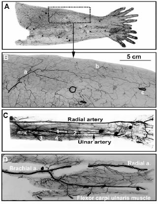
Fig. 3. Angiogram of the left upper limb obtained from
Fig 2D specimen. A,B.
The skin of the left upper limb
showing the territories of individual perforators. C,D.
The deep tissues of the left upper limb. The source
of the 4 musculocutaneous
perforators is marked for
correlation between figures 3A,B
and 3C,D.