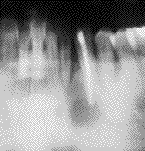
 1A
1A1A
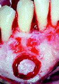 2A
2A
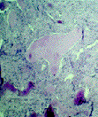 3A
3A
Fig. 1. Caso 1. 1A. Radiografía de la lesión periapical. 2A. Imagen clínica de
la intervención. 3A. Estudio anatomopatológico que muestra el estroma fibroso
y la presencia de material osteoide y calcificaciones distróficas.
Case 1. 1A. X-ray of the periapical lesion. 2A. Clinical aspect of the operation.
3A. Histopathological study that shows the fibrous conective tissue and the
presence of osteoid material and distrophic calcifications.