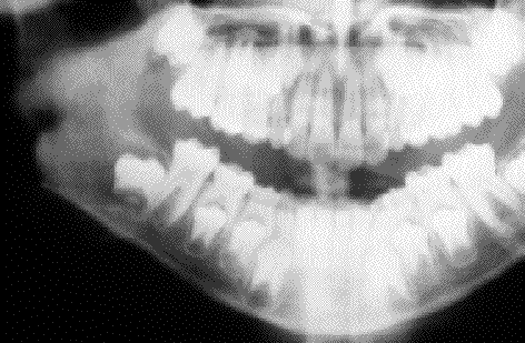
Fig. 1. Caso 9
A. Radiografía panorámica extraoral, en la que se aprecia una lesión radiotransparente
en ángulo y rama ascendente mandibular, de contorno bien delimitado.
A. Oral panoramic X-ray view showing a radiotransparent lesion at the mandibular angle
and ascending ramus, with well delimited contours.
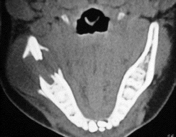
Fig. 1. Caso 9
B. tomografía computarizada, donde se aprecia la lesión ósea, que rompe las corticales lingual y vestibular.
B. Computed tomographic view of the bone lesion, which ruptures the lingual
and vestibular cortical layer.