


My SciELO
 Custom services
Services on Demand
Journal
Article
 text in
  English (pdf)
 Article in xml format
 Article references
 Send this article by e-mail
Indicators
-
 Cited by SciELO
 -
 Access statistics
Related links
-
 Cited by Google
 -
 Similars in
SciELO
Similars in
SciELO -
 Similars in Google
Share

 Permalink
Revista Española de Enfermedades Digestivas
Print version ISSN 1130-0108
Rev. esp. enferm. dig. vol.109 n.3 Madrid Mar. 2017
https://dx.doi.org/10.17235/reed.2016.4071/2015
CASE REPORT
Primary non-functioning neuroendocrine tumor of the extrahepatic bile duct
Tumor neuroendocrino no funcionante primario de la vía biliar extrahepática
Santiago Sánchez-Cabús1,2, Gabriella Pittau2, Mylène Sebagh3 and Daniel Cherqui2
1HPB Surgery and Transplantation Department. Hospital Clínic de Barcelona. Barcelona, Spain.
2HPB Surgery and Transplantation Department. Centre Hépato-Biliaire Paul Brousse. Villejuif, France.
3Histopathology Department. Centre Hépato-Biliaire Paul Brousse. Villejuif, France
ABSTRACT
In contrast to the primary biliary carcinoma or cholangiocarcinoma, other tumors derived from the bile duct are difficult to diagnose preoperatively, mainly because of its low incidence and difficult diagnostic process. However, since cholangiocarcinomas account for about 80% of all primary biliary tumors, it is important to think about other options despite their low frequency when a patient presents with abnormal characteristics. We present a case of a primary neuroendocrine tumor of the bile duct, and a review of the literature on this rare disease.
Key words: Neuroendocrine tumor. Biliary tract. Biliary neoplasia. Periampullary neoplasia. Cholangiocarcinoma.
RESUMEN
En contraste con el adenocarcinoma primario biliar o colangiocarcinoma, otros tumores derivados de la vía biliar son difíciles de diagnosticar preoperatoriamente, debido principalmente a su baja incidencia y el difícil proceso diagnóstico que requieren. Sin embargo, dado que los colangiocarcinomas suponen alrededor del 80% de todas las neoplasias biliares primarias, cuando se trata de un paciente con un tumor biliar con características anormales, es importante pensar en otras opciones, a pesar de su baja frecuencia. En este trabajo presentamos un caso clínico de un tumor neuroendocrino primario de la vía biliar, y una revisión de la literatura acerca de esta rara enfermedad.
Palabras clave: Tumor neuroendocrino. Vía biliar. Neoplasia biliar. Neoplasia periampular. Colangiocarcinoma.
Case report
A 38-year-old male patient consulted for a two-month history of abdominal discomfort associated with obstructive jaundice and asthenia in the last 20 days. CT scan and MRI cholangiogram showed a partially calcified 2 cm mass arising from the extrahepatic bile duct with intense contrast enhancement, causing retrograde dilatation of proximal biliary tree (Fig. 1 A and B), with no signs of local or distant extension. CEA, CA 19.9 and AFP levels were normal.
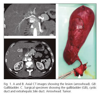
An en-bloc surgical resection of the tumor including the extrahepatic bile duct, cholecystectomy and regional lymphadenectomy was performed (Fig. 1C). The intraoperative examination of the surgical specimen suggested a neuroendocrine tumor (NET) of the common bile duct with clear margins. The postoperative course was uneventful, and the patient was discharged 6 days after surgery.
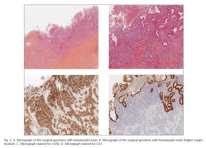
The final pathology report showed a 2 cm tumor with a fibrous stroma in its central part with small and homogeneous cells organized into pseudo-acinar formations. In the periphery, the tumor was composed of large cells with abundant microvacuolized cytoplasm showing rounded nucleus (Fig. 2 A and B). There was also perineural and lymphatic involvement. The final diagnosis was neuroendocrine carcinoma with high expression of CD56 and CK7 (Fig. 2 C and D, respectively), chromogranin and moderate expression synaptophysin, and with an index of Ki67 15% (G2), without lymph node involvement.
Discussion
Primary biliary tree NETs represent 0.2-1% of all digestive NETs (1,2). While functioning NETs of gastrointestinal origin account for 6-10% (3), clinical symptoms resulting from hormonal hypersecretion of biliary NETs are rare, and only a few cases have been reported (4). The most common anatomical site of this neoplasm is the common bile duct, which represents about 60% of cases reported (5). Surgical resection is the primary treatment.
References
1. Godwin JD. Carcinoid tumors. An analysis of 2,837 cases. Cancer 1975;36:560-9. [ Links ]
2. Welch JP, Malt RA. Management of carcinoid tumors of the gastrointestinal tract. Surg Gynecol Obstet 1977;145:223-7. [ Links ]
3. Feldman JM. Carcinoid tumors and syndrome. Semin Oncol 1987;14:237-46. [ Links ]
4. Price TN, Thompson GB, Lewis JT, et al. Zollinger-Ellison syndrome due to primary gastrinoma of the extrahepatic biliary tree: Three case reports and review of literature. Endocr Pract 2009;15:737-49. [ Links ]
5. Modlin IM, Shapiro MD, Kidd M. An analysis of rare carcinoid tumors: Clarifying these clinical conundrums. World J Surg 2005;29:92-101. [ Links ]
![]() Correspondence:
Correspondence:
Santiago Sánchez-Cabús.
HPB Surgery and Transplantation Department.
ICMDiM. Hospital Clínic de Barcelona.
Carrer de Villarroel, 170.
08036 Barcelona
e-mail: ssanche1@clinic.ub.es
Received: 30-10-2015
Accepted: 12-04-2016
