


My SciELO
 Custom services
Services on Demand
Journal
Article
 text in
  English (pdf)
 Article in xml format
 Article references
 Send this article by e-mail
Indicators
-
 Cited by SciELO
 -
 Access statistics
Related links
-
 Cited by Google
 -
 Similars in
SciELO
Similars in
SciELO -
 Similars in Google
Share

 Permalink
Archivos Españoles de Urología (Ed. impresa)
Print version ISSN 0004-0614
Arch. Esp. Urol. vol.62 n.4 May. 2009
Renal cell carcinoma in a horseshoe kidney
Hipernefroma en riñón en herradura
Javier Casasola Chamorro, Sonia Gutiérrez Garcia1 and Aitor Álvarez Dominguez1
Urology and Gynecology Department1. Complejo Hospitalario de Leon. Leon. Spain.
SUMMARY
Objective: We report a clinical case of renal carcinoma in horseshoe kidney and some anatomical particularities during surgery.
Methods: We describe the case of a 54 year old man who presented pain and prostatism. A renal tumor in horseshoe kidney was found in the diagnostic tests.
Results: We performed surgical treatment saving the left kidney.
Conclusions: Horseshoe kidney is the most frequent fusion abnormality of the kidney. Renal carcinoma in this type of renal fusion is similar to those with normal anatomy. Anatomical particularities should be taken into account during surgery.
Key words: Horseshoes kidney. Renal carcinoma.
RESUMEN
Objetivo: Aportar un caso de hipernefroma en un riñón en herradura y valorar el tratamiento revisando la literatura.
Métodos: Describimos el caso de un varón de 54 años que presentó dolor testicular y prostatismo, siendo diagnosticado en el estudio urológico de hipernefroma enriñón en herradura.
Resultados: Se realizó tratamiento quirúrgico conservándose el riñón izquierdo.
Conclusiones: El riñón en herradura es la más frecuente de las anomalías de fusión. El carcinoma renal en este tipo de riñones es similar a los riñones de anatomía normal. Sin embargo las particularidades anatómicas deben tenerse en cuenta en la cirugía.
Palabras clave: Riñón en herradura. Carcinoma renal.
Introduction
Horseshoe kidney occurs 1 to 400 borns with a frecuency 2 to 1 in male(1). It's the most frequent fusion abnormality of the kidney associated with aquired pathology like lithiasis, infection and hydronephrosis secondary to urinary obstruction (1,3), but 33% are asyntomatics. The incidence of renal carcinoma in these abnormal kidneys is similar to those with normal anatomy (1) and 85% are adenocarcinomas.
The surgical treatment is difficult about the isthmus anatomy and the vascular malformations. Us more isthmus parenchyma more difficult surgery, risk of kidney infarct and postoperative fistula.
Case report
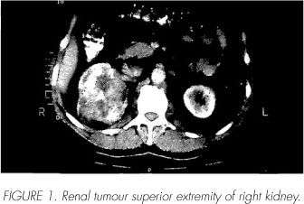
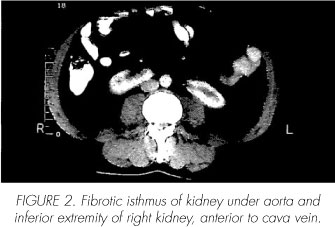
A 54 years old man without known diseases was referred to consultation with left orchialgia and trivial prostatism. The abdomen exploration was normal. There were no megalias. External genitalia were normal, with left testicular pain. Transrectal exploration was normal too. The abdominal ultrasound detect a tumour situated in right kidney. The computerized tomography shows a 8 cm tumour and the isthmus between left and right kidney. Tumour horseshoe kidney diagnosis was done.
Surgical transabdominal treatment right radical nephrectomy by anterior intercostal incision was performed. Arteries of the isthmus were ligated and divided. It was difficult surgery about three renal veins and two renal arteries. Pathological evaluation demostrate Fuhrman grade II renal cell carcinoma. PT2N0MX. Two years after surgery no signs of recurrent disease have been detected.
Discussion
Carcinoma originating in a horseshoe kidney is rare. Numerous vascular anomalies have been reported difficulting surgical treatment that must be us conservative us possible.
The isthmus is more frecuently inferior, and should have important parenchyma volume under inferior mesentery arteria and anterior to big vassels (1). The difficulty in surgical treatment of these tumours is the bilateral affectation of the kidney, no in our patient, the unexpected vascular anatomy in this malformation and the tumour size and localization and the anatomical relations with de big vasa. Vena cava extension (4) of such a tumour and the possibility of abdominal aortic aneurysm (5), more frequent in these patients, do necessary preoperative imaging studies like angiography. Ultrasonogra-phy is useful like screening technique bud urography, arteriography and tridimensional reconstructive compu-tericed tomography (6,7) offert us unexpected vascular anatomy images for preventing surgical complications. We must chose surgical route in order to avoid complications. The lumbotomy permits and easy removal of the kidney whit less infection and bowel complications (3). Transabdominal route is recommended because provides the best access at the isthmus and vascular plane (1,2,6). We must perform conservative surgery in all cases us possible. The accurate surgery and diagnosis of unexpected vascular anatomy offers to the patient best functional and medical prognosis.
Conclusions
Horseshoe kidney's anatomical particularities must be taken account during surgery, because difficult the treatment and must be diagnosed before surgery. We must use all our technology with angiography and computericed tomograghy to report all vascular anormalities in this renal malignancy malformation.
 Correspondence:
Javier Casasola Chamorro
Sto Toribio de Mogrovejo, 70 - 5o Iz.
24006 León. (España).
sguga66@yahoo.es
Accepted for publication: July 3rd, 2008.
References and recomended readings (*of special interest, **of outstanding interest)
**1. Andreú García A, Molina Burgos R, Coronel Sánchez B, Navio Perales J, Botella Almodóvar R, Llamazares Cachá G. Carcinoma renal del istmo en riñon en herradura. A propósito de un caso. Actas Urol Esp. 2008;32(2):249-252. [ Links ]
**2. Otero García JM, Maldonado Alcaraz E, López Sámano V. Carcinoma de células claras en riñón en herradura. Descripción de un caso y revisión de la literatura. Gac Med Méx 2005;141(4):305-307. [ Links ]
**3. Roa Rico M, Roa Luzuriaga M. Hipernefroma en el riñón en herradura: A propósito de dos casos. Ach Esp Urol.1984;37(4):365-372. [ Links ]
*4. Pettus JA, Jameson JJ, Stephenson. Renal cell carcinoma in horseshoe kidney with vena caval involvement. Urol J. 2004;171(1):339. [ Links ]
5. SilversteinAD, Weizer AZ, Anderson EE. Ruptured abdominal aortic aneurysm complicated by horseshoe kidney and renal carcinoma. Urology.2002;60(6):1108. Hellstrom p, Ottenli J, Siniluoto T, Paivansalo M, Kyllonen [ Links ]
6. A. Renal cell carcinoma in horseshoe kidney associated with urner syndrome and caval extension. Urology. 1989;34(1):46-48. [ Links ]
7. Pila Pérez R, Medrano López L, Ochoa Undargarin O, Pila Pelaez R, Ravelo Gutiérrez O. Hipernefroma en el riñón en herradura. Arch Esp Urol.1993;46(10):919-921. [ Links ]
*8. Bahayani SB, Andriole GL. Pure laparoscopic radical heminefrectomy and partial isthmusectomy for renal cell carcinoma in horseshoe kidney: case report and technical considerations. Urology. 2005;66:880-882. [ Links ]

