


My SciELO
 Custom services
Services on Demand
Journal
Article
 text in
  English (pdf)
 Article in xml format
 Article references
 Send this article by e-mail
Indicators
-
 Cited by SciELO
 -
 Access statistics
Related links
-
 Cited by Google
 -
 Similars in
SciELO
Similars in
SciELO -
 Similars in Google
Share

 Permalink
Archivos Españoles de Urología (Ed. impresa)
Print version ISSN 0004-0614
Arch. Esp. Urol. vol.62 n.7 Sep. 2009
Giant cystic degeneration of the rete testis
Degeneración quistica gigante de la rete testis
Luis A. Busto Martin, Daniel López García, lyadh Barghoutti, Venancio Chantada Abal and Luis Busto Castañón1
Hospital and University Complex of La Coruña. 1Urological Clinic Dr. Busto. San Rafael Hospital. La Coruña. Spain.
SUMMARY
Objectives: We present a case of giant cyst of the rete-testis.
Methods/Results: 85 year-old patient on follow -up for prostate cancer with maximum androgen blockade (MAB) treatment consults for a left hemiscrotum increase in size over a 2-month period. We performed bilateral orchiectomy confirming the histopathological diagnosis of cystic dilatation of the rete-testis sized 11x11x9cm.
Conclusions: This case of cystic degeneration of the rete-testis, with a size out of common (11x11x9cm versus medium size in the literature: 3x3x3cm), could be related with an androgen-estrogen misbalance caused by a MAB in a prostate cancer context.
Key words: Cyst of the rete-testis. Degeneration of the rete-testis. Giant testicle cyst.
RESUMEN
Objetivos: Presentar el caso de un quiste gigante de la rete testis.
Método/Resultado: Se trata de un paciente de 85 años que durante el seguimiento por un adenocarcinoma de próstata con bloqueo androgénico completo consulta por aumento del tamaño del hemiescroto izdo de dos meses de evolución. Se realizó una orquiectomía bilateral, confirmando en el estudio histopatológico de dilatación quística de la rete testis de 11x11x9cm de tamaño.
Conclusión: Este caso de degeneración quística de la rete testis, con un tamaño fuera de lo común (11x11x9cm, mientras que en la literatura refieren un tamaño medio de 3x3x3cm) podría estar relacionado con el disbalance andrógeno-estrogénico por el BAC en el contexto de un cáncer de próstata.
Palabras clave: Quiste rete testis. Degeneración rete testis. Quiste gigante de testículo.
Introduction
The rete-testis is a complex organization of interconnected ducts, situated in the upper part of the testicle, covered by a fibrous stroma and the albugineal tunica, frequent location of benign cystic changes. We can also differentiate three segments: the septal portion, formed by the straight tubules, the mediastinic portion, with plain ducts going along the hilium and extratesticular portion, where channels dilatate forming small cavities which end in the efferent ducts.
The benign cystic lesions of the rete testis are classified in four kinds basically: the cystic dysplasia, cystic adenoma, simple cyst and cystic transformation. All four are represented like scrotum mass, painless, palpable and translighted. Cystic dysplasia is a congenital pathology of infancy presentation, with 40 cases described until nowadays, resulting as a defect about the union of the rete testis tubules, originated in the gonad blastula, and the vas deferense, derivated from Wolff's duct (1-3), in 55% of the cases it's associated with ipsilateral kidney agenesis, it supposes a diagnose and therapeutic challenge, tending authors more recently to conservatory surgery of testicular parenchyma. Cystic adenoma of the rete testis is a rare lesion that could possible show up in different periods of life and occasionally it's difficult to distinguish histologically the cystic transformation of the rete testis. Simple cysts happen irregularly associated to hemodialysis, been both cases unusual.
The intrascrotum cysts (5) can be classified in two types: a) intraparenchimal cysts: simple testicle cyst, rete testis cyst, dermoid and epidermoid cysts and cyst from the albugineal tunic and b) extraparenchimal cysts: epi-didymal (congenital and acquired) and vaginal (very rare). The case we present corresponds with a cystic dilatation of the rete testis.
Case report
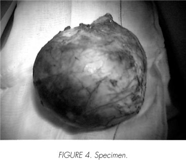
We present an 85 year's age male, with antecedent of prostate adenocarcinoma treated with LHRH analogues, maintaining and adequate biochemistry disease control, who consults for a picture longed 2 for 2 months of evolution, consisted in an increase of the size of the left hemiscrotum (Figure 1), being absent of pain, fever or other clinic. During the physic exploration we see an increased left hemiscrotum. We ask for a blood test, verifying a B-HCG and Alfa-FP inside the reference ranges, a testicular ultrasound (Figures 2 and 3) informed as: "big cystic mass leveled in the left scrotum. 10x8.3 cm sized, that compress and displaces the testicle, compatible with a big cyst dependent of the epididymis, but we can't reject other options like cystic adenoma". With the clinic information and look at the differential diagnosis and the patient situation, he is offered a bilateral orchiectomy, the right one subalbu-gineal and inguinal on the left one, which he accepts. The specimen (Figure 4) had a weight of 550 gr, sized 11x11x9 cm and presented a brown- hemorrhagic; in cuts it was evidence a cystic cavity that compressed the testicular parenchyma. The AP study that included cuts of the wall of the cyst and the rest of the test revealed a cystic lesion covered with flat epithelium, zones with ulceration, hemorrhage and necrotic tissue, testicular parenchyma had intense signs so atrophy, fibrosis and vascular congestion, with an absence of most of the germinal cells in the seminiferal tubules.


The AP diagnose was: a testicular benign cyst compatible with a cystic dilatation of the rete testis.
Discussion
The cystic transformation of the rete testis is a mainly adult injury. It has been found in a 1.6% of the autopsy studies and in surgery pieces obtained casually for other reasons, but rarely appeared clinically as a palpable mass and in occasions the diagnose is incidental in the image tests. Differing from the cystic dysplasia of the rete testis, it is not congenital, but acquired, so it is not associate with Muller's or Wolff's anomalities, and it is related with ambient factors, as malformative like cryptorchism, mechanical like a compression of the epidydimis of the spermatic duct from any cause, including neoplasic, infectious or atrophic changes in epididymal tubules induced to ischemia or hormonal disturbances, like in this case where we have an androgenic-estrogenic misbalance. Sometimes we can't find any cause. Anyway, rete testis's ducts are dilated, reaching a micro and macroscopic ectasia covered with plain epithelium, cubical or columnar methaplasia.
The rete testis's cyst is a benign lesion that can be suspected with a reasonable precision basing in image tests as ecography, MRN1,6, and the clinic data (cystic ectasia usually appears in males, older than 55 years and in 29-69% of the cases it is bilateral and asymmetric), cystic dysplasia and cystic degeneration of the rete-testis also differs in the therapeutic approach: back in the days, cystic dysplasia was treated with radical orchiectomy; nowadays the tendency is more conservatory, including surgery with preservation of the testicular parenchyma and even with an expectant observation in the cases where there is less grade of compression.


Conclusions
The main interest of this case consists on it's size (1 1x1 1x9cm), which rarely exceeds 3x3x3cm, never showing a similar sized in the published literature.
Otherwise we think that it could be produced by the androgen-estrogen misbalance produced by the CAB in the context of a PC.
It is mentioned the need of differential diagnose with other cystic degenerations, associated sometimes to other urogenital malformations
 Correspondence:
Luis Busto Castañón
Clínica Urológica Dr. Busto
Hospital San Rafael La Coruña (Spain)
lbm@urologiabusto.com
Accepted for publication: February 8th, 2009.
References and recommended readings (*of special interest, **of outstanding interest)
1. E. Easley S T, MacLennan G. Benign cystic lesions of the rete testis. J Urol, 2006; 176, 1622-1630 [ Links ]
2. Kajo K, Matoska J, Javorka K, Machalekova K, Tomaskin R, Climent K. Cystic dysplasia of the rete testes. APMIS, 2005; 113: 720-3. [ Links ]
3. Burns J A, Cooper C S, J. Austin C. Cystic dysplasia of the rete testis associated with renal agenesis and contralateral crossed ectopia. Urol, 2002; 60 (2): 344 XIV- 344XV. [ Links ]
4. Mac New H G, Terry N E, Fowler C L. Cystic dysplasia of the rete testis. J. Ped Surg, 2008; 43, 768-770. [ Links ]
**5. García-Morúa A, Triana Vazquez F, Gutierrez-García JD et al. Cistoadenoma de la rete testis: reporte de un caso y revisión de la literatura. Rev Mex Urol 2008; 68(5):296-298 [ Links ]
**6. F. J Amorós Oliveros, E. Cerezo López, J. Lemos Zunzunegui et at. Utilidad de la ecografía en el estudio del escroto. Medicina General 2001, No extraordinario: 97109 [ Links ]
*7. Pascual Mateo C, Fernández González I, Luján Galán M, et al. Ectasia quística de la rete testis. Arch Esp Urol, 2006; 59 (1):203-209. [ Links ]

