


My SciELO
 Custom services
Services on Demand
Journal
Article
 English (pdf)
 Article in xml format
 Article references
 Send this article by e-mail
Indicators
-
 Cited by SciELO
 -
 Access statistics
Related links
 Cited by Google
 -
 Similars in
SciELO
Similars in
SciELO  Similars in Google
Share

 Permalink
Oncología (Barcelona)
Print version ISSN 0378-4835
Oncología (Barc.) vol.27 n.9 Sep. 2004
ORIGINALES
Pronostic value of clinicopathologic factors Ki67, cyclin D1, cyclin D3 and CDK4 in gastric carcinoma
S. Valerdiz Casasola1; M. J. Menéndez Colunga2; O. Aller Millán3; J. M. Martínez Rodríguez3
1 Dept. of Pathology. Hospital de El Bierzo
2 S. of Urgencies. Hospital de El Bierzo
3 Dept. of Animal Pathology. Faculty of Veterinary. Universidad de León
SUMMARY
PURPOSE: To estimate the prognostic value of cyclin D1, Ki67, cyclin D3, cdk4 and classical clinicopathologic features of gastric carcinoma alone and in combination.
MATERIAL AND METHODS: We investigated the expression of cyclin D1, cyclin D3, cdk4 and Ki67 in gastric carcinoma. They were analyzed by immunohistochemical stain in formalin-fixed, paraffin-embedded tissue sections from 74 patients with gastric carcinoma, using automated methods. Immunostains, Ki67 proliferation index, histologic grade and histological type (Lauren classification) were compared with the length of survival.
RESULTS: Positive immunostaining was shown in 97% of the cases for Ki67, in 29% for cyclin D1, in 23% for cyclin D3, and in 35% for cdk4. On multivariate analysis only Ki67 (PI) (p<0.01) and pathologic grade (p<0.03) correlated with the length of survival. Expression of cyclin D3 was related with cdk4 (p<0.001) and Ki67 (PI) (p<0.02), and expression of cyclyn D1 was related with histologic grade (p<0.03).
CONCLUSIONS: Our results suggest that higher proliferation index of Ki67 and histological grade could be useful as markers of poorer prognosis. Over-expression of cyclin D1 could be an important role in cell proliferation. The relations between cyclin D3, cdk4 and Ki67 might be explained by the role of cyclin D3 and cdk4 in the cell cycle and the expression of Ki67 along the cellular cycle.
Key words: Gastric carcinoma. Ki67. Cdk4. Cyclin.
RESUMEN
PROPÓSITO: Conocer el valor pronóstico de la ciclina Dl, ciclina D3, cdk4 y Ki67, estudiados por métodos inmunohistoquímicos, junto con las características clinicopatológicos de los carcinomas gástricos.
MÉTODOS Y RESULTADOS: Realizamos estudio inmunohistoquimicos de material incluido en parafina para ciclina D1, ciclina D3, cdk4 y Ki67 en 74 pacientes con carcinoma gástrico. Las inmunotinciones para ciclinas D1, D3 y cdk4 así el índice de proliferación de Ki67, el grado histológico y el tipo histológico (según la clasificación de Lauren) se compararon con la supervivencia. El 97% de los casos eran Ki67 positivos, el 29% para ciclina D1, el 23% para ciclina D3 y el 35% para cdk4. El análisis multivariante sólo mostró correlación entre el Ki67 (PI) (p<0,01) y la supervivencia. En el análisis univariante el grado histológico también se correlaciona con la supervivencia (p<0,03). La expresión de ciclina D3 se relaciona con cdk4 (p<0,001) y Ki67 (PI) (p<0,02) y la expresión de ciclina D1 con el grado histológico (p<0,03).
CONCLUSIONES: Nuestros resultados sugieren que un índice de proliferación elevado de Ki67 y el grado histológico son marcadores de mal pronóstico. La sobreexpresión de la ciclina D1 podría tener un importante papel en la proliferación celular. La relación entre ciclina D3, cdk4 y Ki67 podrían explicarse por su papel a lo largo del ciclo celular.
Palabras clave: Ki67. Carcinoma gástrico. Ciclinas.
Introducción
Gastric adenocarcinoma is one of the main causes of cancer mortality in the world. It is still the more prevalent worldwide despite it's incidence is decreasing in Western countries. The world distribution is variable with an average prevalence of 45/10000 in Japan, Latin America and Eastern Europe; 15/100000 in USA, Australia and New Zealand, and Spain being in an intermediate situation.
From the histological and epidemiological point of view, gastric adenocarcinoma has been classified in two main typcs. Intestinal adenocarcinoma, well differentiated and expansive, is prevalent in populations with high rates of gastric carcinoma. On the contrary, diffuse or infiltrating adenocarcinoma is more frequent in populations with low incidence of gastric cancer. The etiology of the intestinal type is mainly associated with environmental factors, usually appearing as the result of a large multifactorial process with dietetic, social and economic factors being involved (Correa, 1992). In contrast, diffuse carcinoma is more dependent on genetic factors. Despite the amount of information on environmental factors and gastric cancer, little is know about genetic factors. Some authors have suggested that genetic susceptibility would predispose to gastric cancer to a minority of the population while environmental factors would accelerate tumor progression.
Cyclins are family of proteins that have been recognized. that plays, a role in the development of neoplasia. Under normal conditions, the progress of mammalian cells through the stages of the cell cycle is precisely coordinated by a group of related cyclindependent kinases and cyclins1-3. In general, cell cycle transitions are controlled by cdks4. Those holoenzymes contain both regulatory (cyclin) and catalytic (cdk) subunits but likely exist as higher order complexes that include additional proteins. Restriction point control is mediated by two families of enzymes, the cyclin-D and Edependent kinases. Whereas cdk4 and cdk6 are relatively long-lived proteins, the D type cyclins are unstable, and their induction, synthesis, and assembly with their catalytic partners, all depend upon persistent mitogenic signaling5. The D-type cyclins act as growth factor sensors, forming active kinases in response to extra cellular cues. The D-type cyclins reach maximal levels of expression and form functional kinases complexes with cdk4 or cdk6 during the mid-G1 phase6.
The D cyclin binds to cyclin-dependent kinases and proliferating cell nuclear antigen, by which the complexes formed are strangely implicated in the control of cell proliferation7. Particularly cyclin D1, expressed in early G1, and shown to be amplified, or over expressed in a wide variety of tumors7-11. In gastric cancer has been proved over expression of cyclin E, cyclin A and cyclin B1, but not cyclin D12.
Protein Ki67 (MIB1) is an antigen of proliferating cells and may prove a more robust marker of cell proliferation than PCNA (proliferating cell nuclear antigen)13. That expression occurs during G1, increases during the cycle cell and the rapidly declines after mitosis13. Among previously published studies of proliferations markers in gastric cancer, Ki67 labelling rate correlate with large tumor size, peritoneal metastases and advanced clinical stage14 or not15.
In order to increase the knowledge on the correlations between clinicopathologic and genetic factors in gastric cancer, we analysed, using clinical samples, the expression levels of nuclear markers associated with the cell cycle (cyclin D1, D3, cdk4, Ki67). Our aim is to establish the prognostic value of these markers. This work is part of a major project in which the molecular characterization of the tumour samples will be performed.
Materials and methods
Patients and tumor specimens
Samples from primary gastric carcinoma diagnosed between 1994 and 1999 were obtained from the files of Dept. of Pathology at Hospital de El Bierzo. The total patient number was 74, 51 (68.9%) men and 23 (31.08%) women with an average age of 68.2 years (range 35-91 years). All the material consisted of endoscopy biopsy. We examined hematoxylin and eosin-stained slides from each case to evaluate the tumour type according to the Lauren classification16 and the histological grade. Clinical data was obtained on each patient from the tumour registries.
Immunohistochemical analysis
Paraffin sections from each case with representative neoplastic tissue were carefully selected. Immunohistochemical staining procedures were performed on the Dako Autostainer (Dako, Carpinteria, CA) using antibodies listed in Table I. Sections were deparaffinized, rehydrated, and pretreated with buffer, pH 6.0. Primary antibody incubation time was 30 minutes. The detection system used was the LSAB+ kit from the Dako Corporation. Staining used was visualized with diaminobenzidine-cobalt chloride, resulting in a brown end product sections then wore counterstained with ethyl green pH 4.0

Proliferation index (PIS) were calculated by Ki67 (MIB-1) as percentage of positive nuclear cells among more the 1000 cells counted. Scoring of immunostaining was categorized as follows: zero, <10% cells stained; 1, 10-49% cells stained and 2, >50% cells stained. For cyclin D1, cyclin D3 and cdk4 the scoring of immunostained was categorized as follows: -, <10% cells stained and +, >10% cells stained.
Statistical analysis
The correlation of the immunostaining of each of markers was assessed with each of the others| markers and clinicopathological parameters, using the Fisher's exact test. Univariate and multivariate analyses for the prediction of length of survival by marker, histological type, histological grade, age, sex were performed using the Cox proportional hazards model. The impact of each prognostic variable on survival was also studied using the method of Kaplan and Meier. P<0,05 was assumed to be statistically significant.
Results
The majority of tumours, 67 (81,9%) were of intestinal type, whereas only 7 tumours (18,9%) were of the diffuse type. Twelve tumours (16%) were well-differentiated, 55 (74,6%) moderately differentiated and 7 (9,4%) poor differentiated. The follow-up time ranged from 92 days to 351 days, with a mean time of 252 days (Fig. 1). The mean age of the patients was 68 years (range, 35-91 years). Fifty-one (68,9%) were men, and 23 (31,08%) were women. By the time this study was performed, twelve patients were alive (16,2%), 57 (77%) had died and 5 (6,8%) failed to follow-up (Table II).


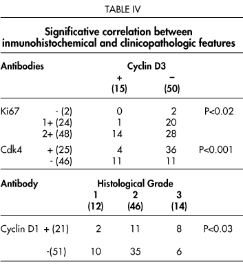
Of the 74 tumours studied, 97,3% positively expressed Ki67, 29,2% cyclin Dl, 23,1% cyclin D3 and 35, 3% cdk4 (Fig. 2 and 3). Ki67 (PI) was negative (score 0) for 2 tumours (2,7%), score 1 for 24 tumours (32,4%) and score 2 for 48 tumours (64,9%) (Table III).


Expression of CD3 were related with cdk4 (p<0,001) and Ki67 (p<0, 02). Significantly, correlation between other antibodies was not found. The patients with well differentiated gastric carcinoma revealed a relatively better prognosis with moderately and poor differentiated gastric carcinoma (p<0,02). Interestingly, all the patients who were alive at the end of this study showed well-differentiated adenocarcinomas (p<0,02). Age, histological type and sex were not related with survival. The expression of CD1 was significantly related with the histological grade (p<0,03) (Table 4).
Multivariate survival analysis was performed based on clinicopathologic factors and immunohistochemical expression of CD1, CD3, cdk4 and Ki67. The statistical results revealed that only Ki67 expression (PI) was independent and significant prognostic factor (p<0,01) (Fig. 4). On univariate analysis histological grade (p<0,03) (Fig. 5) was correlated with length of the survival.


Discusion
Gastric carcinoma occurs in patients throughout the world, although the incidence varies geographically. In Spain, gastric carcinoma accounts for approximately 3% of cancer deaths. Gastric adenocarcinomas can be divided histologically in two types, intestinal and diffuse16. The intestinal type can resemble colorectal carcinoma, being composed of tubular structures, with areas of solid growth or papillary pattern. The diffuse type is composed of individual cells or small groups of cells that infiltrate the gastric wall in a diffuse fashion. Cells of the diffuse type often have a signet ring appearance. Survival tends to be worse in the diffuse type, probably related to advanced stage at presentation. When matched stage for stage, however, there is no difference in survival between the two histological type12. The prognosis largely depends on the extent of penetration into or through the gastric wall, extension into adjacent structures, and the presence or absence of lymph node and/or distant metastases. As with histological type, neither size nor location is useful in predicting survival17.
In the present study, histological grade was correlated (in univariate analysis) with survival, when the tumour was classified into well, moderately and poor differentiation type (p<0,03). In addition, the histological grade was correlated with clinical situation of the end of study (p<0,02). The relationship between histological grade and survival is controversial. In two different studies using the same classification (well, moderately and poor differentiated) the correlation with survival is different. Shiu MH et al.17 considered that there is significantly correlation with survival, but in study of Roder J et al.18 there is not correlation. Likewise, in our study to be demonstrated that the majority of patients live at end of study are patients with diagnosed of welldifferentiated gastric carcinoma.
Multivariate analysis revealed that only Ki67 was independent prognostic factor. Prognostic significance of Ki67 has been reported in various cancers19. In gastric carcinomas, other studies15, 20 do not in agreement with our results. The difference with these studies may be explained by the difference in immunohistochemical and evaluation procedures.
Growth factors and cytokine positively or negatively regulate cell proliferation, cell differentiation and cell death or apoptosis though cell cycle operated by the CDKs and CDK inhibitors. Many positive growth factors are finally linked with cyclin D, whereas negative growth factor may act as a major inhibitor of cell proliferation though a cyclinCDK inhibitor p27Kip131,32 or p21Waf/1cip1. The D-type cyclins reach maximal levels of expression and form functional complexes with CDK4 during the mid-G1 phase. Therefore, the biological role of the D3-cyclin had been related that a possible dual role in cell proliferation and induction and/or maintenance of terminal differentiation21. In gastric cancers, cyclin D1 overexpression was associated with a nonsignet ring phenotype and one study22, with no pathological features in another study23 and with a signet ring cell phenotype and poor differentiation in other24. Our study demonstrated significant relation between cyclin-D1 and histological grade. Probably, because the cyclin D1 is more important in the cellular proliferations that other cyclin D type. Therefore, it is probable that expression of cyclin D3 is later to the cyclin D121 and would explain to statistical relation between cyclin CD3 and Ki67 since this later is expressed throughout the cellular cycle except G phase. The same relation between cyclin CD3 and cdk4 could be explained the role of the cyclin D3 in the maintenance of terminal differentiation and because cdk4 is relative long-lived protein with cyclins-D.
References
1. Matsushine H, Ewen ME, Strom DK, Kato JY, Hanks SK, Rosussel MF et al. Identification and properties of an atypical catalytic subunit (p34 psk-J3/cdk4) for mammalian D type G1 cyclins. Cells 1992; 71:323-34. [ Links ]
2. Sherr CJ. Mamalian G1 cyclins. Cell 1993; 73:1059-65. [ Links ]
3. Dunphy WG. The decision to enter mitoses. Trends Cell Biol 1994; 4:202-7. [ Links ]
4. Pardee HB. G1 events and regulation of cell proliferation. Science (Washington DC) 1989; 246:603-8. [ Links ]
5. Scherr ChJ. The Pezcoller Lecturer: Cancer cells cycles revisited. Cancer Research 2000; 60:3689-95. [ Links ]
6. Ohbu M, Kobayashi N, Okayasu I. Expression of cell cycle regulatory proteins in the multistep process of aesophageal carcinogenesis: stepwise over-expression of cyclin E and p53, reduction of p21WAF/C1P1 and dysregulation of cyclin D1 and p27KIP1. Histopathology 2001; 39:589-96. [ Links ]
7. Tahara E. Genetic alterations in human gastrointestinal cancers. The application to moleculardiagnosis. Cancer 1995; 75:1410-7. [ Links ]
8. Jiang W, Zhang YJ, Kahn SM, Hollstein M, Santela RM, Lu SH, et al. Altered expresion of the cyclin D1 and the retinoblastoma genes in human aesophageal cancer. Proc Nath Acad Sci USA 1993; 90:9026-30. [ Links ]
9. Leach FS, Elledge SJ, Sherr CJ, Wilson JKV, Markowitz S, Kinzler KW, et al. Amplification of cyclin genes in colorectal carcinomas. Cancer Res 1993 ;53:1986-9. [ Links ]
10. Giménez-Conti IB, Collet AM, Lanfranchi H, Itoiz ME, Luna M, Xu Hy, et al. P53, Rb and cyclin D1 expression in human oral verrucous carcinomas. Cancer 1996; 78:17-23. [ Links ]
11. Ito K, Sasano H, Yoshida Y, Sato S, Yajima A. Immunohistochemical study of cyclins D and E and dependent kinase (cdk) 2 and 4 in human endometrial carcinoma. Anticancer Res 1998; 18:1661-4. [ Links ]
12. Brien TP, Depowsky PL, Sheehan Ch, Ross JS, Mckenna B. Prognostic factors in gastric cancer. Mod Pathol 1998; 11:870-7. [ Links ]
13. McKormi K, Chong H, Hobbs C, Datta C, Hall PA, et al. Detection of the Ki67 antigen in fixed and waxembedded sections with the monoclonal antibody MIB1. Histopathology 1993; 22: 355-60. [ Links ]
14. Kimura H, Yonemura Y, Miyazaki I. Proliferative activity in gastric cancer determined with the cell cycle-related monoclonal antibodies Ki67 and plO5: analysis by flow cytometry. J Surg Oncol 1992; 51:174-8. [ Links ]
15. Broll R, Mahke C, Best R, Schimmelpenning H, Strik MW, Schiedeck T, Bruch HP, Duchrow M. Assessment of the proliferation index in gastric carcinomas with the monoclonal antibody MIB-1. J Cancer Res Clin Oncol 1998; 124:49-54. [ Links ]
16. Lauren P, The two histological main typcs of gastric carcinoma: diffuse and socalled intestinal type carcinoma. Acta Pathol Microbiol Scand 1965; 64:31-49. [ Links ]
17. Shiu MH, Perrotti M, Brennan MF. Adenocarcinoma of the stomach: a multivariate analysis of clinical, pathological and treatment factors. Hepatogastroenterology 1989; 36:7-12. [ Links ]
18. Roder J, B6ttcher K, Siewert R, et al. Prognostic factors in gastric carcinoma. Results ofthe German Gastric Carcinoma Study 1992. Cancer 1993; 72:2089-97. [ Links ]
19. Brown DC, Gatter KG. Ki67 protein: The immaculate deception? Histopathology 2002; 40:2-11. [ Links ]
20. Ramires M, David L, Leiato D, Seixas M, Sansonetty F, Sobrinho-Simoes M. Ki67 labelling index in gastric carcinomas. An immunohistochemical study using double staining for the evaluation of the proliferative activity of diffuse-type carcinomas. J Pathol 1997; 182;62-7. [ Links ]
21. Bartkova J, Lukas J, Strauss M, Bartek J. Cyclin D3: requirement for G1/S transition suggest a dual role in proliferation and differentiation. Oncogene 1998; 17:1027-37. [ Links ]
22. Müller W, Noguchi T, Wirtz H, et al. Expression of cell-cycle regulatory proteins cyclin D 1, Cyclin E and their inhibitor p21 WAF/CIP in gastric cancer. J Pathol 1999; 89:186-93. [ Links ]
23. Takano Y, Kato Y, Masuda M, et al. Cyclin D2, but not cyclin D1, overexpression closely correlats with gastric cancer progresión and prognosis. J Pathol 1999; 189:194-200. [ Links ]
24. Feakins RM, Nickols CD, Bidd H, Walton SJ. Abnormal expression of pRb, p16, and cyclin D1 in gastric adenocarcinoma and lymph node metastases:relationship with pathological features and survival. Hum Pathol 2003; 34:1276-82. [ Links ]
 Correspondence to
Dr. S. Valerdiz Casasola
Anatomía Patológica
Hospital de El Bierzo
E-24400 Ponferrada (León)
E-mail: svc@usuarios.retecal.es
Recibido: 20.04.04
Aceptado: 16.06.04

