My SciELO
Services on Demand
Journal
Article
Indicators
-
 Cited by SciELO
 -
 Access statistics
Related links
 Cited by Google
 -
 Similars in
SciELO
  Similars in Google
Share
Revista Española de Enfermedades Digestivas
Print version ISSN 1130-0108
Rev. esp. enferm. dig. vol.97 n.2 Madrid Feb. 2005
| PICTURES IN DIGESTIVE PATHOLOGY |
Bronchobiliary fistula secondary to biliary stricture after hepatectomy
J. A. Lucero Pizones, A. Iglesias López1, M. Alcázar Iribarren Marín1 and J. L. Márquez Galán
Departments of Gastroenterology and 1Radiology. Hospital Universitario Virgen del Rocío. Sevilla, Spain
CASE REPORT
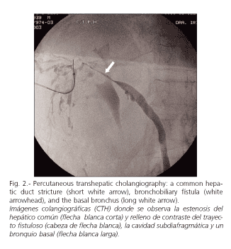
A 63-year-old woman with compensated HCV-induced liver cirrhosis underwent a right hepatectomy through thoracophrenolaparotomy due to a giant hydatid cyst (segments 6, 7, 8) in 1977, which developed an external biliary fistula complication from 1980 on. Twenty-six years after hepatectomy, at the time of external biliary fistula close, she showed expectoration of bile, and bronchoscopy discovered bile in the bronchial tree. Magnetic resonance cholangiography and percutaneous transhepatic cholangiography showed a common hepatic duct stricture, intrahepatic duct dilation, and bronchobiliary fistula. Due to its benign character, we decided to treat the stricture with dilation and placement of a bile drainage, thus solving the situation.


Bronchobiliary fistula is an uncommon condition usually seen in endemic regions as a complication of hydatid or amoebic disease (4). In Western society, bile obstruction, usually secondary to hepatectomy, is one of the most frequent causative factors (1,3). It may develop from days to years after hepatectomy (1). Bilioptysis is a very specific symptom. On computed tomography and magnetic resonance images, we may usually see both the fistula and the obstruction. Endoscopic retrograde cholangiopancretography and percutaneous transhepatic cholangiography may also allow therapeutic management, so they are considered the techniques of choice (1). The treatment strategy that is common for patients with bronchobiliary fistula and obstruction is the reposition of bile drainage into the duodenum, which allows the fistula to heal by reducing intrabiliary pressure. Surgical approaches should be considered only after failure of less invasive techniques. This is because reoperative procedures tend to be complicated with significant morbidity and mortality (1). Various techniques capable of bile drainage reestablishment are the placement of a nasobiliary drainage (2), endoscopic sphinterotomy, placement of a biliary endoprosthesis (1,3), dilation of stenotic bile duct, and embolization of biliary fistula with diverse embolic materials (coils, Histoacryl) (4).
REFERENCES
1. Rose DM, Rose AT, Chapman WC, Wright JK, López RR, Pinson CW. Management of bronchobililary fistula as a late complication of hepatic resection. Am Surg 1998; 64: 873-6.
2. Patrinou V, Dougenis D, Kriticos N, Polydorou A, Vagianos C. Treatment of postoperative bronchobiliary fistula by nasobiliary drainage. Surg Endosc 2001; 15: 758.
3. Oettl C, Schima W, Matz-Schimmed S, Fugger R, Mayrhofer T, Herold CJ. Bronchobiliary fistula after hemipatectomy: cholangiopancreaticography, computed tomography and magnetic resonance cholangiography findings. Eur Journal Radiol 1999; 32: 211-5.
4. Memis A, Oran I, Parilder M. Use of histoacryl and a covered nitinol stent to treat a bronchobiliary fistula. J Vasc Interv Radiol 2000; 11: 1337-40.


 text in


