


My SciELO
 Custom services
Services on Demand
Journal
Article
 English (pdf)
 Article in xml format
 Article references
 Send this article by e-mail
Indicators
-
 Cited by SciELO
 -
 Access statistics
Related links
-
 Cited by Google
 -
 Similars in
SciELO
Similars in
SciELO -
 Similars in Google
Share

 Permalink
Revista Española de Enfermedades Digestivas
Print version ISSN 1130-0108
Rev. esp. enferm. dig. vol.101 n.3 Madrid Mar. 2009
LETTERS TO THE EDITOR
Capsule endoscopy "retention" permits diagnosis of eosinophilic esophagitis
La "retención" de la cápsula endoscópica permite el diagnóstico de esofagitis eosinofílica
Key words: Capsule endoscopy. Complications. Eosinophilic esophagithis. Diagnosis.
Dear Editor,
Eosinophilic esophagitis (EE) is an inflammatory disease of the esophagus characterized by the presence of high number of eosinophils in the esophageal mucosal layer. Capsule endoscopy is a technology that allows direct visualization of the entire small bowel (1).
Case report
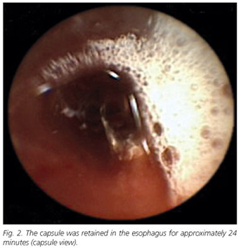
A 24-year-old man without a significant medical medical history developed insidious abdominal pain and diarrhea. He was initially treated empirically for traveler's diarrhea (ciprofloxacin); however, his symptoms continued to worsen. After fifteen days he was referred to the Gastroenterology Department to the refractory nature of his condition. A single stool culture and parasite examination and serologic evaluation for neuroendocrine tumors were negative; subsequent colonoscopy and upper endoscopy were normal. Two days after a capsule endoscopy (M2A capsule, Given Imaging) was performed to evaluate the small bowel. Immediately after ingestion of capsule endoscopy presented dysphagia followed by vomiting and sialorrhea. An upper endoscopy revealed a capsule retained in the distal esophagus (Fig. 1) for approximately 24 minutes (Fig. 2; Video 1). When tried to remove it with a Roth Net® (foreign body retriever) it progressed to the stomach, resulting in a small laceration in esophageal mucosa. Subsequent esophageal biopsies in the normal mucosa permitted the diagnosis of eosinophilic esophagitis.

Discussion
Eosinophilic esophagitis is a chronic inflammatory disorder of the esophagus in which the only presenting symptom in adults is a history of attacks of acute and recurrent dysphagia. Endoscopic findings are very subtle such as granularity, absent vascular markings, vertical furrowing, whitish exudates resembling fungal infection and sometimes esophageal strictures and rings are found, but generally no endoscopic sign is found (2). The diagnosis can be established only by microscopic examination: presence of more than 20 eosinophilic leukocytes per high-power field (3). EE lesions are not endoscopically specific and histological investigation is essential to the diagnosis. To our knowledge, this is the first report of capsule retention presumed secondary to eosinophilic esophagitis: follow-up studies demonstrated no other anatomic or pathologic cause for retention of the capsule endoscopy.
R. Ramos, J. Mascarenhas, P. Duarte, C. Vicente and C. Casteleiro
Department of Gastroenterology. Covilhã University Hospital. Covilhã, Portugal
References
1. Baichi MM, Arifuddin RM, Mantry PS. What we have learned from 5 cases of permanent capsule retention. Gastrointest Endosc 2006; 64(2): 283-7. [ Links ]
2. Khan S, Orenstein SR, Di Lorenzo C, et al. Eosinophilic esophagitis: strictures, impactions, dysphagia. Dig Dis Sci 2003; 48(1): 22-9. [ Links ]
3. Norvell JM, Venarske D, Hummell DS. Eosinophilic esophagitis: an allergist's approach. Ann Allergy Asthma Immunol 2007; 98(3): 207-14; quiz 214-7, 238. [ Links ]

