


My SciELO
 Custom services
Services on Demand
Journal
Article
 text in
  English (pdf)
 Article in xml format
 Article references
 Send this article by e-mail
Indicators
-
 Cited by SciELO
 -
 Access statistics
Related links
-
 Cited by Google
 -
 Similars in
SciELO
Similars in
SciELO -
 Similars in Google
Share

 Permalink
Revista Española de Enfermedades Digestivas
Print version ISSN 1130-0108
Rev. esp. enferm. dig. vol.102 n.2 Madrid Feb. 2010
PICTURES IN DIGESTIVE PATHOLOGY
Ectopic sebaceous glands in the esophagus
Ectopia esofágica de glándulas sebáceas
E. Marín-Serrano1, M. Jaquotot-Herranz1, L. Casanova-Martínez1, R. Tur-González2 and J. M. Segura-Cabral1
Services of 1Digestive Disease and 2Pathology. Hospital Universitario La Paz. Madrid, Spain
The discovery of sebaceous glands in ectoderm-derived tissues such as the oral cavity, parotid glands, and external genitalia is a frequent finding. On the contrary, the presence of sebaceous glands in the esophagus, which is an organ of endodermal origin, is rare: there are fewer than thirty cases reported, most of them affecting the esophagus in a patchy pattern.
Case report
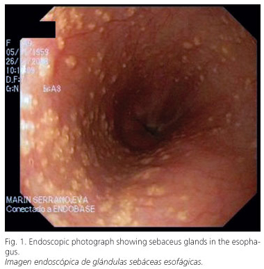
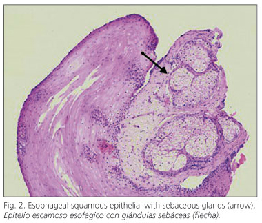
A 50-year-old woman suffering from heartburn and acid regurgitation was referred to our department for endoscopic examination. Endoscopy demonstrated more than one hundred tiny, rounded, elevated, white-yellowish lesions distributed throughout the entire esophagus (Fig. 1), which were identified during the histopathological examination as sebaceous glands (Fig. 2).
Discussion
As of now, the histogenesis of ectopic sebaceous glands in the esophagus is unknown; whilst it could be a congenital abnormality, a majority of authors defined it like an acquired metaplastic process.
From a practical standpoint, it is an incidental finding that on occasion, as in our case, has been associated with gastroesophageal reflux disease, not requiring treatment or endoscopic follow-up and, and needing a differential diagnosis from Candida infection and glycogen acanthosis.
Recommended references
1. Bae JY, Chon CY, Kim H. Sebaceous glands in the esophagus. J Korean Med Sci 1996; 11: 271-4. [ Links ]
2. Nakanishi Y, Ochiai A, Shimoda T, Yamaguchi H, Tachimori Y, Kato H, et al. Heterotopic sebaceous glands in the esophagus: histopathological and immunohistochemical study of a resected esophagus. Pathol Int 1999; 49: 364-8. [ Links ]
3. Tak AM, Scott RD, Khan NA. Sebaceous glands in esophagus in gastroesophageal reflux disease. Indian J Gastroenterol 2007; 26: 36. [ Links ]
4. Thalheimer U, Wright JL, Maxwell P, Firth J, Millar A. Sebaceous glands in the esophagus. Endoscopy 2008; 40 (Supl. 2): E57. [ Links ]
5. Wei IF, Chang CC, Fang CL, Hsieh CR, Wang JJ, Lou HY, et al. Education and imaging. Gastrointestinal: ectopic sebaceous glands in the esophagus. J Gastroenterol Hepatol 2008; 23: 338. [ Links ]

