


My SciELO
 Custom services
Services on Demand
Journal
Article
 English (pdf)
 Article in xml format
 Article references
 Send this article by e-mail
Indicators
-
 Cited by SciELO
 -
 Access statistics
Related links
-
 Cited by Google
 -
 Similars in
SciELO
Similars in
SciELO -
 Similars in Google
Share

 Permalink
Revista Española de Enfermedades Digestivas
Print version ISSN 1130-0108
Rev. esp. enferm. dig. vol.105 n.7 Madrid Aug. 2013
https://dx.doi.org/10.4321/S1130-01082013000700007
PICTURES IN DIGESTIVE PATHOLOGY
Retroperitoneal neurogenic tumor diagnosed by endoscopic ultrasonography
Tumor neurogénico retroperitoneal diagnosticado por ultrasonografía endoscópica
Nuno Veloso1, Pedro Figueiredo1, Pedro Pinto-Marques1, Ana Reis1, María José Brito2 and João Freitas1
Departments of 1Gastroenterology and 2Pathology. Hospital Garcia de Orta. Almada, Portugal
Introduction
The retroperitoneum can host a wide spectrum of pathologies, ranging from rare benign tumors to malignant neoplasms (primary or metastatic). Malignant tumors are four times more frequent than benign, being neurogenic tumors one of the most common benign pathologies (1).
Case report
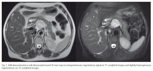
A 57-year-old woman presented to our department with a several-months history of right upper quadrant pain. Physical examination and laboratory evaluation was unremarkable. On abdominal ultrasound, a retroperitoneal solid mass adjacent to the posterior surface of the pancreatic neck was shown. To further characterize the lesion, an abdominal magnetic resonance imaging was performed which revealed a well-demarcated round 35 mm mass, located in retroperitoneum, below the emergence of the celiac trunk, shaping the posterior surface of the pancreatic neck (Fig. 1).
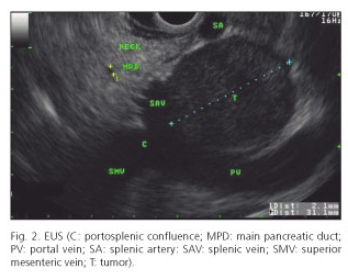
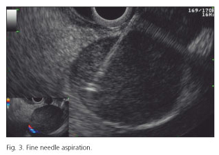
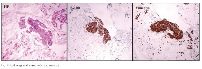
The patient was referred for an endoscopic ultrasonographic fine needle aspiration (EUS-FNA). A well delineated, Doppler negative, retroperitoneal 30 mm solid mass was seen, adjacent to posterior surface of the pancreatic neck at the level of the portosplenic confluence (Fig. 2). A EUS-FNA was performed (Olympus EZ-ShotTM; 22G; 4 passes) (Fig. 3). Cytology showed clusters of eosinophilic cells, spindle-shaped, uniform nuclei, without mitoses. Immunohistochemistry performed on the cell block was positive for S-100 and vimentin, compatible with neural tissue (Fig. 4).
Discussion
Tumors of the retroperitoneum are rare, and neurogenic tumors comprise only 1-10 % (2). Benign neurogenic tumors are discovered as an incidental finding during imaging for unrelated symptoms (3). There are no specific tumor markers or characteristic imagiology. EUS-FNA could be an effective and minimal invasive choice for the definitive diagnosis and tissue sample. Radiological surveillance in asymptomatic patients or surgical resections in symptomatic patients are the options for treatment.
References
1. Van Roggen JF, Hogendoorn PC. Soft tissue tumours of the retroperitoneum. Sarcoma 2000;4:17-26. [ Links ]
2. Lane RH, Stephens DH, Reiman HM. Primary retroperitoneal neoplasms: CT findings in 90 cases with clinical and pathologic correlation. AJR Am J Roentgenol 1989;152:83-9. [ Links ]
3. Strauss DC, Hayes AJ, Thomas JM. Retroperitoneal tumours: Review of management. Ann R Coll Surg Engl 2011;93:275-80. [ Links ]

