My SciELO
Services on Demand
Journal
Article
Indicators
-
 Cited by SciELO
 -
 Access statistics
Related links
-
 Cited by Google
 -
 Similars in
SciELO
 -
 Similars in Google
Share
Revista de Osteoporosis y Metabolismo Mineral
On-line version ISSN 2173-2345Print version ISSN 1889-836X
Rev Osteoporos Metab Miner vol.7 n.2 Madrid Apr./Jun. 2015
https://dx.doi.org/10.4321/S1889-836X2015000200005
Stress fracture in metatarsals: concerning two cases
Fractura de estrés en metatarsos: a propósito de dos casos
Del Río Martínez P.S.1, Moreno García M.S.1, Casorrán Berges M.P.1 and Baltanás Rubio P.2
1 Servicio de Reumatología - Hospital Clínico Universitario "Lozano Blesa" - Zaragoza
2 Servicio de Anestesia y Reanimación - Hospital Clínico Universitario "Lozano Blesa" - Zaragoza
SUMMARY
Stress fractures occur when a bone with normal elastic strength is subjected to higher loads than its mechanical strength. Although they may occur in any location they are more frequent in the metatarsals, these being the areas subject to greatest load. The clinical presentation for stress fractures is highly non-specific, which means that a detailed history is key to a suspected diagnosis. X-rays may be normal in the first stages, with gammagraphy and magnetic resonance being the gold standards for diagnosis in the initial stages. It is recommended that a study of possible underlying causes which may have contributed to the fracture is carried out. Generally the treatment is conservative, although in some cases, such as those occurring in the 5th metatarsal, surgical treatment may be necessary.
Key words: fracture, stress, metatarsals.
RESUMEN
Las fracturas de estrés se producen cuando un hueso con resistencia elástica normal es sometido a cargas superiores a su resistencia mecánica. Aunque pueden producirse en cualquier localización, son más frecuentes a nivel de metatarsos, al ser zonas anatómicas sometidas a mayor carga. La clínica de las fracturas de estrés es muy inespecífica, por lo que una historia detallada es clave para el diagnóstico de sospecha. La radiología puede ser normal en los primeros estadíos, siendo la gammagrafía y la resonancia magnética los gold standards para el diagnóstico en etapas iniciales. Es recomendable realizar un estudio de posibles causas subyacentes que hayan podido contribuir a la fractura. Generalmente el tratamiento es conservador, aunque en algunos casos, como en las localizadas en el 5o metatarsiano, puede ser necesario tratamiento quirúrgico.
Palabras clave: fractura, estrés, metatarsianos.
Introduction
Stress fractures occur when a bone with a normal elastic strength is subject to repeated force by tension or compression. We need to differentiate between stress fractures and fractures due to insufficiency, which are those which are produced by physiological tensions on a bone with reduced bone strength.
Stress fractures may appear in any location, being more common in the metatarsals (MTT), mainly in the neck of the 2nd and 3rd MTT. The clinical presentation and physical examination permit a suspected diagnosis, confirmed with an X-ray. However, in the initial stages the X-ray may be normal or inconclusive, which means that the carrying out of a CT, NMR or bone gammagraphy is necessary. Below, we describe two cases of stress fractures.
Case 1
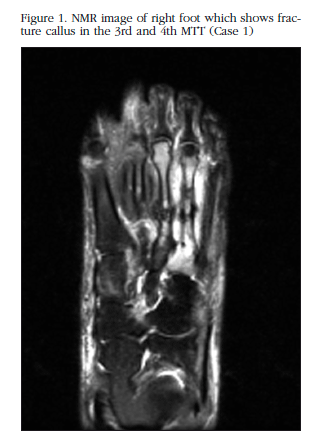
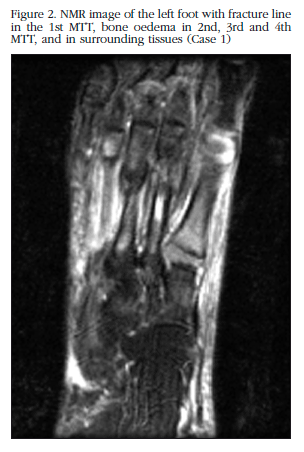
We describe the case of a female patient of 47 years of age being monitored due to psoriatic spondyloarthritis and fibromyalgia, in treatment with leflunomide, celecoxib and gabapentin, without history of interest and who maintained a regular menstrual cycle. She reported no toxic habits and her body mass index (BMI) was normal. In a routine examination she reported pain in her feet and ankles, with no history of trauma, with a mechanical rhythm, which had increased progressively until claudication occurred, with modification of the foot statics due to the pain. The examination highlighted the inflammation of the ankles and feet with pain from the pressure and bilateral fovea. An echography was carried out at the surgery which showed up a very marked inflammation of the subcutaneous cell tissue (SCT) with no signs of synovitis or Doppler signal.An X-ray was requested of the feet, which showed no pathological signs. Due to the significant SCT oedema, the patient was referred to angiology for assessment. From this service a lymphography was requested which confirmed severe bilateral lymphatic insufficiency. With the persistence of the symptoms of intense pain with claudication, the carrying out of an NMR of the feet was initiated, which showed in the right foot a fracture callus in the 3rd and 4thMTT and oedema in the 2ndMTT (Figure 1); and in the left foot a fracture line in the 1stMTT, and oedema in the 3rd and 4thMTT and in the surrounding tissue (Figure 2). Given the findings of the NMR, the patient was assessed by the traumatology service which indicated conservative treatment with non-weight-bearing and rehabilitation (magnet therapy). Due to the finding of multiple stress fractures, the study proceeded in our clinics, with analysis of renal function, calcium in blood and urine, ionic calcium, magnesium and PTH being carried out, which were normal. Only vitamin D was confirmed to be 19.5 ng/ml, for which treatment for supplements was indicated.
Assessing the case of this patient as a whole, we suggested, as a predisposing factor to the appearance of stress fractures, the significant antalgic alterations in foot statics which had developed due to the pain produced by the severe lymphatic insufficiency the patient had suffered.
Case 2
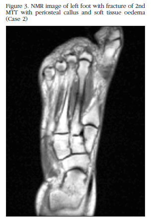
We describe the case of a 58 year old female patient. Her history includes a hysterectomy at 42 years of age due to metrorrhagia secondary to myoma. Two years before, she had been assessed by the gynaecology department due to densitometric lumbar osteoporosis (T-score in L1-L4 of -3, with normal figures in the femoral neck), treated with denosumab and vitamin D supplements. No toxic habits or personal or family history of fracture were reported, and her BMI was normal. She attended the clinic due to mechanical pain in the left foot, acute onset, without inflammation or triggering cause, which had increased in intensity until it became refractory to NSAIDs. In the examination there were no notable findings, except pain on the movement of the left forefoot. No alterations in the foot statics were observed. The patient was given an X-ray of the feet with showed no pathological signs. An NMR of the left foot was requested, which revealed a stress fracture in the 2nd MTT with periosteal callus and soft tissue oedema (Figure 3). An analytic study was carried out, which highlighted an increase in levels of PTH and vitamin D. (103.7 pg/ml and 272 ng/ml, respectively), attributed to an excess in the supplementation of vitamin D. Renal function and calcium in blood andurine were normal. Treatment with vitamin D supplements and denosumab were stopped. Assessed by the traumatology service, conservative treatment was indicated, with non-weight-bearing, relative rest, NSAIDs and magnet therapy, with progressive improvement. Due to the age of the patient, 58 years, and the predominance of osteoporosis in the lumbar region, the patient was considered an appropriate candidate for treatment with SERMs (bazedoxifene), associated with supplements of calcium with vitamin D. One year later, in the same month in which the pain started in the earlier episode, the patient again reported the same symptoms in the left foot, without a triggering cause. An X-ray was requested which showed a callus from an old fracture in the 2nd MTT due to an earlier stress fracture, with no other findings. An NMR was carried out of the left foot to complete the study, which showed oedema in the 1st and 3rd MTT, cuneiform, scaphoid and astragalus bones, and posterior tibial tenosynovitis. A new bone densitometry was requested which showed a T-score in the lumbar spine of -3.5. The patient had not been taking bazedoxifene and vitamin D continuously, so the importance of resuming them was emphasised given that the levels of bone mineral density had worsened. The fractures were treated with rehabilitation and non-weight-bearing with progressive improvement.
Assessing the case overall, we suggest osteoporosis as the predisposing factor, since the patient was not obese, nor had she presented trauma or other risk factors. The fact that the two episodes of pain started in the same month (coinciding with a change of season) with a year's difference, appears to us striking. The patient reported no change in her habits or in her state of physical activity (sedentary) at these times, which is why we consider that the change in type of footwear may have put an overload on the left foot causing the appearance of new stress fractures.
Discussion
The first description of stress fractures are attributable to Dr Briethaupt, who studied pain in the feet of recruits which worsened with standing and training. Stress fractures are located in the MTTs in 25% of cases, this being the area of greatest load [1]. Those located in the 2nd, 3rd or 4th MTT are considered to be low risk because they usually respond to conservative treatment, while those in the 5th MTT are of high risk [2,3] since they may require more aggressive treatment [4,5]. Although different causes have been suggested risk factors are considered to be [6,7,8]: anatomical anomalies (flat feet, dorsiflexion or plantar flexion of MTT, contracted gastrocnemius, excessively long 2nd MTT); physical anomalies, obesity, osteoporosis and related diseases, lack of exercise, muscular insufficiency and external factors (footwear, changes in the intensity or amount of training, change in the training surface).
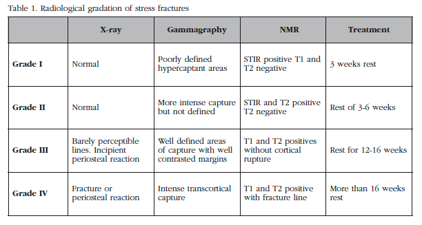
The diagnosis is based on a detailed clinical history which includes data on working and sporting habits. A stress fracture should be suspected in a case of foot pain which is poorly located and worsens gradually after the start of a new activity or very hard training weeks before the start of the pain. The radiological evidence does not usually appear before 2-6 weeks, cortical stretching and periosteal thickening and hypertrophy being the initial radiological signs. The degree of bone lesion may be of different intensity: bone contusion, cortical microfracture, extended periosteal microfracture and macroscopic transcortical fracture [9]. Gammagraphy and NMR are the "gold standards" for the initial diagnosis of cases, those in which X-ray tests may show normal findings. The classification of Arendt [10] correlates the histopathological studies with the imaging tests and treatment (Table 1). Grades I and II correspond to the stage of medullar oedema, grade III corresponds to periosteal changes and bone stress, and grade IV to clear cortical fracture.
The treatment is initially conservative, although in some cases, especially in fractures in the 5th MTT, surgical treatment may be necessary [11].
![]() Correspondence:
Correspondence:
Pilar del Río Martínez
Servicio de Reumatología
Hospital Clínico Universitario "Lozano Blesa"
Avda. San Juan Bosco, 15
50009 Zaragoza (España)
e-mail: psdelrio@yahoo.es
Date of receipt: 25/04/2015
Date of acceptance: 26/06/2015
Bibliography
1. Raghavan P, Christofides E. Role of teriparatide in accelerating metatarsal stress fracture healing: a case series and review of literature. Clin Med Insights Endocrinol Diabetes 2012;5:39-45. [ Links ]
2. Schmoz S, Voelcker AL, Burchhardt H, Tezval M, Schleikis A, Stürmer KM, et al. Conservative therapy for metatarsal 5 basis fractures-retrospective and prospective analysis. Sportverletz Sportschaden 2014;28:211-7. [ Links ]
3. DeVries JG, Taefi E, Bussewitz BW, Hyer CF, Lee TH. The fifth metatarsal base: anatomic evaluation regarding fracture mechanism and treatment algorithms. J Foot Ankle Surg 2015;54:94-8. [ Links ]
4. Lee KT, Park YU, Jegal H, Kim KC, Young KW, Kim JS. Factors associated with recurrent fifth metatarsal stress fracture. Foot Ankle Int 2013;34:1645-53. [ Links ]
5. Perron AD, Brady WJ, Keats T.A. Management of common stress fractures; when to apply conservative therapy, when to take an aggressive approach. Postgrad Med 2002;111:95-106. [ Links ]
6. Childers RL Jr, Meyers DH, Turner PR. Lesser metatarsal stress fractures: a study of 37 cases. Clin Podiatr Med Surg 1990;7:633-44. [ Links ]
7. Pegrum J, Dixit V, Padhiar N, Nugent I. The pathophysiology, diagnosis, and management of foot stress fractures. Phys Sportsmed 2014;42:87-99. [ Links ]
8. Shindle MK, Endo Y, Warren RF, Lane JM, Helfet DL, Schwartz EN, et al. Stress fractures about the tibia, foot, and ankle. J Am Acad Orthop Surg 2012;20:167-76. [ Links ]
9. Hatch RL, Alsobrook JA, Clugston JR. Diagnosis and management of metatarsal fractures. Am Fam Physician 2007;76:817-26. [ Links ]
10. Arendt EA, Griffiths HJ. The use of MR Imaging in the assessment and clinical management of stress reaction of bone in high-perfomance athletes. Clin Sports Med 1997;16:291-306. [ Links ]
11. Zwitser EW, Breederveld RS. Fractures of the fifth metatarsal; diagnosis and treatment. Injury 2010;41:555-62. [ Links ]


 text in


