


My SciELO
 Custom services
Services on Demand
Journal
Article
 text in
  English (pdf)
 Article in xml format
 Article references
 Send this article by e-mail
Indicators
-
 Cited by SciELO
 -
 Access statistics
Related links
-
 Cited by Google
 -
 Similars in
SciELO
Similars in
SciELO -
 Similars in Google
Share

 Permalink
Revista de Osteoporosis y Metabolismo Mineral
On-line version ISSN 2173-2345Print version ISSN 1889-836X
Rev Osteoporos Metab Miner vol.12 n.3 Madrid Jul./Sep. 2020 Epub Jan 25, 2021
https://dx.doi.org/10.4321/s1889-836x2020000300006
IMAGES IN OSTEOLOGY
Fibrous dysplasia mimicking rib metastasis
2Nuclear Medicine Service. University Hospital Virgen Macarena. Seville (Spain)
We present the diagnostic images of a 30-year-old woman, an asymptomatic BRCA1 mutation carrier and undergoing clinical-radiological follow-up for bilateral mammary fibroadenomas. The control MRI (Figure 1) highlighted the appearance of a nodular lesion posterior to the right breast prosthesis, relatively well defined and with lobulated contours. Given the suspicion of metastatic bone disease, a positron emission tomography (PET/CT) with 18F-fluorodeoxyglucose (18F-FDG) was carried out to assess its metabolic activity and extent of the disease. This was the only active lesion, with a 2.6 cm diameter and high metabolic activity, located in the fourth right costal arch (Figure 2). In this context, the lesion was excised to rule out neoplastic etiology. Pathology studies showed it was fibrous dysplasia, a benign and slowly progressive pseudotumoral disease, which represents less than 5% of bone tumors.
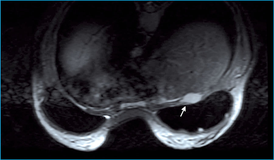
Figure 1. MRI showing nodular lesion in the right anterior thoracic wall (arrow), posterior to the breast prosthesis, with intense enhancement with contrast, intense rapid uptake and early lavage, compatible with a possible primary or secondary neo-formative nature, without ruling out other diagnostic options
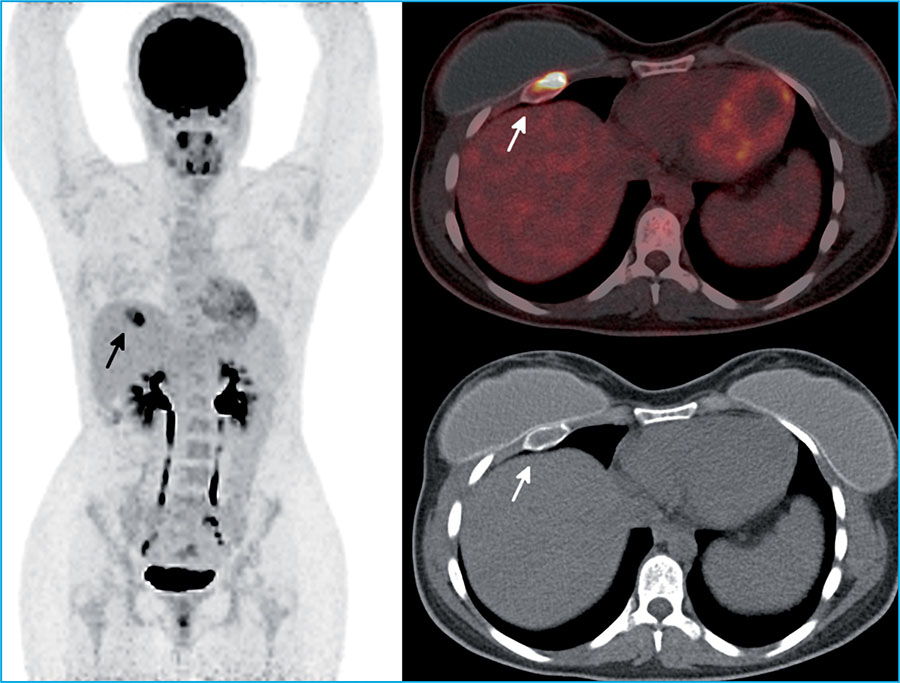
Figure 2. PET/CT with 18F-FDG, which highlights a single hypermetabolic lesion circumscribed to the anterior third of the fourth right costal arch (arrow), with very high metabolic activity (SUVmax:15.5)
Fibrous dysplasia is characterized by the replacement of normal bone tissue by osteofibrous connective tissue, adopting a sclerotic, cystic-lytic or mixed pattern1,2. The disease is due to an imbalance in the function of osteogenic cells, triggering expansive osteolytic lesions that affect adjacent normal bone and fibrous tissue1. The monostotic variant accounts for 70% of cases, and can be asymptomatic and detected incidentally2. In monostotic forms, the bones most frequently affected are, in decreasing order, the maxillae, the proximal femur, the tibia, the humerus, the ribs, the skull, the radius and the iliac1,2.
In its diagnosis, diagnostic imaging techniques will be jective determination of the extension, metabolic activity useful. As these lesions present a high rate of bone turno-and predicting the evolution of the disease3,4. When there ver, they will show high uptake in both bone scintigraphy are doubts concerning the diagnosis, a bone biopsy or exand PET/CT, making them crucial techniques in the ob-cision with mutational study may be carried out.
Bibliografía
1 Lacoma Latre EM, Sánchez Lalana E, Bescós Marín JM. Fibrous rib dysplasia. Med Clin (Barc). 2017;148(9): e51. [ Links ]
2 Florez H, Peris P, Guañabens N. Fibrous dysplasia. Clinical review and therapeutic management. Med Clin (Barc). 2016;147(12):547-553. [ Links ]
3 Collins MT, Kushner H, Reynolds JC, Chebli C, Kelly MH, Gupta A, et al. An instrument to measure skeletal burden and predict functional outcome in fibrous dysplasia of bone. J Bone Miner Res. 2005;20:219-226. [ Links ]
4 Tuncel M, Kiratli PO, Gedikoglu G. SPECT-CT imaging of poliostotic fibrous dysplasia. Rev Esp Med Nucl Imagen Mol. 2012;31:47-48. [ Links ]
Received: August 02, 2020; Accepted: September 15, 2020
 Este es un artículo publicado en acceso (Open Access) abierto bajo la licencia Creative Commons Attribution Non-Commercial, que permite su uso, distribución y reproducción en cualquier medio, sin restricciones siempre que sin fines comerciales y que el trabajo original sea debidamente citado.
