


 Custom services
 English (pdf)
 Article in xml format
 Article references
 Send this article by e-mail
 Cited by SciELO
  Cited by Google
  Similars in
SciELO
Similars in
SciELO  Similars in Google
 Permalink
Dear Editor,
Hepatic mucinous cystic neoplasms represent a challenging pathology due to their very low frequency, insidious presentation and unclear demography due to the limited cases reported to date. Surgical treatment is mandatory when possible to prevent recurrence and malignant transformation 1,2,3. The case presented herein has a peculiarity, mainly an increased level of CA 19-9 which has not been previously reported.
Case report
A 26-year-old female with a recurrent hepatic cyst and persistent abdominal pain had undergone two previous surgical interventions due to the cyst. Laboratory findings showed an increased level of CA 19-9 (94.6 U/ml); the rest of parameters, including liver function tests and blood count, were within the normal limits. An abdominal computed tomography (CT) scan revealed a hepatic cystic lesion of 176 x 297 x 208 mm in the left hepatic lobe, with an irregular and thickened wall and the presence of internal septa.
The patient underwent a left hemihepatectomy with the following intraoperatory findings: hepatic cyst which involved the II, III and IV segments. The histopathological study reported a hepatobiliary cystadenoma with mucinous epithelium and the presence of ovarian-like stroma. The patient was discharged three days after surgery.
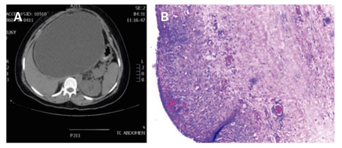
Fig. 1 A. CT scan showing a hepatic cyst measuring 176 x 297 x 208 mm in the left hepatic lobe with internal septa and a thickened wall. B. Microscopic view of hepatobiliary cystadenoma. The image shows the epithelium with a mucinoid appearance and stroma with submucosal increased cellularity that resemble ovarian stroma.
Discussion
Due to the initial suspicion, it was possible to establish the recommended treatment, which resulted in a remission of the symptoms and a recurrence free period of 12 months. Thus, this diagnosis must be taken into account in any patient with a recurrent hepatic cyst after surgical treatment. An imaging test, preferably a CT or magnetic resonance imaging (MRI), should be performed not only for their diagnostic value but also to aid the surgical plan. We also recommend to routinely measure CA 19-9 levels as this has been shown to be a valuable marker for the differential diagnosis with other hepatic cystic lesions.

