SciELO - Scientific Electronic Library Online

 
vol.12 issue3Vestibular rehabilitation in patients with vertigo and personality disorderTreatment of Dysphagia Oculopharyngeal muscular dystrophy. Systematic review author indexsubject indexarticles search
Home Pagealphabetic serial listing  

My SciELO

Services on Demand

Journal

Article

Indicators

Related links

  • On index processCited by Google
  • Have no similar articlesSimilars in SciELO
  • On index processSimilars in Google

Share


Revista ORL

On-line version ISSN 2444-7986

Rev. ORL vol.12 n.3 Salamanca Jul./Sep. 2021  Epub May 02, 2022

https://dx.doi.org/10.14201/orl.25399 

REVIEW

Frontal sinus foreign body. A systematic review

Cuerpo extraño en seno frontal. Revisión sistemática

Mahomed Sidique Abdul Cadar Dadá1  , Zulaikhah Mahomed Sidique Dadá2  , Abdul Habib Mahomed Dadá2 

1Servicio de Anatomía Humana. Facultad de Medicina. Universidad Eduardo Mondlane. Maputo. Mozambique.

2Instituto Superior de Ciências e Tecnologia de Moçambique (ISCTEM)

SUMMARY:

Introduction and objective:

Accidents with foreign bodies of all types have been described in most parts of the body. The presence of foreign bodies (FB) are common complaints in urgent and otorhinolaryngology consultations. The most common places are the natural head and neck cavities. The objective of this study is to know the situation of the foreign body in front sinus.

Method:

We conducted a search in PubMed, Cochrane, Scopus and WoS, selecting those articles in which the existence of foreign bodies in the frontal sinus was mentioned.

Results:

Of the 72 articles identified by means of information sources, 34 were excluded because they are not clinical cases, determining a selection of 38 clinical cases. Of these, 35 excluded for not complying with inclusion criteria. 3 Full-text articles assessed for eligibility. Of this, 2 Records excluded for not being related to the topic under study, only one case remaining.

Discussion:

Of the articles found there are few studies about foreign bodies located in frontal sinus. One of the first published cases dates from 1967, in Bristol, England.

Conclusions:

Accidents with foreign bodies are common in ENT clinical practice, however, in sinus location are very rare. The maxillary sinus is the most common site. Although the frontal sinus is more prominent on the face. Foreign bodies are rare. Most of them result from trauma by accident, gunshot or surgery or dental treatment. Accidents with foreign bodies are common in ENT clinical practice, however, in sinus location are very rare.

KEYWORDS: Frontal sinus; foreign body; systematic review

RESUMEN:

Introducción y objetivo:

Se han descrito accidentes con cuerpos extraños de todo tipo en la mayor parte del cuerpo. La presencia de cuerpos extraños (CE) es frecuentes en las consultas de urgencia y otorrinolaringología. Los lugares más comunes son las cavidades de cabeza Objetivo: Conocer las características de los cuerpos extraños en seno frontal.

Método:

Realizamos una búsqueda en PubMed, Cochrane, Scopus y WoS, seleccionando aquellos artículos en los cuales se menciona la existencia de cuerpos extraños en seno frontal.

Resultados:

De los 72 artículos identificados mediante fuentes de información, 34 fueron excluidos por no ser casos clínicos, determinando una selección de 38 casos clínicos. De estos, 35 excluidos por no cumplir con los criterios de inclusión. 3 artículos de texto completo evaluados para elegibilidad. De este, 2 Registros excluidos por no estar relacionados con el tema en estudio, solo queda un caso.

Discusión:

De los artículos encontrados existen pocos estudios sobre cuerpos extraños localizados en seno frontal. Uno de los primeros casos publicados data de 1967, en Bristol, Inglaterra.

Conclusiones:

Los accidentes con cuerpos extraños son frecuentes en la práctica clínica ORL, sin embargo, la localización sinusal es muy rara. El seno maxilar es el sitio más común. Aunque el seno frontal es más prominente en la cara. Los cuerpos extraños son raros. La mayoría de ellos son el resultado de un traumatismo por accidente, disparo o cirugía o tratamiento dental.

PALABRAS CLAVE: Seno frontal; cuerpo extraño; revisión sistemática

INTRODUCTION

Accidents with foreign bodies of all types have been described in most parts of the body. The presence of foreign bodies (FB) are common complaints in urgent and otorhinolaryngology consultations. The most common places are the natural head and neck cavities.

FB in the paranasal sinuses are extremely rare in clinical practice [1]. More than 50% of the sinus foreign bodies are located in the maxillary sinus. The incidence of a foreign body in the frontal, ethmoid and sphenoid sinuses is nearly equal [1]. Foreign bodies in the frontal sinuses are rare [2].

The foreign bodies found in the sinuses are retained roots of teeth, fragments of wood or bamboo, pieces of cotton or gauze, bullets, shrapnel fragments, knife blade and glass fragments [1].

The most frequent cause of the presence of FB in the paranasal sinuses is maxillofacial trauma (about 70 percent), followed by trauma resulting from dental treatment [1], displacement of tooth roots inwards of the maxillary sinus [3] and self-inoculation [4].

Due to its prominent location on the head, the frontal sinus is frequently involved in facial trauma [5], but despite its vulnerable position, the frontal sinus is not a frequent site of lodgment of foreign bodies [1, 6].

Foreign body penetration on frontal sinus is of particular apprehension because of the close proximity of the frontal lobe and duramater to the posterior sinus wall [5].

The existing literature on frontal sinus foreign body is limited [6].

The presence of foreign bodies should be suspected in lacerations of frontal region in cases of maxillofacial trauma [1], and so, in the case of trauma to the frontal sinus, it is important to carefully explore the cavity in search of any fragment of FB [7, 5].

The presence of frontal sinus FB can be asymptomatic and go unnoticed for years or have recurrent infections due to obstruction of the nasofrontal canal [6].

Frontal sinusitis can result from the entry of microorganisms together with the FB or by blocking the nasofrontal canal[8]

Access to the frontal sinus for removal of a foreign body typically needs an external ethmoidectomy approach or osteoplastic flap [8].

MATERIAL AND METHODS

Type of study: systematic review of the topic.

Research question: How foreign bodies appear in the human frontal sinus and which foreign bodies are more frequent?

Eligibility criteria: For the selection of studies, the following inclusion criteria were used:

  • Language: Portuguese, English or Spanish.

  • Publication date: 2010 to 2020.

  • Availability: full text.

  • Article type: case report.

  • Traumatism or iatrogenic events in human resulting in foreign bodies in the frontal sinus.

Exclusion criteria:

  • Repeated studies.

  • Publications out of context.

Information sources: The studies were retrieved from PubMed (https://www.ncbi.nlm.nih.gov/pubmed/), Cochrane (https://es.cochrane.org/es), Scopus (https://www.scopus.com/sources.uri) and Wos (https://www.recursoscientificos.fecyt.es/), Scielo (Scientific Electronic Library Online), from 01 January 2010 to 18 November 2020 (last date of search). The search included Portuguese, English and Spanish studies, following the PRISMA guidelines for systematic reviews (https://www.equatornetwork.org/reporting-guidelines/prisma/).

Period: Information sources were consulted between 24 December 2020.

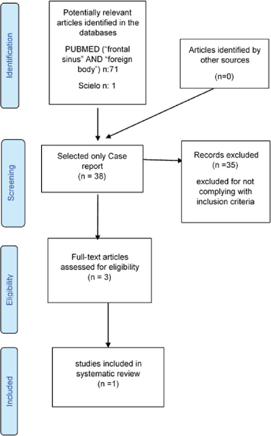
The search strategy and article selection are summarized in Figure 1.

Figure 1:  General description of the systematic literature review process. 

Search: "Frontal sinus" AND "Foreign body"

Study selection:

1st Stage - the titles and abstracts of the references identified through the search strategy were evaluated and the potentially eligible studies were pre-selected.

2nd Stage - the full text evaluation of the pre-selected studies was carried out to confirm the eligibility.

Data collection process:

Data were extracted by reviewer and checked by the remaining reviewers for accuracy. We used Rayyan® (Mourad Ouzzani, Hossam Hammady, Zbys Fedorowicz, and Ahmed Elmagarmid. Rayyan — a web and mobile app for systematic reviews. Systematic Reviews (2016) 5:210, DOI: 10.1186/s13643-016-0384-4; https://rayyan.qcri.org/welcome) for the selection and for the duplication extraction process.

Data items: The variables for which data were sought age, sex, symptoms, etiology and type of foreign bodies (Human, Sinusal trauma, Foreign body).

Risk of bias across studies: Since only 1 study were found eligible for this review, we considered there is a publication bias in this review and any conclusion from this study will only reflect this one scenario.

Ethical considerations: Being a research based on the literature review, it did not need prior ethical permission.

Weaknesses and limitations: Limited number of studies founded.

We used the PRISMA checklist when writing our report [9].

RESULTS

Of the 72 articles identified by means of information sources, 34 were excluded because they were not clinical cases, determining a selection of 38 clinical cases. Of these, 35 were excluded for not complying with inclusion criteria. 3 Full-text articles assessed for eligibility. Of this, 2 records excluded for not being related to the topic under study, only one case remaining.

DISCUSSION

Of the articles found there are only one study about foreign bodies located in frontal sinus from Ear, Nose & Throat Journal [10]. One patient of 34-year-old man with headache and fever for the past 7 days was diagnosed as having a comminuted fracture of the frontal sinus, where he had had a cranioplasty 15 years earlier with Methyl methacrylate to obliteration of the frontal sinus. The MRI suggested an abscess in the left frontal sinus (iatrogenic event). Methyl methacrylate is a monomer of acrylic resin usually used in a variety of medical, dental, applications.

The major limitation of this research is the number of articles found in a vast period of 10 years in three languages. However, in the gray literature we find many clinical cases.

CONCLUSIONS

Accidents with foreign bodies are common in ENT clinical practice, however, in sinus location are very rare. The maxillary sinus is the most common site. Although the frontal sinus is more prominent on the face. Foreign bodies are rare. Most of them result from trauma by accident, gunshot or surgery or dental treatment.

REFERENCES

1 Garces SM, Norris CW. Unusual frontal sinus foreign body. J Laryngol Otol. 1972;86(12):1265-8. [ Links ]

2 F Payne BR. Case reports Foreign bodies in the frontal sinus. Vol. 40, Br. J. Radial. 1967. [ Links ]

3 Morais HHA, Rocha NS, Gondim DGA, Melo AR. Corpo estranho no seio maxilar: relato de caso atípico. Rev Cir Traumatol Buco-Maxilo-Fac. 2007;7(1):65-70. [ Links ]

4 Lima MM, Moreira CA, da Silva VC, de Freitas MR. 34 Self-inflicted foreign bodies in the maxillary sinus. Braz J Otorhinolaryngol. 2008;74(6):948. [ Links ]

5 Kaplan AS, Green JDJ, McCaffrey TV. Unsuspected foreign body in the frontal sinus and anterior cranial fossa. Ann Emerg Med. 1989; 18(9):988-90. [ Links ]

6 Cukurova I, Demirhan E, Gumussoy M, Yalcin Y, Yigitbasi OG. Foreign body in frontal sinus: case report. Turk Otolarengoloji Arsivi/Turkish Arch Otolaryngol. 2013;51(4):123-6. [ Links ]

7 Cukurova I, Demirhan E, Gumussoy M, Yalcin Y, Yigitbasi OG. Foreign body in frontal sinus: case report. Turk Otolarengoloji Arsivi/Turkish Arch Otolaryngol. 2013;51(4):123-6. [ Links ]

8 Aydin S, Şanli A, Eken M, Hardal Ü. Glass particles in the frontal sinus. Turkish J Med Sci. 2009;39(2):313-5. [ Links ]

9 Calhoun KH, Peters BR, Stiernberg CM, Quinn FB. Magnet extraction of frontal sinus foreign body. Otolaryngol Neck Surg. 1988;99(1):76-8. [ Links ]

10 Moher D, Liberati A, Tetzlaff J, Altman DG, The PRISMA Group. Preferred reporting items for systematic reviews and meta-analyses: the PRISMA statement. PLOS Med. 2009 Jul 21;6(7):e1000097. [ Links ]

RELATED LINKS

Mourad Ouzzani, Hossam Hammady, Zbys Fedorowicz, and Ahmed Elmagarmid. Rayyan — a web and mobile app for systematic reviews. Systematic Reviews (2016) 5:210, DOI: 10.1186/s13643-016-0384-4; https://rayyan.qcri.org/welcomeLinks ]

Received: January 05, 2021; Accepted: January 27, 2021; preprint: September 01, 2021

motiar786@gmail.com

Conflict of interest: The authors declare no conflicts of interest

Images: The authors declare that the images have been obtained with the permission of the patients

Rights policy and self-archive: the self-archive of the post-print version (SHERPA / RoMEO) is allowed

Creative Commons License License CC BY-NC-ND. Creative Commons Attribution-Noncommercial-NoDerivate Works 4.0 International University of Salamanca. Its commercialization is subject to the permission of the publisher