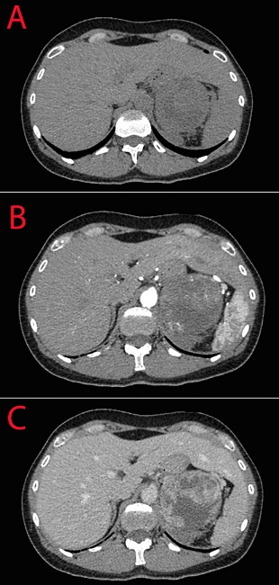
Anatomical variations of the liver are relatively understudied, while they deserve special attention in the day-to-day clinical practice [1, 2]. Beaver tail liver (also known as sliver of liver, sabre-shaped liver or falx-like liver) is a normal anatomical variation of liver development when an elongated left liver lobe extends laterally contacts and/or surrounds the spleen. The true incidence of this developmental variation is not known. It is seen more predominantly in females and usually encountered incidentally during routine ultrasound or computed tomography (CT) evaluation of the abdomen. However, it is a difficult imaging picture in setting of an acute abdomen or trauma. Due to similar density and echogenicity, the two organs may be hard to differentiate during examination. We present a clinical image of a 42-year old female admitted to the emergency department with abdominal pain irradiating to the back that began abruptly with no apparent reason. Her laboratory examination revealed moderate leukocytosis and anemia (leukocytes 12*109/l [normal range 4-9*109/l], erythrocytes 3,2*1012/l [normal range 3,8-5,2*1012/l], hemoglobin 101 g/l [normal range 120-150 g/l]). Her abdominal CT revealed adrenal mass with signs of intraparenchymal hemorrhage and beaver tail liver. Figure 1 demonstrates the difficulties in differentiation of the two organs that seem similar in the native and portal phases of examination and are different only in the arterial phase of the study. The patient undergone supraselective angiography of the adrenal artery, which demonstrated extravasation of contrast material. We performed selective catheterization of adrenal arteries and embolization of her adrenal mass. The postoperative period was uneventful and she was discharged 10 days after the procedure. Further evaluation revealed normetanephrine value 1180 mcg/d (normal value <600 mcg/d), she was consulted by an endocrinologist and scheduled for a planned adrenalectomy, which confirmed adrenocortical carcinoma. Beaver-like liver is an important anatomical variation as it can be mistaken for perisplenitis, subcapsular hematoma, and splenic mass [3, 4]. Abdominal trauma in the left hypochondrum region that usually lead to splenic injury can in turn affect the left lobe of the liver [5].


