INTRODUCTION
In the first half of the XX century, there was little interest in the spleen. However, in 1911 Micheli F. performed the first splenectomy for the treatment of hemolytic anemia, and in 1916, Kaznelson performed the same operation for the treatment of autoimmune thrombocytopenic purpura [1, 2]. These two operations give a new rise for the study of this organ, including its anatomy. Splenectomies are becoming widely practiced, but they have not always been performed according to strict guidelines. For decades 20-40% of splenectomies in large medical centers have been performed without proper clinical indications [3].
The spleen is located in the upper part of the abdominal cavity. As an organ, the spleen can have various shapes and size. The volume and mass of the spleen varies depending on the sex, the type of body constitution, the hematopoietic activity and the amount of blood stored in the spleen. The shape and size of the spleen change during ontogenesis. This is partially due to the pressure exerted by the adjacent organs on the spleen [4, 5].
Abdominal surgery is founded on a profound understanding of the anatomy including developmental variations [4]. To avoid some operative complications, we must take into account the diversity of shapes, surfaces, boundaries and dimensions of the spleen and its relationships with adjacent organs [5, 6]. Different splenic shapes and variations can lead to misdiagnosis particularly in patients with abdominal traumas.
MATERIAL AND METHODS
The human spleen was studied according to sex belonging to a group of 273 cadavers (154 men and 119 women), who did not have diseases of the spleen. The organs were fixed in a 10% formalin solution for seven days and then carefully dissected. The study was conducted according to the ethical laws of the institution and was approved by the ethical commission (19.08.2018 nr. 80). The shape of the spleen was analyzed based on splenic index (SI), which was defined as: width of the spleen divided by length of the spleen and multiplied by 100% (proposed by Inakov A.K., 1985). When the SI is less than 63% the spleen has elongated shape. If the SI is between 63% and 76% the shape is intermediate. When the SI is more than 76% the shape is round [11]. We also documented the shape of the spleen according to Michels classification (Figure 1) and other rare shapes (Figure 2) [5]. Statistical analysis was performed with the assistance of SPSS version 20 with the uses of modules of the program. The difference between means was assessed suing t-test. A p less than 0.05 was regarded as statistically significant.
RESULTS
Based on the available data (Table 1), the most common shape of the spleen in men is the elongated one. It was encountered in 79 (51.3%) observations out of 154 cases. In women, the most common shape was the intermediate. It was encountered in 51 (42.9%) of the 119 cases. The elongated shape was more common in males than in females (Table 1, p<0.05). There was no difference for intermediate and round shape between groups (Table 1, p>0.05).
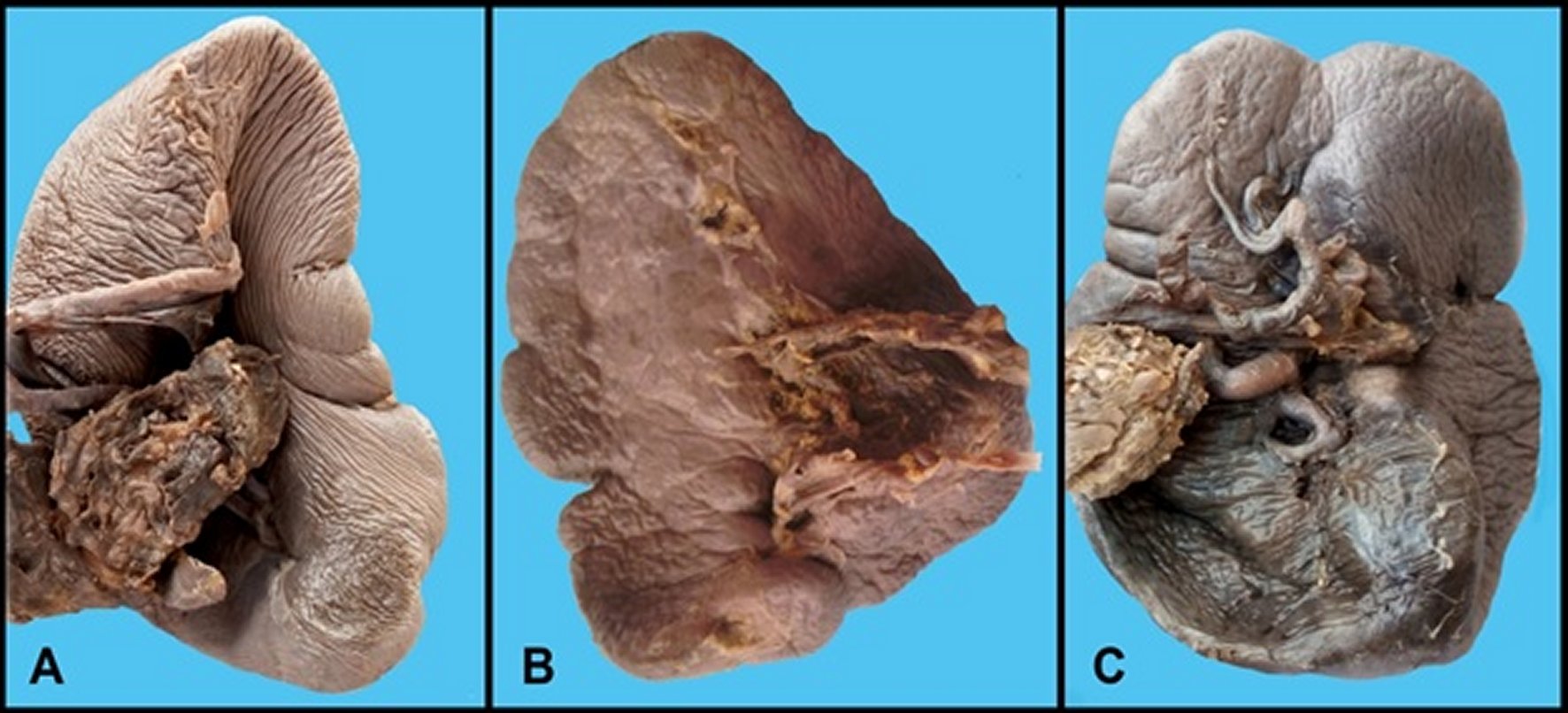
Based on Michels classification the clinoid (wedge) shape was encountered in 102 (37.74%) cases, triangular in 59 (21.83%) and tetrahedral in 30 (11.1%). There was no difference between sex (p>0.05). In 30.26% the shape of the spleen couldn't be classified according to Michels classification. These shapes encountered in our study are demonstrated in figure 2. In 21 cases (7.77%) the spleen had a flat shape; in 27 (9.99%) – dome-shaped; in 1 case (0.37%) – Z-shape; in 18 (6.66%) – round shape; in 6 (2.22%) – irregular shape; in 2 (0.66%) - shape with a node in the hilum; in 1 (0.37%) – rhomboid shape, in 2 (0.74%) – bilobed shape and in 4 cases (1.48%) – lobular shape.
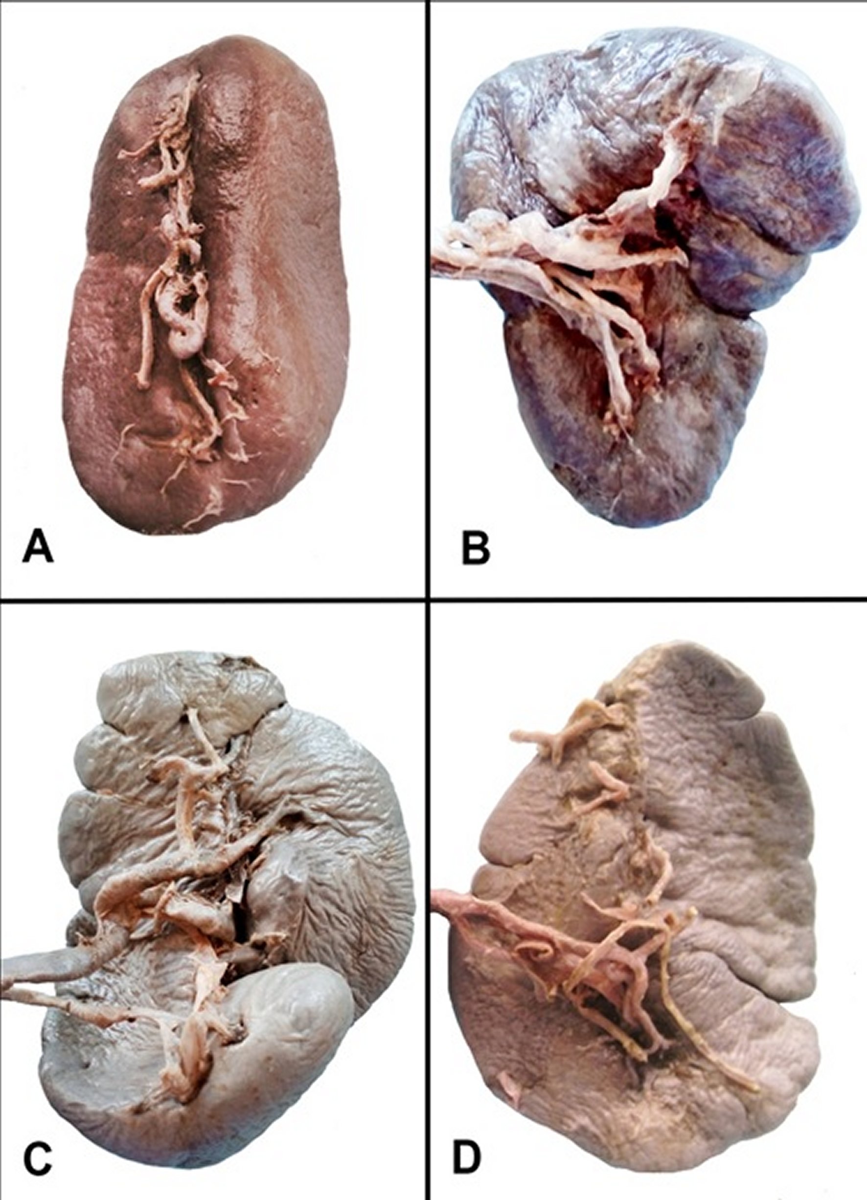
In our study, undertaken on 273 specimens, the splenic fissures located on the upper edge of the organ were found in 81 (29.91%) cases, and also on the lower edge - in 41 (14.02%) cases. In 13 (4.67%) cases fissures were encountered on both sides. In 148 (51.4%) cases the spleen had no fissures on its surface. The variability of splenic fissures is demonstrated in Figure 3.
DISCUSSION
Splenic variations and anomalies are important to consider for avoiding diagnostic pitfalls [7]. The vast introduction of imaging techniques leads to increased discovery of various anomalies and variations the often require further evaluation [8].
Different shapes of the spleen have been described in the literature. Inakov A.K. in 1985 proposed a formula and classification of the splenic shapes. He identified three main shapes based on the dimensions of the spleen. While studying the shape of the spleen in newborns, infants and children, he considered the shape of the spleen changes during lifetime. In the neonatal period, the intermediate shape of the spleen is the most common (76%), and the less common are the elongated shape (18%) and round shape (6%). During infancy, the prevalence of the intermediate shape decreases to 46%. At the same time, the elongated and round shapes are more common (36% and 18% of cases, respectively). During early childhood, the elongated shape of the spleen is more common (53%) and less common is the round shape (8%). At this age, the intermediate shape of the spleen was detected in 39% of cases [9, 10]. We also found that the round shape of the spleen is less common compared to elongated and intermediary.
Michels describes three shapes of the spleen: wedge, tetrahedral, and triangular and other authors support his findings. It seems that three splenic shapes are described in several studies: clinoid (44%), tetrahedral (42%) and triangular (14%) (Figure 1) [5, 11]. Similarly, we found the three most common shapes described by Michels. However, we also encountered several rare shapes. Authors report other splenic shapes which are encountered less frequently such as irregular shape (12.5%), dome-shaped (10%), oblong shape (7.5%) and a completely round shape (5%) [12]. Besides, the classical wedge, triangular and tetrahedral shapes of the spleen; we also encountered a flat shape, dome-shaped, Z-shaped, round shape, irregular shape, rhomboid shape, shape with a node, bilobed shape and lobular shape (Figure 2). Some of these shapes may be mistaken as tumors, lacerations and other pathological conditions, therefore requiring additional investigations.
The spleen with multiple lobes is a normal shape of the spleen and is not associated with any pathological condition. Deep fissures, sometimes called crenae splenis, can reach 2-3 cm in depth and are located mainly on the diaphragmatic surface and especially on the upper pole [7].
Splenic fissures located on the upper pole of the organ are found in 70-98% of cases. However, it is important to note that, although they are common, they have a different depth and morphology [13]. Splenic fissures located on the lower pole are less common and are seen in 2-32% of cases [7, 13]. We found similar results that fissures on the upper pole are almost two times more common than on the inferior surface. Sometimes, in patients with abdominal trauma, these lobulations can be misinterpreted as splenic lacerations. For this reason, it should be in mind that the anatomical shape of the splenic lobulation may be confused with a rupture of the spleen [14]. We do not consider accessory spleens as a distinct splenic shape as this is a separate morphological variation. However, accessory spleens seem to arise due to prominent lobulations and in the areas with deep fissures.
Such variations in the morphology of the spleen are remnants of the channels that initially separated the splenic lobes during fetal development. Rarely, splenic lobules may be associated with other congenital forms, such as the short pancreas, or may be misdiagnosed as renal or gastric tumors [9, 10, 15].
In healthy subjects the spleen is rarely palpable. However, it can enlarge in different conditions such as infectious (malaria), autoimmune (sarcoidosis), hematological (leukemia) and other. In this state it becomes palpable and in case of prominent lobulations seems as a multilobular thick mass [12]. Moreover, prominent splenic fissures can be misdiagnosed as lacerations in patients with abdominal trauma [16].
The lobulations and irregularity encountered in shape of some spleens might be attributed to the development of the spleen that arises from proliferation of the mesenchymal cells in-between the two layers of middle portion of dorsal mesogastrium. This divides the mesogastrium into two portions. Ventral part connecting the spleen with the stomach is called gastrosplenic ligament while the dorsal part called lienorenal ligament is shifted from the midline to the left side and connects it with the left kidney [17].
Most often, splenic abnormalities are noted incidentally during imaging studies performed for another indication. Therefore, clinician's knowledge of normal and abnormal findings helps to prevent unnecessary intervention or work-up for incidental or benign lesions [18].
CONCLUSIONS
The spleen has various shapes beyond the classical wedge, triangular and tetrahedral. There are various classifications based on dimensions and visual interpretation of the organ. All of these shapes do not represent a pathological finding but in certain situation may require further analysis and interpretation depending on the imaging technique and experience of the physician. Some of the variations of developmental can be mistaken as lacerations in trauma patients therefore require careful evaluation during diagnostic procedures.


