Mi SciELO
Servicios Personalizados
Revista
Articulo
Indicadores
-
 Citado por SciELO
 -
 Accesos
Links relacionados
-
 Citado por Google
 -
 Similares en
SciELO
 -
 Similares en Google
Compartir
Revista Española de Enfermedades Digestivas
versión impresa ISSN 1130-0108
Rev. esp. enferm. dig. vol.103 no.9 Madrid sep. 2011
https://dx.doi.org/10.4321/S1130-01082011000900011
LETTERS TO THE EDITOR
Diagnosis, treatment and follow-up of gastric carcinoid tumors. Analysis of 14 cases
Diagnóstico, tratamiento y seguimiento de los tumores carcinoides del estómago. Estudio de 14 casos
Key words: Gastric carcinoids. Diagnosis. Treatment. Follow-up.
Palabras clave: Carcinoide gástrico. Diagnóstico. Tratamiento. Seguimiento.
Dear Editor,
Gastric carcinoids represent 3 per thousand of all types of gastric cancer. They are classed as type I, associated with chronic atrophic gastritis (CAG) or pernicious anaemia; type II, associated with multiple endocrine neoplasia (MEN) or Zollinger-Ellison syndrome; and type III, sporadic. Between 9-30% of patients with type I and II carcinoids develop metastasis, while in type III the rate is 54-66% (1). Further, it is important to follow-up patients with CAG and pernicious anaemia with hypergastrinaemia, given the relatively high risk of developing synchronous or metachronous carcinomas (4-5% of cases), and polips or carcinoids (4-11%) in the stomach (2,3).
The objective of this letter is to analyse retrospectively the clinical characteristics and progression of 14 patients (7 men and 7 women) with carcinoid tumours in the stomach, examined between 1996 and 2009 in our unit. In all the patients a pathological study was performed, by immunohistochemistry, including analysis using chromogranin, synaptophysin and Ki-67 the proliferation marker.
Cases
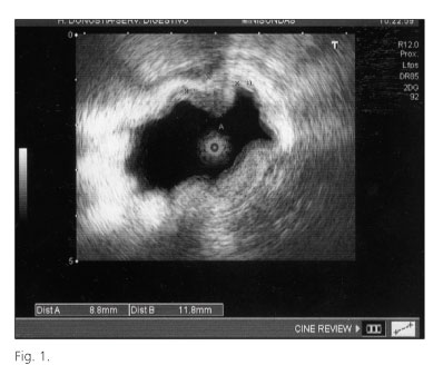
The mean age of patients in the series was 59 years and 4 months (range 42-82 years). The most common clinical finding was dyspepsia, with onset around 15 years earlier (10 patients). Anaemia and/or melena were observed in two patients, dysphagia in one and carcinoid syndrome with asthenia and weight loss in another. Three patients had history of hypothyroidism, suprarenal adenoma and multinodular goitre respectively. The most relevant blood test result was pernicious anaemia in four of the patients (29%). Gastroscopy and endoscopic ultrasound (7.5 MHz linear array and 12 and 20 MHz radial mini-probes) were used as diagnostic tests in 14 and 10 patients, respectively. Carcinoids developed in 13 patients who had chronic atrophic gastritis with intestinal metaplasia but no H. pylori. The gastroscopy showed that in 9 cases there were multiple masses (5 with all masses less than 1 cm in diameter and 4 with at least one of 1-2 cm), and 5 cases a single mass (3 with a mass larger than 1 cm, 1 with a mass of 1.5 cm and another with one of 7 cm, corresponding to a patient with sporadic carcinoid with liver and peritoneal metastases), all located in the fundus and/or body of the stomach. In four cases there were ulcers and in another case a hyperplastic polyp of 4 cm. Endoscopic ultrasound confirmed that the lesions were hyperechogenic and involved the gastric mucosa and submucosa (Fig. 1).
Mucosectomy was performed in 12 patients, using the rubber-band technique or a polypectomy snare. Of these, two patients underwent mucosal resection using a cap-fitted endoscope due to new lesions being observed during the follow-up. Gastric resection was performed in four cases given the large number and size of the carcinoids or suspicion of local and regional involvement. No invasion of neighbouring structures was detected by laparotomy. The follow-up period ranged from 1 to 10 years. Other carcinoids were found during the endoscopic follow-up of patients at 16 and 66 months, respectively.
Discussion
In 1923, von Askanazy reported the first case of carcinoid tumours of the stomach (4). Their known prevalence has increased in recent decades due to endoscopic monitoring carried out in people aged above 50 years old with atrophic chronic gastritis. In a recent American study, based on 120,000 gastroscopies carried out over one year period, the relative prevalence of gastric carcinoids was 0.58% (46 cases). Of these cases, half of the patients had chronic gastritis and/or intestinal metaplasia (47.8 and 52.2%, respectively), four were associated with hyperplastic polyps and another one with an inflammatory fibroid polyp (5).
Carcinoids develop from enterochromaffin-like cells (ECL) that regulate the production of hydrochloric acid by the secretion of histamine. In subjects with CAG and pernicious anaemia there is permanent hypo or achlorhydria that stimulates antral G cells, which produce gastrine. Hypergastrinaemia acts on the ECL cells causing, first, simple hyperplasia, then adenomatous hyperplasia and, finally, intramucosal carcinoid tumours. Types I and II are associated with hypergastrinaemia and tend to be multiple, while cases of type III without elevated levels of gastrin are seldom multicentric (6).
The size and number of lesions, the depth of invasion of the gastric wall, vascular and lymphatic involvement, mitotic index, the proliferation marker Ki-67, and the histological grade are determinants when assessing the suitability of resection of these tumours using gastroscopy or surgery. Endoscopic mucosal resection is recommended in patients with type I and II carcinoids that have hypergastinaemia and lesions less than 1 cm in diameter, while for lesions larger than 2 cm and if there is local involvement, total gastrectomy or just antrectomy (type I, pernicious anaemia) are advisable. There is no consensus on 1-2 cm tumours and both treatments are used. In type III, with a single 1-2 cm tumour with carcinoid syndrome and tendency to develop metastasis, surgery is generally advised. Endoscopic mucosal resection is performed using a variety of techniques, namely, the strip-off biopsy, polypectomy snares, the rubber-band technique and resection using a tube or cap-fitted endoscope (7). The 5-year survival among those with type I carcinoids is very similar to that of the general population, while in type II cases it is similar to patients with gastrinoma/MEN-1 (60-75% of the cases) and in type III is less than 50%.
To conclude, in this series there was a predominance of type I gastric carcinoids, and there was only one case with type III carcinoids with widespread metastasis. In most patients, endoscopic mucosectomy using various different techniques was carried out at diagnosis or during the follow-up period.
Alazne Aguirre1, Ángel Cosme1*, Luis Bujanda1*, José E. Navascués2,
Santiago Larburu2, Mikel Larzabal3, Inés Gil1 and José Ignacio Asensio2
Department of 1Gastroenterology, 2Surgery and 3Pathology. Hospital Donostia. San Sebastian.
*CIBEREHD. Universidad del País Vasco. San Sebastián, Guipúzcoa. Spain
References
1. Xie SD, Wang LB, Song XY, Pan T. Minute gastric carcinoid tumor with regional lymph node metastasis: A case report and review of literature. World J Gastroenterol 2004;2461-3. [ Links ]
2. Kokkola A, Sjöblom SM, Haapiainen R, Sipponen P, Puolakkainen P, Järvinen H. The risk of gastric carcinoma and carcinoid tumors in patients with pernicious anaemia. A prospective follow-up study. Scand J Gastroenterol 1998;3:88-92. [ Links ]
3. Chan JCW, Liu HSY, Kho BCG, Sim JPY, Lau TKH, Luk YW, et al. Pernicious anemia in chinese. A study of 181 patients in a Hong Kong Hospital. Medicine (Baltimore) 2006;85:129-38. [ Links ]
4. Von Askanazy M. Zur Pathogenese der Magenkarzinoide und inhren gelegentlichen Ursprung aus angeborenen epithelialen Keimen in der Magenwand. Dtsch Med Wochenschr 1923;49:642-7. [ Links ]
5. Carmack SW, Genta RM, Schuler CM, Saboorian HS. The current spectrum of gastric polyps: a 1-year National Study of over 120.000 patients. Am J Gastroenterol 2009;104:1524-32. [ Links ]
6. Moriayama T, Matsumoto T, Hizawa K, Eski M, Iwai K, Yao T, et al. A case of multicentric gastric carcinoids without hypergastrinemia. Endoscopy 2003;35:86-8. [ Links ]
7. Muro N, Cosme A, Múgica F, Alzate LF, Bujanda L. Tratamiento de un tumor carcinoide gástrico tipo I mediante resección mucosa endoscópica con cabezal. Gastroenterol Hepatol 2009;32:533-4. [ Links ]


