Mi SciELO
Servicios Personalizados
Revista
Articulo
Indicadores
-
 Citado por SciELO
 -
 Accesos
Links relacionados
-
 Citado por Google
 -
 Similares en
SciELO
 -
 Similares en Google
Compartir
Revista Española de Enfermedades Digestivas
versión impresa ISSN 1130-0108
Rev. esp. enferm. dig. vol.108 no.1 Madrid ene. 2016
ORIGINAL PAPERS
Comparative study between open and minimally invasive approach in the surgical management of esophageal leiomyoma
Comparativa entre el abordaje abierto y el mínimamente invasivo en el tratamiento quirúrgico del leiomioma esofágico
Diego Ramos, Pablo Priego, Magdalena Coll, María de los Ángeles Cornejo, Julio Galindo, Gloria Rodríguez-Velasco, Francisca García-Moreno, Pedro Carda and Eduardo Lobo
Division of Esophagogastric, Bariatric and Minimally Invasive Surgery. General Surgery Department. Hospital Unviersitario Ramón y Cajal. Madrid, Spain
ABSTRACT
Introduction: Leiomyomas are the most common benign tumors of the esophagus. Although classically surgical enucleation through thoracotomy or laparotomy has been widely accepted as treatment of choice, development of endoscopic and minimally invasive procedures has completely changed the surgical management of these tumors.
Material and methods: We performed a retrospective review of all esophageal leiomyoma operated at Hospital Universitario Ramón y Cajal (Madrid, Spain) between January 1986 and December 2014, analyzing patients' demographic data, symptomatology, tumor size and location, diagnostic tests, surgical data, complications and postoperative stay.
Results: Thirteen patients were found within that period, 8 men and 5 women, with a mean age of 53.62 years (range 35-70 years). Surgical enucleation was achieved in all patients. In 8 cases (61.54%) a thoracic approach was performed (4 thoracotomies and 4 thoracoscopies), and in 5 cases (38.56%) an abdominal approach was performed (3 laparotomies and 2 laparoscopies); enucleation was carried out through a minimally invasive approach in 6 patients (46.15%). There were no cases of endoscopic resection alone. Surgery mean length was 174.38 minutes (range 70-270 minutes) and median postoperative stay was 6.5 days (range 2-27 days). There was neither mortality nor cases of intraoperative complications were described. No postoperative major complications were reported; however one patient presented important pain in his right hemithorax that required management and long term follow-up by the Pain Management Unit. With a mean follow-up of 165.57 months (median 170; range 29-336 months) no recurrences were reported.
Conclusion: Enucleation is the treatment of choice for the majority of esophageal leiomyomas. In our experience, duration of the surgical procedure through minimally invasive approach was longer than surgery through open approach; however, postoperative stay was shorter in the first group. Paradoxically, incision pain after surgery (thoracic neuralgia) was found to be higher in the minimally invasive approach group. Nevertheless, none of the results obtained in the study reached statistical significance, probably due to the small simple size.
Key words: Esophageal leiomyoma. Enucleation. Laparoscopy. Thoracoscopy. Endoscopy.
RESUMEN
Introducción: el leiomioma es el tumor benigno más frecuente del esófago. Aunque clásicamente, el tratamiento de este tipo de tumores ha consistido en la enucleación por medio de una laparotomía o toracotomía, el auge de las técnicas endoscópicas y mínimamente invasivas ha revolucionado totalmente el manejo terapéutico de este tipo de tumores.
Material y métodos: realizamos un estudio retrospectivo de todos los leiomiomas esofágicos intervenidos en el Hospital Universitario Ramón y Cajal entre el 1 de enero de 1986 y el 31 de diciembre de 2014, analizando características demográficas de los pacientes, sintomatología, localización tumoral, pruebas diagnósticas, datos quirúrgicos, complicaciones y estancia hospitalaria.
Resultados: encontramos un total de 13 pacientes, siendo 8 varones y 5 mujeres, con una edad media de 53,62 años (rango 35-70 años). El tratamiento quirúrgico fue en todos los casos una enucleación. En 8 casos (61,54%) se realizó un abordaje torácico (4 toracotomías y 4 toracoscopias) y en 5 casos (38,56%) el abordaje fue abdominal (3 laparotomías y 2 laparoscopias). La enucleación se llevó a cabo a través de un abordaje mínimamente invasivo en 6 pacientes (46,15%). No hubo ningún caso de resección puramente endoscópica. La media de duración de la cirugía fue de 174,38 minutos (rango 70-270 minutos) y la mediana de estancia hospitalaria de 6,5 días (rango 2-27 días). No se describió ningún caso de mortalidad ni complicación intraoperatoria, aunque un paciente presentó importante dolor en hemitórax derecho que requirió manejo y seguimiento por la unidad del dolor. Con un seguimiento medio de 165,57 meses (mediana 170; rango 29-336 meses), no se han observado recidivas.
Conclusiones: la enucleación constituye el tratamiento de elección en la mayor parte de los leiomiomas esofágicos. En nuestra experiencia, la duración de la cirugía es mayor tras cirugía mínimamente invasiva (CMI) que tras cirugía abierta (CA), sin embargo, la estancia media hospitalaria es menor. Paradójicamente, en valores absolutos, las complicaciones relacionadas con el dolor de la herida quirúrgica (neuralgia torácica) son mayores en el grupo de CMI. Sin embargo, ninguno de los resultados obtenidos en el trabajo es estadísticamente significativo, seguramente debidos al escaso tamaño muestral.
Palabras clave: Leiomioma esofágico. Enucleación. Laparoscopia. Toracoscopia. Endoscopia.
Introduction
Esophageal leiomyomas are the most common benign tumors of the esophagus (1). Incidence is variable, fluctuating from 0.005 to 5.1%, based on different necropsy series (2,3). They are more often found in men, usually between 20 to 50 years old, and are usually located in the lower two-thirds of the esophagus (4).
Traditionally, they have been uniformly classified with other tumors as gastrointestinal stromal tumors (GISTs), but late evidence in the immunochemistry field has shown that they are two different entities (5).
Symptoms, usually non-specific and long-lasting, do not seem to be related to tumor size. Most of them are asymptomatic and, when symptoms appear, the most common are dysphagia, heartburn and retrosternal pain (3,4).
The treatment of these tumors is, in most cases, surgical enucleation, which would be indicated in big or symptomatic tumors, or tumors that show growth after initial observation (6). Traditionally, surgical excision has been performed through an open approach (thoracotomy or laparotomy) (7); however, the boom of endoscopic and minimally invasive procedures has completely changed the therapeutic management of these tumors (6,8-10).
The aim of this study is to perform a retrospective analysis of our experience at Hospital Universitario Ramón y Cajal in the treatment of these tumors, comparing the results between open and minimally invasive approaches, and trying to set a management algorithm for this kind of tumors.
Material and Methods
A retrospective study of all operated esophageal leiomyomas at Hospital Universitario Ramón y Cajal (Madrid, Spain) from January 1986 to December 2014 was performed, analyzing the patients' demographic data, symptomatology, tumor size and location, diagnostic evaluation, surgical data, complications and postoperative stay.
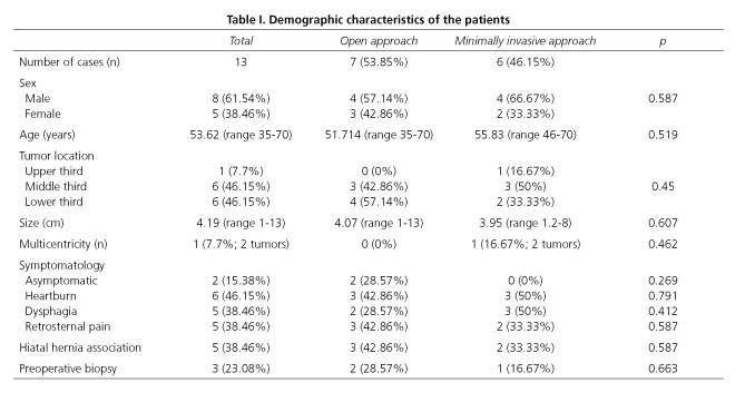
Patients were divided in two groups depending on the surgical approach, either open surgery (OS) or minimally invasive surgery (MIS). Patients' demographic data are shown in table I, being both groups homogeneous and comparable, with no statistically significant differences in any of the variables studied.
Twenty esophageal leiomyomas were diagnosed within that period of time. Five of them were subcentimeter and asymptomatic tumors, accidentally found in diagnostic procedures performed for other causes, and observation with regular follow-up was decided. Among the 15 esophageal leiomyomas surgically excised, one was excluded because the tumor was an accidental discovery in the pathology specimen after total gastrectomy for gastric stump cancer, and another one was also excluded when the definitive pathologic diagnosis reported an esophageal duplication cyst.
Surgical procedure, both for OS and MIS was already described by the authors in previous articles (11,12).
In the present study, a therapeutic algorithm is proposed (Fig. 1), based in our institution's experience and results previously published by other authors. Hereby, treatment of esophageal leiomyoma is indicated in tumors larger than 1 cm and in all symptomatic cases; endoscopic management for small size tumors is also proposed as an alternative to surgery (and particularly in tumors that arise from the muscularis mucosae) and only for experienced groups. We do not have much experience in endoscopic submucosal resection at our institution, so surgical enucleation was performed in all cases.
All statistical analyses were performed using IBM SPSS Statistics 22.0 (IBM SPSS Inc., Chicago, IL). Categorical data are presented as frequencies, and were compared by χ2 tests; when expected frequencies fell below 5 in any of the contingency tables, Fisher's exact test was performed. Continuous data are described by the arithmetic mean and the range, or by the median for asymmetric distributions with extreme scores; they were compared using the Mann-Whitney U test for independent groups, as the sample showed a non-normal distribution. p < 0.05 was considered statistically significant.
Results
A total of thirteen patients were analyzed, 8 men (61.54%) and 5 women (38.46%), with a male-female ratio of 1.6:1. Mean age was 53.62 years, ranged from 35 to 70 years old.
Regarding to the tumors' location, leiomyomas were found in one patient in the upper esophageal third (7.7%), in 6 patients in the middle esophageal third (46.15%), and in the other 6 patients in the lower esophageal third; thus, the most common location was the distal two-thirds of the esophagus.
Mean size of the tumors was 4.19 cm in their long axis (range 1-13 cm).
Definitive anatomopathological study showed esophageal leiomyoma in all 13 cases, with no evidence of leiomiosarcoma degeneration areas in any of the specimens. Single lesion tumors were found in 12 patients (92.3%); meanwhile, only one patient (7.7%) presented two simultaneous lesions (both of them in the same esophageal area).
Clinically, only 2 patients (15.38%) were completely asymptomatic and the tumors were found during diagnostic procedures for other diseases (peptic ulcer and rectorrhagia); on the other hand, the remaining 11 patients (84.62%) presented with different kind of symptoms, being the most frequent heartburn (46.15%), dysphagia (38.46%) and retrosternal pain (38.46%).
All patients were studied with an esophagogastroduodenoscopy (EGD); indeed, most of them were also studied with an upper gastro-intestinal endoscopy (PES) (12 patients, 92.31%), and a thoraco-abdominal computerized tomography (CT) (8 patients, 91.54%). Endoscopic ultrasound was performed in five patients (38.46%), becoming more usual within the group of patients diagnosed during the last years. Other diagnostic studies performed were abdominal ultrasound (23.07%), pulmonary function tests (23.07%; only among patients who underwent thoracic approach), manometry, and 24 hour pH monitoring (23.07% and 15.38% respectively, in patients who suffered gastro-esophageal reflux symptoms).
Biopsy was performed prior to surgery in three cases (23.08%), two of them ultrasound-guided and the remaining one CT-guided, showing no evidence of tumor in any of the samples obtained.
Five patients presented simultaneously a hiatal hernia (38.46%).
Regarding to surgical treatment, eight patients (61.54%) underwent a thoracic approach (4 thoracotomies and 4 thoracoscopies), and the other five patients underwent an abdominal approach (3 laparotomies and 2 laparoscopies). There were no conversion cases to open surgery (OS) in the thoracic approach group; on the other hand, one patient who presented a 13x4x2.5 cm distal-esophageal leiomyoma and whose procedure started through laparoscopy had to be converted to OS (laparotomy) due to technical difficulties in the tumor dissection caused by its size. Thus, conversion rate to OS was 14.29% (1 patient within a total of 7 attempts at performing a MIS procedure). There were no endoscopic resection alone cases.
When surgical approach evolution of esophageal leiomyoma at our institution was analyzed, a new trend was clearly observed for MIS. Thus, prior to year 2000, 100% of esophageal leiomyoma (4 cases) underwent OS, while 80% of the cases after that year underwent MIS procedures, the tumor excision being achieved though that approach in all cases except the one previously explained.
Tumor enucleation was achieved in 100% of the cases, with no need of esophageal resections. No cases of mucosal injury during enucleation occurred; mucosal integrity was intraoperatively confirmed in six cases by lumen insufflation with air (with the esophagus submerged underwater) or methylene blue, and in five cases performing an intraoperative endoscopy.
Muscular layer was re-approximated in all cases with non-absorbable interrupted suture.
An antireflux procedure was associated in 5 cases (38.46%); a Dor fundoplication was performed in three cases, and a Nissen fundoplication in the remaining two cases.
Mean length of surgical procedure was 174.38 minutes (range 70-270 minutes), with a mean length of 160 minutes (range 70-205 minutes) in the OS group and 217.5 minutes (range 165-270 minutes) in the MIS group. No statistically significant differences were observed between both groups (p = 0.402).
Median postoperative stay was 6.5 days (range 2-27 days), with a median postoperative stay of 7 days (range 3-14 days) in the OS group, and 4 days (range 2-27 days) in the MIS group, although no statistically significant differences were observed between both groups (p = 0.469).
There were neither intraoperative complications (0%) nor perioperative deaths (0%).
Regarding to postoperative complications, one case of wound infection was described after a thoracoscopic approach (7.7%) that eventually developed a local neuralgia and required long-term follow-up by the Pain Management Unit. As a late complication after an OS procedure (laparotomy), one patient required re-operation months later due to a small-bowel obstruction caused by abdominal adhesions.
However, no difference was observed in postoperative major complications or mortality in any of the groups (0%).
With a mean follow-up of 165.15 months (median 170; range 29-336 months) no tumor recurrence was reported (Table II).
Discussion
Leiomyomas are the most common benign esophageal tumors. They show variable incidence, fluctuating from 0.005 to 5.1% based on different necropsy series (2,3).
They are more frequent in men, by a ratio of 2:1, usually between 20 and 50 years old at diagnosis, and can be multifocal in 3-10% of the patients (4). The results obtained in our series were similar to those previously published, with a mean age of 53.62 years at diagnosis and a male predominance of 1.6:1; moreover, multifocal tumors were detected in 7.7% of the patients.
Due to their origin in smooth-muscle cells they are much more frequent in the distal two-thirds of the esophagus and more unusual in the upper third, where the muscular layer is predominately skeletal in origin. These facts were also confirmed in our series, with 92.31% of the cases in the lower two-thirds, and a mean size of 4.19 cm.
Clinically, leiomyomas are slow-growing tumors and, in many cases, asymptomatic (15-50%), what leads in the vast majority of the cases to a late diagnosis after years of evolution (13); however, with the increasing spread of radiologic tests and endoscopic procedures, the number of diagnosed cases is growing up, mainly as accidental discoveries in asymptomatic patients (14). In our series 15.38% of the patients were completely asymptomatic at the moment of diagnosis. Of all symptomatic patients, the most frequent symptoms were heartburn (46.15%), dysphagia (38.46%), and retrosternal pain (38.46%), showing similar frequencies to those reported by Seremetis in his 838-cases series (4). Other less common symptoms are dyspepsia, vague retrosternal discomfort, regurgitations and, nearly exceptional, gastrointestinal bleeding secondary to erosion through the mucosa or weight loss caused by dysphagia (7). Even though there are series that find significant relationship between tumor size and symptomatology, we have not found that relationship (3,4).
Although other clinical entities such as epiphrenic diverticula or gastroesophageal reflux can appear at the same time, it stands by its frequency the simultaneous presence of hiatal hernia, which appears in our series in 38.46% of the cases, slightly higher than other published series, with frequencies from 4.5 to 23% (3,15).
Regarding to diagnosis, there are many possible diagnostic procedures, both radiologic and endoscopic (3); however, we do believe that the essential tests are esophagogastroduodenoscopy, upper gastrointestinal endoscopy and endoscopic ultrasound. An EGD should be the initial test, not only because of its high sensitivity but also due to its non-invasive nature, followed by PES, which can easily locate the tumor and differentiate it from other lesions such as esophageal polyps and cancer based on certain characteristics already described by Postlethwait (2). However, it cannot differentiate leiomyomas from other submucosal lesions or external compression of the esophageal wall; for this, EUS is very useful, not only to show the lesion within the esophageal wall, but also accurately estimate the nature, size, location (muscularis mucosae or muscular propria layer) and its relation to the surrounding organs, which is of great clinical value to determine an optimum treatment depending on these characteristics (16).
Anyway, definitive diagnosis is anatomopathological, and it is only possible through histological examination of the excised specimen. Preoperative biopsy is not exempt from morbidity and in many cases, due to the tumor's intramural location, cannot provide enough material to establish an accurate diagnosis. In our series 3 preoperative biopsies were performed (23.08%), most of them in the earlier cases, and none achieved histopathological diagnosis of the tumor. Fortunately, and even though complications related to biopsy have been described, such as infection, bleeding, increased intraoperative esophageal perforation rate (3,6,15) and increased technical difficulties in surgical dissection to perform an extramucosal enucleation due to mucosal and submucosal scarring (17), no complications appeared in our series. In our opinion, preoperative biopsy should only be performed in situations such as diagnostic doubt, previous history of malignancy or suspicion of unresectable disease to determine the need of other neoadjuvant therapies.
Indication of surgical treatment in these tumors remains controversial. Surgical excision seems to be clear in symptomatic tumors, increase of tumor size, mucosal ulceration, to reach a definitive histopathological diagnosis and to facilitate other surgical procedures (3,6). However, there are different opinions about patients with asymptomatic tumors who, as previously mentioned, can be as many as 50% of the patients. Some authors recommend observation and follow-up in these cases, specially with lesions smaller than 5 cm (3,7,18). On the other side, other authors, among whom we are placed, recommend their excision not only in symptomatic lesions, but also in those asymptomatic sized between 1 and 5 cm, not only due to the rare possibility of malignant degeneration, but also to confirm histopathological diagnosis and differentiate them from GISTs. What also seems clear is the surgical abstention with asymptomatic tumors smaller than 1cm, because of the high difficulty to locate them in the surgical field (19-21). In such cases annual or biannual follow-up with endoscopic and/or radiologic procedures is recommended (Fig. 1).
Traditionally, surgery has been the treatment of choice of esophageal leiomyomas since Sauerbruch (22) performed the first resection in 1932 and, barely one year later, Ohsawa (23) described the first enucleation. Since then, enucleation has become the gold standard procedure (3,15). In our study, and although one patient presented a 13 cm leiomyoma, all cases underwent enucleation, with no need of any esophageal resection.
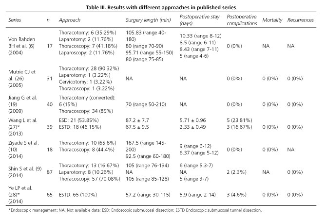
Classically, thoracotomy has been the most common approach, either a right thoracotomy for tumors of the upper and middle third of the esophagus, or a left thoracotomy or transhiatal approach through laparotomy for lower third and esophagogastric junction (3,7), showing a high success and low complications rates. However, since the first thoracoscopic enucleations performed by Everitt (24) and Bardini (25) in 1992, minimally invasive procedures have rapidly increased, and many studies have demonstrated that with MIS shorter postoperative stay, better pulmonary re-expansion with less pulmonary complications, reduced wound related pain and reduced postoperative discomfort are achieved (6,8-10). In our opinion, if the tumor is located in the upper/middle third of the esophagus, a thoracic approach is recommended, preferably a thoracoscopic approach, and if the tumor locates in the distal third of the esophagus, a conventional transhiatal laparoscopic approach is preferred. In our series a shorter postoperative stay is observed with MIS (4 days vs. 7 days), although no statistical significance is reached, probably due to our small simple size. Some authors defend open approaches with tumors larger than 5cm or if malignancy is not ruled out (3); however, we think that with enough experience in MIS the vast majority of tumors could be correctly excised, offering the advantages of this kind of surgery. We also believe that esophageal resection should be limited to great size tumors with important dissection difficulties (even larger than the traditional limit of 8 cm) or cases with extensive damage of the esophageal wall and with high risk of postoperative leaks (Table III).
Complementary procedures have been described to facilitate extramucosal enucleation using intraluminal dispositives, such as endoscopic devices with balloon dilators that assist the tumor expulsion and dissection (29,30), or simultaneous usage of flexible endoscopy to localize the tumor and help to identify the dissecting plane by a transillumination effect and provides control over the integrity of esophageal mucosa (31). In our study, flexible endoscopy has been used in 2 cases to facilitate intraoperative tumor location, without complications; in series published by other authors no associated complications were described. We think that these procedures could be useful during enucleation, but are clearly not essential to accomplish surgical excision.
We do also believe that systematic checking of esophageal mucosal integrity is not necessary, although is advisable. Different procedures can be performed, such as lumen insufflation with air (with the esophagus submerged underwater), use of methylene blue through a nasogastric tube, or performing an intraoperative endoscopy. In our series it was checked in 6 cases, with no evidence of mucosal damage in any of them.
Another controversial issue is whether to re-approximate or not the myotomy edges after enucleation. Although there are authors that suggest that it could be left open without subsequent complication, the majority, among whom we are placed, recommend to close the muscular layer following enucleation with non-absorbable interrupted suture, in order to repair the esophageal wall and preserve the propulsive activity of the esophageal body (3,6,10,15,19-21,26), and to avoid mucosal bulging and formation of pseudodiverticula which can cause postoperative dysphagia, which has been described by many authors. Large tumors' enucleations have been associated with muscle atrophy and large extramucosal defects, not allowing a tension-free suture, which can require tissue flaps with pleural films, diaphragm, omentum or pericardium (20).
It stands by its frequency and its specific management the association with hiatal hernia (which presented in our series in 38.46% of the cases). We agree with Bonavina that antireflux surgery should only be performed during the same procedure if there exist previous confirmed gastroesophageal reflux symptoms and if due to the tumor's characteristics an important dissection of the diaphragmatic crura is needed (through and abdominal approach) (15). In case that gastroesophageal reflux symptoms appear during the follow-up, a conventional laparoscopic fundoplication could be performed. In our series, simultaneous fundoplication was performed in five cases (all through an abdominal approach), and was later performed (months after the first procedure) in other two cases who firstly underwent a thoracic approach.
Another alternative which has recently become more popular is the endoscopic approach alone, initially developed as a treatment for early gastric cancer and whose indications have expanded to other kind of lesions and locations. Thus, submucosal injection of different solutions (such as glicerol or hyaluronic acid) a cushion beneath the lesion is created, separating the mucosa from the muscularis propia and enabling the tumor excision through a endoscopic submucosal dissection (ESD) (16,32), which safely allows enucleation of lesions that arise in the muscularis mucosae and the muscularis propia layer up to 3 cm. Endoscopic submucosal tunnel dissection (ESTD) has also been recently described, showing similar results and a lower risk of complications (27,28). We do not have enough experience in this kind of procedures, but we think they could be an available management alternative for experienced groups and small tumors, according to the published results (Fig. 1).
To summarize, in this review we have shown a single institution's experience in surgical management of esophageal leiomyoma, an infrequent entity with some controversial issues. This study has nevertheless several limitations, with data retrospectively collected and reviewed, and a small simple size, which hampers its power to reach statistically significant conclusions.
Conclusion
Enucleation stands as the gold standard treatment for most of esophageal leiomyiomas. In this study, duration of the surgical procedure was longer in the MIS group than in the OS groups; nevertheless, postoperative stay was found to be shorter in the MIS group.
Paradoxically, incision pain after surgery (thoracic neuralgia) was found to be higher in the MIS group, although the major complications rate remains similar as the OS group. Nevertheless, none of the results obtained in the study reached statistical significance, probably due to the small simple size.
References
1. Choong CK, Meyers BF. Benign esophageal tumors: Introduction, incidence, classification, and clinical features. Semin Thorac Cardiovasc Surg 2003;15:3-8. DOI: 10.1016/S1043-0679(03)70035-5. [ Links ]
2. Postlethwait RW, Musser AW. Changes in the esophagus in 1,000 autopsy specimens. J Thorac Cardiovasc Surg 1974; 68:953-6. [ Links ]
3. Lee LS, Sinighal S, Brinster CJ, et al. Current management of esophageal leiomyoma. J Am Coll Surg 2004;198:136-46. DOI: 10.1016/j.jamcollsurg.2003.08.015. [ Links ]
4. Seremetis MG, Lyons WS, De Guzman VC, et al. Leiomyomata of the esophagus. An analysis of 838 cases. Cancer 1976;38:2166-77. DOI: 10.1002/1097-0142(197611)38:5<2166::AID-CNCR2820380547> 3.0.CO;2-B. [ Links ]
5. Zhu X, Zhang XQ, Li BM, et al. Esophageal mesenchymal tumors: Endoscopy, pathology and immunohistochemistry. World J Gastroenterol 2007;13:768-73. DOI: 10.3748/wjg.v13.i5.768. [ Links ]
6. Von Rahden BH, Stein HJ, Feussner H, et al. Enucleation of submucosal tumors of the esophagus: Minimally invasive versus open approach. Surg Endosc 2004;18:924-30. DOI: 10.1007/s00464-003-9130-9. [ Links ]
7. Punpale A, Rangole A, Bhambhani N, et al. Leiomyoma of esophagus. Ann Thorac Cardiovasc Surg 2007; 13:78-81. [ Links ]
8. Kent M, d'Amato T, Nordman C, et al. Minimally invasive resection of benign esophageal tumors. J Thorac Cardiovasc Surg 2007;134:176-81. DOI: 10.1016/j.jtcvs.2006.10.082. [ Links ]
9. Shin S, Choi YS, Shim YM, et al. Enucleation of esophageal submucosal tumors: A single institution's experience. Ann Thorac Surg 2014;97:454-9. DOI: 10.1016/j.athoracsur.2013.10.030. [ Links ]
10. Ziyade S, Kadioğlu H, Yediyildiz Ş, et al. Leiomyoma of the esophagus: open versus thoracoscopic enucleation. Turk J Med Sci 2014;44:515-9. DOI: 10.3906/sag-1303-93. [ Links ]
11. Priego P, Lobo E, Rodríguez G, et al. Surgical treatment of esophageal leiomyoma: an analysis of our experience. Rev Esp Enferm Dig 2006;98:350-8. DOI: 10.4321/S1130-01082006000500005. [ Links ]
12. Priego P, Lobo E, Rodríguez G, et al. Endoscopic treatment of oesophageal leiomyoma: Four new cases. Clin Transl Oncol 2007;9:106-9. DOI: 10.1007/s12094-007-0020-9. [ Links ]
13. Tsai SJ, Lin CC, Chang CW, et al. Benign esophageal lesions: Endoscopic and pathologic features. World J Gastroenterol 2015;21:1091-8. DOI: 10.3748/wjg.v21.i4.1091. [ Links ]
14. Lewis RB, Mehrotra AK, Rodríguez P, et al. From the radiologic pathology archives: Esophageal neoplasms: radiologic-pathologic correlation. Radiographics 2013;33:1083-108. DOI: 10.1148/rg.334135027. [ Links ]
15. Bonavina L, Segalin A, Rosati R, et al. Surgical therapy of esophageal leiomyoma. J Am Coll Surg 1995; 181:257-62. [ Links ]
16. Xu GQ, Qian JJ, Chen MH, et al. Endoscopic ultrasonography for the diagnosis and selecting treatment of esophageal leiomyoma. J Gastroenterol Hepatol 2012;27:521-5. DOI: 10.1111/j.1440-1746.2011.06929.x. [ Links ]
17. Wang Y, Zhang R, Onyang Z, et al. Diagnosis and surgical treatment of esophageal leiomyoma. Zhonghua Zong Liu Za Zhi 2002; 24:394-6. [ Links ]
18. Glanz I, Grunebaum M. The radiological approach to leiomioma of the oesophagus with a long-term follow-up. Clin Radiol 1977;28:197-200. DOI: 10.1016/S0009-9260(77)80103-7. [ Links ]
19. Jiang G, Zhao H, Yang F, et al. Thoracoscopic enucleation of esophageal leiomyoma: A retrospective study on 40 cases. Dis Esophagus 2009; 22:279-83. DOI: 10.1111/j.1442-2050.2008.00883.x. [ Links ]
20. Sun X, Wang J, Yang G. Surgical treatment of esophageal leiomyoma larger than 5cm in diameter: A case report and review of the literature. J Thorac Dis 2012;4:323-6. DOI: 10.1097/JTO.0b013e3182381515. [ Links ]
21. Pinheiro FA, Campos AB, Matos JR, et al. Videoendoscopic surgery for the treatment of esophagus leiomyoma. Arq Bras Cir Dig 2013;26:234-7. DOI: 10.1590/S0102-67202013000300015. [ Links ]
22. Sauerbruch F. Presentations in the field of thoracic surgery. Arch Klin Chir 1932;173:457. [ Links ]
23. Ohsawa T. Surgery of the esophagus. Arch Jpn Chir 1933;10:605. [ Links ]
24. Everitt NJ, Glinatsis M, McMahon MJ. Thoracoscopic enucleation of leiomioma of the oesophagus. Br J Surg 1992;79:643. DOI: 10.1002/bjs.1800790715. [ Links ]
25. Bardini R, Segalin A, Ruol A, et al. Videothoracoscopic enucleation of esophageal leiomioma. Ann Thorac Surg 1992;54:576-7. DOI: 10.1016/0003-4975(92)90463-E. [ Links ]
26. Mutrie CJ, Donahue DM, Wain JC, et al. Esophageal leiomyoma: A 40-year experience. Ann Thorac Surg 2005;79:1122-5. DOI: 10.1016/j.athoracsur.2004.08.029. [ Links ]
27. Wang L, Ren W, Zhang Z, et al. Retrospective study of endoscopic submucosal tunnel dissection (ESTD) for surgical resection of esophageal leiomyoma. Surg Endosc 2013;27:4259-66. DOI: 10.1007/s00464-013-3035-z. [ Links ]
28. Ye LP, Zhang Y, Mao XL, et al. Submucosal tunneling endoscopic resection for small upper gastrointestinal subepithelial tumors originating from the muscularis propia layer. Surg Endosc 2014;28:524-30. DOI: 10.1007/s00464-013-3197-8. [ Links ]
29. Izumi Y, Inoue H, Endo M. Combined endoluminal-intracavitary thoracoscopic enucleation of leiomyoma of the esophagus. Surg Endosc 1996;10:457-8. DOI: 10.1007/BF00191641. [ Links ]
30. Mafune K, Tanaka Y. Thoracoscopic enucleation of an esophageal leiomyoma with balloon dilator assistance. Surg Today 1997;27:189-92. DOI: 10.1007/BF02385915. [ Links ]
31. Pross M, Manger T, Wolff S, et al. Thoracoscopic enucleation of benign tumors of the esophagus under simultaneous flexible esophagoscopy. Surg Endosc 2000;14:1146-8. DOI: 10.1007/s004640000258. [ Links ]
32. Fernández-Esparrach G, Calderón A, de la Peña J, et al. Endoscopic submucosal dissection: Sociedad Española de Endoscopia Digestiva (SEED) clinical guideline. Rev Esp Enferm Dig 2014;106:120-32. DOI: 10.4321/S1130-01082014000200007. [ Links ]
![]() Correspondence:
Correspondence:
Pablo Priego Jiménez.
Division of Esophagogastric, Bariatric
and Minimally Invasive Surgery.
General Surgery Department.
Hospital Unviersitario Ramón y Cajal.
Ctra. de Colmenar Viejo, km. 9,100.
28034 Madrid, Spain
e-mail: papriego@hotmail.com
Received: 13-05-2015
Accepted: 14-07-2015


 texto en


