Mi SciELO
Servicios Personalizados
Revista
Articulo
Indicadores
-
 Citado por SciELO
 -
 Accesos
Links relacionados
-
 Citado por Google
 -
 Similares en
SciELO
 -
 Similares en Google
Compartir
Medicina Oral, Patología Oral y Cirugía Bucal (Internet)
versión On-line ISSN 1698-6946
Med. oral patol. oral cir.bucal (Internet) vol.11 no.2 mar./abr. 2006
Metastatic tumors to the jaws: A report of eight new cases
Lipa Bodner 1, Netta Sion-Vardy 2, David B. Geffen 3, Michael Nash 4
(1) Associate Professor and Chairman, Department of Oral and Maxillofacial Surgery
(2) Lecturer and Chairman, Department of Pathology
(3) Senior Lecturer, Department of Oncology
(4) Senior Lecturer, Department of Otolaryngology Head and Neck Surgery, Soroka University Medical Center,
Faculty of Health Sciences, Ben Gurion University of the Negev, Beer Sheva, Israel
ABSTRACT
Purpose: The purpose of the article is to present 8 new cases of metastatic tumors occurring in the jawbones, their clinical features, diagnostic workup and management.
Patients and methods: The records of 8 patients with metastatic jaw lesions were reviewd. Demographic data, presenting symptoms, primary tumor site, radiographic findings, bone scintigraphy , histopathology and clinical management were analyzed.
Results: The patients, ranged in age from 44 to 80 years, with a mean of 64.5 years. The primary malignant sites were: the lung , the breast, the rectum, the thyroid, the uterus and the parotid gland. The mandible was the site of oral involvement in seven cases and the maxilla in one. There was no gender difference with respect to the oral site affected. The clinical jaw presentations were: exophytic soft tissue mass, paresthesia of the lower lip and a periapical lesion The provided treatment protocols were: chemotherapy , radiotherapy and chemotherapy, surgery and chemotherapy and supportive care only. In one case the jaw lesion was the first indication of an unknown malignancy at a distant primary site.
Conclusions: Metastatic jaw lesions are uncommon. Paresthesia of the lower lip and the chin is a sinister sign for patients with a metastatic jaw lesion. In view of these cases it can be said that meticulous work-up of of jaw lesions suspected of being metastatic, may be life saving or extend the patients survival period.
Key words: Jaw, mouth, neoplasm, metastases.
Introduction
Metastatic tumors to the oral region are uncommon, comprising only 1%-3% of all malignant oral neoplasms. These tumors, however, are of great clinical significance, as their appearance may be the first indication of an undiscovered malignancy at a distant primary site, or the first evidence of dissemination of a known tumor from its primary site. Metastatic lesions may occur in the oral soft tissues, in the jawbones or in both osseous and soft tissue. The common primary sources of tumors metastatic to the oral region are the breast, lung and kidney. The lung is the most common source of metastases to the oral soft tissues, whereas the breast is the most common source for metastatic tumors to the jawbones. In the jawbones the mandible is the most common location for metastases, with the molar area being the most frequently involved site (1-4).
The diagnosis of a metastatic lesion in the oral region is challenging , both to the clinician and to the pathologist. The clinician must recognize the possibility that a lesion may represent a metastasis, and the pathologist must determin the site of tumor origin.
The purpose of this article is to present eight new cases of metastatic tumor (MT) of the jaws , their clinical features and management.
Patients and Methods
During the eight year period (1996-2004) eight patients with the diagnosis of MT to the jawbones were treated at the Department of Oral and Maxillofacial Surgery at the Soroka University Medical Center. We conducted a retrospective review of their charts, with attention to patient demographics, presenting symptoms, history of other tumors, radiographic and clinical findings, histopathology and treatment. The findings are presented, along with a discussion of MT to the jawbones.
Results
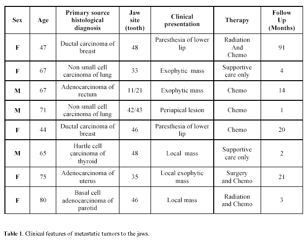
The clinical fearures of the eight cases of MT to the jawbones are presented in Table 1. Three of the patients were males and five were females. Ages ranged from 44 to 80 years, with a mean of 64.5 years.
The primary malignant sites were: the lung (2 cases), the breast (2 cases), the rectum, the thyroid, the uterus and the parotid gland (1 case each). The mandible was the main site of oral involvement in seven cases and the maxilla in one case (Figure 1).
In the mandible five cases were on the right side whereas two cases were on the left side. There was no gender difference with respect to the oral site affected. The clinical presentations were : exophytic soft tissue mass in five cases, paresthesia of the lower lip in two cases and a periapical lesion in one case. The jaw metastasis were diagnosed an average of 28.5 months (range 0 - 48 months) after initial tumor diagnosis at the primary site. The provided treatment protocols were: chemotherapy (in 3 cases), radiotherapy and chemotherapy (in 2 cases) , surgery and chemotherapy (in 1 case) and supportive care only (2 cases) (Table 1).Treatment protocols were determined by the hospital tumor board or in consultation with a senior oncologist, based on the systemic condition of the patient at the time of diagnosis of the metastatic lesion.In one patient the jaw lesion was the presenting feature and a solitary MT, whereas in seven patient the jaw lesions were part of a more extensive metastatic process. Patients were followed for an average of 19.5 months (range 1- 91 months), which represents the time period between the discovery of the jaw MT and their death . The cause of death was widespead metastatic disease in all cases.
Discussion
The oral cavity, and the jawbones are occasionally the site for MT from primary malignant tumors elsewhrere in the body. It is reported that MT account for only 1%-3% of all malignant neoplasms presenting in the oral region. However, as the jaws are not routinely examined at autopsy, the true frequency of MT in the jaws may posssibly be higher (1). The typical tumors that metastasize to the jaws in order of decreasing frequency are : breast, lung, kidney, colon, prostate and thyroid. MT to the jaw bones is a long term process. The time difference between the initial diagnosis of the malignancy at the primary site and the diagnosis of the jaw MT , in our cases was an average of 28.5 months, which correspnds to the time frame of discovery of general bone metastasis, namely, 1-5 years (5).
As reported by others (1,4,6) , MT to the jaws more often occur in the mandible than in the maxilla , and most often in posterior mandible. This site preference exists despite the fact that the mandible and maxilla share a common blood supply, the maxillary artery.
Paresthesia of the lower lip and the chin was found in two of our patients. As reported by others, this should be considered an ominous sign for metastatic lesions to the mandible, as this signifies deep invasion of the tumor into the bone and involvement of the inferior dental or mental nerves. When seen in a patient with a known malignancy, mental nerve neuropathy or the "numb chin syndrome", in the absensce of other causes, should be considered to be due to mandibular metastses until proven otherwise (7-9).
The radiological changes in the jaw bones, found in our series, depend mainly on the mineral loss in the area of tumor, as compared to the adjacent bone. Although most of the metastatic lesions to the jaw bones are osteolytic, some of the lesions, particularly prostatic metastates, are more likely to be osteoblastic (10,11). In some of the reported autopsy cases, metastses were found on histologic examination of in the mandible despite the fact that no radiological changes were detectable (12). Thus, lack of radiographic changes does not exclude the possible presence of a small metastatic lesion in the jaw bone. Unlike the oral soft tissues , where a potentially metastaic lesion can be easily recognized, the presence of an early focus of tumor metastasis in the jawbone may be overlooked. Bone scintigraphy was performed in all our patients, and in ageement with others (13,14) it was found to be an important tool for the detection of relatively small lesions.
The criteria by which one can consider a malignant jaw lesion to be a MT include:
(a) histologic verification – namely finding that the primary tumor and the jaw lesion are identical from a histologic standpoint, including special staining and other studies such as EM.
(b) the fact that the MT is not found in a site typical to primary oral tumors.
(c) the fact that the possibility of direct extension to the jawbones from a primary oral tumor can be exluded (1,3).
(d) genetic analysis - namely, the identical cytogenetic findings in both the primary tumor and in the metastatic jaw lesion, can be an important contribution to the histopathologic diagnosis of the lesion, being a metastatsis (15).
The exact mechanism of tumor metastasis from distant sites to the jawbones is not fully understood. However, autopsy records indicate that tumors tend to be site-specific in their patterns of metastases. For many tumors the nearest anatomic site encountered will be the most common site for metastatic-colony formation. Other tumors are more "selective" and by-pass nearby proximal organs and selectively colonize in a specific distal organ (16). The primary tumors that metastatize to the oral region and the jawbones probably belong to the group of "more selective" tumors. The common routes of metastases by distant tumors to the oral region and/or the jawbones are via the lymphatic channels or by hematogenous spread.
Comment
We have presented eight cases of metastatic tumors to the jawbones. The presentation of a malignant lesion in the oro-facial region may be the first indication of the existence of an unknown malignancy at a distant primary site. Lack of radiographic changes in the jawbones in the presence of suggestive symptoms does not absolutely exclude the possible presence of a MT. The presence of altered sensation in the area of the lower jaw and lip/chin region in a patient with a known non-head and neck malignancy should alert the clinician to the possibility of metastatic malignant disease and the appropriate investigation should be conducted.
References
1. Meyer I, Shklar G. Malignant tumors metastatic to the mouth and jaws. Oral Surg Oral Med Oral Pathol 1965;20:350-62. [ Links ]
2. Nishimura Y, Yakata H, Kawasaki T T, Nakajima T. Metastatic tumors of the mouth and jaws. A review of the Japanese literature. J Oral Maxillofac Surg 1982;10:253-8. [ Links ]
3. Zachariades N. Neoplasms metastatic to the mouth, jaws and surrounding tissues. J Cranio Maxillofac Surg 1989;17:283-90. [ Links ]
4. Hirshberg A, Leibovich P, Buchner A. Metastatic tumors to the jawbones: analysis of 390 cases. J Oral Pathol Med 1994;23:337-41. [ Links ]
5. Coleman RE, Rubens RD. The clinical course of bone metastses from breast cancer. Brit J Cancer 1987;55:61-6 [ Links ]
6. Carmichael FA, Mitchell DA, Dyson DP. Two contrasting radiological presentations of prostatic adenocarcinoma in the jaws. Dentomaxillofac Radiol 1996;25:283-6. [ Links ]
7. Penarrocha Diago M, Bagan Sebastian JV, Alfaro Giner A, Escrig Orenga V. Mental nerve neuropathy in systemic cancer. Oral Surg Oral Med Oral Pathol 1990;69:48-51. [ Links ]
8. Lossos A, Siegal T. Numb chin syndrome in cancer patients: etiology, response to treatment and prognostic significance. Neurology 1992; 42:1181-4. [ Links ]
9. Gaver A, Polliack G, Pilo R, Hertz M, Kitai E. Orofacial pain and numb chin syndrome as the presenting symptoms of a metastatic prostate cancer. J Postgrad Med 2002;48:283-4. [ Links ]
10. Ciola B. Oral radiographic manifestations of a metastaic prostatic carcinoma. Oral Surg Oral Med Oral Pathol 1981;52:105-8. [ Links ]
11. Koutsilieris M. Osteoblastic metastasis in advanced prostate cancer. Anticancer Res 1993;13:443-9. [ Links ]
12. Hashimoto N, Kurihara K, Yamasaki H, Ohba S, Sakai H, Yoshida S. Pathological charachteristic of metastatic carcinoma in human mandible. J Oral Pathol 1987;16:362-7. [ Links ]
13. Lewis-Jones HG, Rogers SN, Beirne JC, Brown JS, Woolgar JA. Radionuclide bone imaging for detection of mandibular invasion by squamous cell carcinoma. Brit J Radiol 2000;73:488-93 [ Links ]
14. Carranza Pelegrina D, Lomena Caballero F, Soler Peter M, Berini Aytes L, Gay Escoda C. The diagnostic possibilities of Positron Emission Tommography (PET). Med Oral Patol Oral Cir Bucal 2005;10:331-42 [ Links ]
15. Manor E, Sion-Vardy N, Bodner L. Cytogenetic and fluorescence in situ hybridization analysis of basal cell adenocarcinoma of the mandible. Cancer Genet Cytogenet 2006 (in press). [ Links ]
16. Zetter BR. The cellular basis of site-specific tumor metastsis. N Engl J Med 1990;332:605-12. [ Links ]
![]() Correspondence
Correspondence
Prof. Lipa Bodner,
Department of OMF Surgery,
Soroka University Medical Center,
P.O. Box 151,
Beer-Sheva 84101, Israel.
Fax: 972-8-6403651
E-mail: lbodner@bgu.ac.il
Received: 1-08-2005
Accepted: 5-11-2005


