


Mi SciELO
 Servicios personalizados
Servicios Personalizados
Revista
Articulo
 texto en
  Inglés (pdf)
 Articulo en XML
 Referencias del artículo
 Enviar articulo por email
Indicadores
-
 Citado por SciELO
 -
 Accesos
Links relacionados
-
 Citado por Google
 -
 Similares en
SciELO
Similares en
SciELO -
 Similares en Google
Compartir

 Permalink
Archivos Españoles de Urología (Ed. impresa)
versión impresa ISSN 0004-0614
Arch. Esp. Urol. vol.62 no.2 mar. 2009
Papillary renal carcinoma presenting as hydronephrosis
Hidronefrosis como cáncer papilar renal
Juan Carlos Regueiro López, Jesús Ruiz García, Manuel Leva Vallejo and Antonio López Beltran1
Urology Department and Pathological Anatomy1 Service. Surgery Department. Faculty of Medicine. Cordoba. University Regional Reina Sofia Hospital. Cordoba. Spain.
SUMMARY
Objective: We describe a new case of incidental renal papillary carcinoma. We perform a bibliographic review.
Methods: The papillary renal cell carcinoma is a variant of renal carcinoma. They classify in two subtypes that have relation with their prognosis. We presented one case of renal papillary carcinoma in a male of 76 years, play-acting as severe hydronephrosis.
Results/discussion: We describe the findings and final pathological result associated with a synchronic metastasis in the ipsilateral ureter.
Keywords: Papillary renal cell carcinoma. Renal carcinoma.
RESUMEN
Objetivo: Describir un nuevo caso de carcinoma papilar incidental. Revisión de la literatura.
Métodos: El carcinoma papilar renal (CRP) es una variante dentro del carcinoma renal. Se clasifican en dos subtipos que tienen relación con su pronóstico. Presentamos un caso de CRP en un varón de 76 años, simulando una hidronefrosis evolucionada.
Resultado/conclusiones: Se describen los hallazgos y el posterior resultado histológico asociado de una metástasis sincrónica en el uréter ipsilateral.
Palabras clave: Carcinoma renal papilar. Carcinoma renal.
Introduction
The papillary renal carcinoma (CRP) is a histological subtype 1 established of the renal carcinoma. The clinical and histological details were described in 1976 (1). It represents 7-15% in the total amount of renal cell carcinoma. The tumor predominates in males in 2:1 proportion. His age of presentation places himself between the 50 and 55 years (2, 3). Use to be multi-focal. Generally we found trisomies in 7, 16 and 1, with loss of the Y chromosome (4).
Presentation
Literatures have described histological variants that include trabecular, tubular, sclerosed, high grade and solid (5,6). His association with renal chronic insufficiency has been described (7). Carcinoma is named also chromofilic for the cytoplasm shown basofilia. They have defined two histological patterns, based at genetic abnormalities, tumor stage, number of mitosis and the rates of cell proliferation. The tumors use to be delimited, with eccentric location in the cortex, with hemorrhage and necrosis associated.
Case report
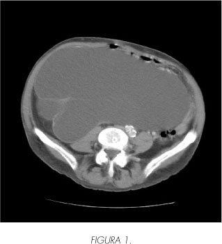
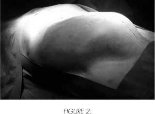
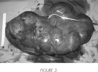
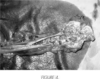
Male of 76 years without family record neither personal of concern, that we attended in urgencies for picture of diffuse abdominal pain of six months of evolution accompanied of sensation of abdominal mass, slow loss of weight but progressive. No urological symptoms associated. The physical exploration did not show abdominal peritonism, but a 20 cm's mass approximately that you locate in straight hypochondrium, overtaking the halfline. His analytical (hemogram with formulate and score, general biochemistry, study of coagulation) do not show abnormalities of entrance. The radiological basic study, the ultrasound scan shows an anechoic mass that occupies 37 cm's in right hemiabdomen, that displaces liver, pancreas and vena cava (Figure 1). The intervening findings confirm CT themselves, giving consideration to stenosis of ureter suspicion evolved with massive hydronephrosis without parenchyma working (Figure 2). We operated making right nephroureterectomy without data of renal or external infiltration. We did not find adenopathies. The histological diagnosis was papillary carcinoma of renal cells type 2 with height nuclear grade (Fuhrman 3) that substitute the parenchyma of complete form, infiltrating focally the renal capsule and perirrenal fat (pT3a) (Figure 3). The study of his ureter (Figure 4) evidenced metastasis of papillary carcinoma of renal cells that infiltrated extensively the muscular layers. Posoperative without incidences.
Discussion and conclusion
This neoplasia's type is correlated with other types of carcinoma of renal cells. His architecture is papillary or tubular-papillary with epithelial arranged stratified cells on fine vascular stems. Exist two types of CRP defined by Delahunt and Eble 8, like type 1 and 2. The type 1 is the most frequent with small cells, with scarce cytoplasm, being little his nuclei as well as nucleoles (grades 1 and 2 of Fuhrman ). The type 2 shows more irregular papillas with more big cells, with big nuclei and prominent nucleoles (grade 3 of Fuhrman). It is a classification that is correlated with the stage and evolution, being the type 2 the one belonging to worse prognosis.
Classically the CRP has associated with a most favorable prognosis than the Carcinoma of renal cells (1,9). There are contradictory results in another series themselves. The basic chromosome anomalies have found in localized tumors and no infiltrative, while the most aggressive forms in his presentation have more genetic correlated abnormalities, as it happens in the type 2 (6). The tumor stage upon the diagnosis is a important parameter. The actual tumor shows sign factors unfavorable prognosis: Invasion of perinephric fat, synchronic ureteral metastasis, nuclear grade 3 of Fuhrman, subtype 2, superior mass to 10 centimeters. The beneficial effect of the radical surgery like only treatment gets in patients with an only metastatic focus itself resectable and less than 60 years, with a level 0-1's ECOG. His best-suited treatment continues to be the radical surgery because does not seem to respond to quimio-immunotherapy. Literature have described ureteral and bladder metastasis of renal carcinoma of clear cells 10, but we have not found in literature the description of metastasis in ureteral having the origin in a CRP.
 Correspondence:
Juan Carlos Regueiro López
Avda. Lagartijo, 22 - 3o puerta 1.
14005 Córdoba. (Spain).
jcregueo@ono.com
Accepted for publication: October 18th, 2008.
References and recomended readings (*of special interest, **of outstanding interest)
*1. Mancilla-Jiménez R, Stanley R, Blath R. Papillary renal cell carcinoma. A clinical, radiologic, and pathologic study of 34 cases. Cancer 1976; 38: 246980. [ Links ]
2. Kletscher BA, Qian J, Bostwick DG, Andrews PE, Zincke H. Prospective analysis of multifocality in renal cell carcinoma: influence of histological pattern, grade, number, size, volume and deoxyribonucleic acid ploidy. J Urol 1995; 153: 904-06. [ Links ]
**3. Kovacs G: Papillary renal cell carcinoma. A morphologic and cytogenetic study of 11 cases. Am J Pathol 1989; 134:27-34. [ Links ]
4. American Joint Committee on Cancer. In: Bearhrs O, Henson D, Hutter R, Kennedy B. eds. Manual for Staging of Cancer, 4th ed. Philadelphia: JB Lippincott 1992; 201-04. [ Links ]
*5. Lager D J, Huston, BJ, Timmerman TG, Bonsib SM. Papillary renal tumors. Cancer 1995; 76: 669-73. [ Links ]
*6. Espinosa Bravo R, Lemourt Oliva M, Pérez Cárdenas JC, Véliz Medina PA y Lorenzo Cruz Ortega R. Carcinoma papilar renal quístico. A propósito de un caso. Arch Esp Urol 2003; 56:946-48. [ Links ]
7. Tickoo SK, de Peralta-Venturina MN, Harik LR et al. Spectrum of epitelial neoplasm in end-stage renal disease: an experience from 66 tumors-bearing kidneys with emphasis on histologic patterns distinct from those in sporadic adult renal neoplasia. Am J Surg Pathol 2006; 30: 141-53. [ Links ]
8. Delahunt B, Eble JN. Papillary renal cell carcinoma: a clinicopathologic and inmunohistochemical study of 105 tumors. Mod Pathol 1997; 10: 537-44. [ Links ]
**9. Boczko S, Fromowitz FB, Bard RH. Papillary adenocarcinoma of kidney. A new perspective. Urology 1979; 14: 491-95. [ Links ]
10. Kamota S, Harabayashi T, Suzuki S, Takeyama Y et al. Ureteral and bladder metastases of renal cell carcinoma following synchronous renal cell carcinoma and bladder cáncer; a case report. Nippon Hinyokika Gakkai Zasschi 2003; 94:705-08. [ Links ]

