


Mi SciELO
 Servicios personalizados
Servicios Personalizados
Revista
Articulo
 texto en
  Inglés (pdf)
 Articulo en XML
 Referencias del artículo
 Enviar articulo por email
Indicadores
-
 Citado por SciELO
 -
 Accesos
Links relacionados
-
 Citado por Google
 -
 Similares en
SciELO
Similares en
SciELO -
 Similares en Google
Compartir

 Permalink
Archivos Españoles de Urología (Ed. impresa)
versión impresa ISSN 0004-0614
Arch. Esp. Urol. vol.62 no.5 jun. 2009
Genitourinary tuberculosis
Tuberculosis urogenital
Xavier Ruiz Plazas, Marta de la Cruz Ruiz, Diego Alonso Rodríguez, Lorena Fernández Barranco, Jaime de Oleza Simó and Mariano Ozonas Moragues.
Department of Urology. Son Dureta Universitari Hospital. Palma de Mallorca. Spain.
Tuberculosis, caused by the Koch's bacillus, is one of the diseases with more prevalence and mortality worldwide. It is considered that the genitourinary affectation is between 2,7% and 8% of the total, being the most frequent extrapulmonary manifestation.
In Spain, between 10,000 and 12,000 new annual cases are being diagnosed with an increasing trend due to the arrival of population from countries with high prevalence of the disease. Therefore, it is important to detect the most characteristic radiological images in order to suspect and diagnose it.
We present a case of a 65-years-old male with renal tuberculosis antecedents diagnosed at the age of 3 years old, presenting with recurrent colic pain on the right flank.
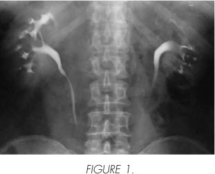
The intravenous urography shows a filling defect with shell shape calcifications on the upper pole of the left kidney. A "shriveled flower" image can be observed on the left kidney (Figure 1).
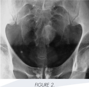
Both seminal vesicles are calcified. Note that the calcifications are intraluminal in contrast with calcifications found on diabetics, which are usually intramural (Figure 2).
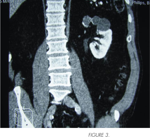
Scanner coronal reconstruction shows cluster shape cystic lesions located on the upper pole of the left kidney, with absence of viable parenchyma and cyst wall calcifications (Figure 3).
 Correspondence:
Xavier Ruiz Plazas
Servicio de Urología
Hospital Universitari Son Dureta
C/ Andrea Doria 55
07014 Palma de Mallorca. (Spain).
xarupl@gmail.com

