


Mi SciELO
 Servicios personalizados
Servicios Personalizados
Revista
Articulo
 texto en
  Inglés (pdf)
 Articulo en XML
 Referencias del artículo
 Enviar articulo por email
Indicadores
-
 Citado por SciELO
 -
 Accesos
Links relacionados
-
 Citado por Google
 -
 Similares en
SciELO
Similares en
SciELO -
 Similares en Google
Compartir

 Permalink
Revista Española de Enfermedades Digestivas
versión impresa ISSN 1130-0108
Rev. esp. enferm. dig. vol.103 no.5 Madrid may. 2011
https://dx.doi.org/10.4321/S1130-01082011000500007
PICTURES IN DIGESTIVE PATHOLOGY
Tuboovarian abscess as unusual presentation of tubarian fistula secondary to sigmoid diverticulitis
Absceso tuboovárico como presentación inusual de fístula tubárica secundaria a diverticulitis sigmoidea
Nuria Fernández-García, Alicia Mesa-Álvarez, Juan Calvo-Blanco, Ana Álvarez-Vázquez, Covadonga Pereira-Menéndez and Ana María Benítez-Vázquez
Department of Radiodiagnosis. Hospital Universitario Central de Asturias. Oviedo, Asturias. Spain
Case report
A 80-year-old female with hypertension and aortic valve disease, as a past medical history, who went to the emergency department with abdominal pain located in right lower quadrant and leukocytosis.
The sonographic exploration identified a formation adjacent to the uterus of 44 x 36 mm, heterogeneous, probably related to a right adnexal mass, so she was admitted onto the gynecology service.
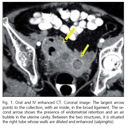
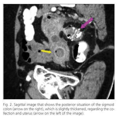
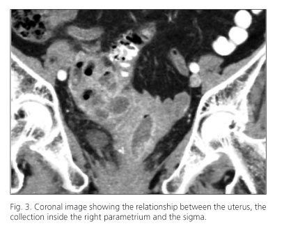
A multidetector computed tomography (TCM) revealed a collection with air inside, adjacent to the uterus, and a distended and enhancement of the right tube's walls. It caused endometrial retention and there was an air bubble in the uterine cavity. These findings correspond to salpingitis and tuboovarian abscess. There was a segment of the adjacent sigma with walls slightly thickened and diverticula.
The patient was diagnosed with perforated diverticulitis with an abscess in the broad ligament. The collection underwent drainage and intravenous antibiotics were prescribed with clinical and radiology improvement.
Discussion
Diverticular disease is a common entity whose incidence increases with age, representing, the 50-60% of the people who are 80 or over. The stage II classification of Hinchey et al. shows the formation of retroperitoneal or pelvic abscess secondary to diverticular perforation. In the literature there have been found complications of diverticular disease with fistula formation colovesical (50%), colovaginal (25%) and coloenteric (7%). In men have also been reported to prostate and seminal vesicles.
The sensitivity of MDCT for the diagnosis of acute diverticulitis varies around 90-97%, with high specificity and a false positives rate of 7-21%. It is useful for early detection of acute diverticulitis complicated by abscesses, it is also helpful predicting which patients will not respond to medical treatment and it provides guidance for percutaneous drainage of collections.
Recommedded references
1. Telepak RJ, Huggins TJ, Bova JG. Tuboovarian abscess simulating giant colonic diverticulum. Gastrointest Radiol 1984;9(4):369-71. [ Links ]
2. Bahadursingh AM, Virgo KS, Kaminski DL, Longo WE. Spectrum of disease and outcome of complicated diverticular disease. Am J Surg 2003; 186(6):696-701. [ Links ]
3. Pompeo L, Heller DS, Hameed MR, Sama J, Cracchiolo BM. Unilateral chronic tubo-ovarian abscess secondary to ruptured colonic diverticulum presenting as a brain abscess. A case report. J Reprod Med 2000;45(2):145-8. [ Links ]

