


Mi SciELO
 Servicios personalizados
Servicios Personalizados
Revista
Articulo
 texto en
  Inglés (pdf)
 Articulo en XML
 Referencias del artículo
 Enviar articulo por email
Indicadores
-
 Citado por SciELO
 -
 Accesos
Links relacionados
-
 Citado por Google
 -
 Similares en
SciELO
Similares en
SciELO -
 Similares en Google
Compartir

 Permalink
Revista Española de Enfermedades Digestivas
versión impresa ISSN 1130-0108
Rev. esp. enferm. dig. vol.105 no.8 Madrid sep. 2013
https://dx.doi.org/10.4321/S1130-01082013000800012
LETTERS TO THE EDITOR
Spinal meningioma diagnosis based on transesophageal endoscopic ultrasound-guided fine-needle aspiration (EUS-FNA)
Diagnóstico de meningioma espinal por punción aspiración con aguja fina guiada por ultrasonografía endoscópica (PAAF-USE)
Key words: Colorectal carcinoma. Meningioma. Transesophageal endoscopic ultrasound (EUS). Echoendoscopy.
Palabras clave: Cáncer de colon. Meningioma. Ultrasonografía endoscópica (USE). Ecoendoscopia.
Dear Editor,
The role of transesophageal endoscopic ultrasound-guided fine-needle aspiration (EUS-FNA) in the diagnostic staging of different malignancies (1-3) has been clearly established. However, the role of EUS-FNA in the diagnosis of other neoplasias is under investigation (4). Here, we present a case of spinal meningioma diagnosed by EUS-FNA during the staging of a cecum adenocarcinoma.
Case report
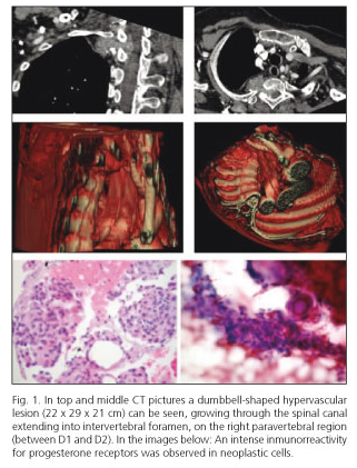
An 80-year-old woman with a history of mitral valve disease, pulmonary hypertension and atrial fibrillation was studied by asthenia and ferropenic anaemia. Colonoscopy revealed a polylobulated vegetant lesion occupying 60-70 % of the cecum, with a histopathological diagnosis of moderately differentiated adenocarcinoma. TAC showed a marked thickening of the cecum wall (5.8 x 4.9 x 3.7 cm) without any signs of obstruction. In the right paravertebral region, between D1 and D2, an hourglass-shaped lesion of 22 x 29 x 21 mm was observed, which was growing through the spinal canal and widening the intervertebral foramen (Fig. 1). By EUS-FNA, the lesion was accessed from the back side of the oesophagus, observed in a right para-spinal arrangement a hyperechogenic and echogenic fat rounded mass (30 mm diameter); 3 passes were conducted on this mass with a 25-gauge needle (ECHO-25-HD Cook©). The puncture was analysed by an in situ pathologist and was made by slow in-and-out stylet movements, instead by syringe suction. A sufficient amount of material was obtained to establish a meningioma diagnosis (intense immunoreactivity for progesterone receptor; negative expression for TTF-1 and CK AE1/AE3) (at the bottom of Fig. 1) from samples showing medium-sized round cells without nuclear atypia but with pseudoinclusions and psammomas bodies nuclear modifications that formed lobules surrounded by collagenous septa.
A right hemicolectomy was performed following a diagnosis of cecum adenocarcinoma without involvement of the 11 removed nodes or risk factors (pT2, pN0, Mx). No signs of local or distant relapse were observed during a 3-year follow-up period.
Discussion
Although the association between meningioma and colon adenocarcinoma has been described (5), the histopathological confirmation of the dorsal lesion was necessary in this case to most appropriately manage the cecum adenocarcinoma. Spinal meningiomas account for 12 % of all meningiomas and between 25-45 % of intradural spinal tumours (6). Management of spinal meningiomas has been extrapolated from intracranial meningiomas (7). In this case, we dismissed surgery as an option based on the absence of symptoms, patient comorbidity and the high risk of recurrence after surgery.
Since the introduction of EUS-FNA in 1992, its use, beyond the diagnosis of gastrointestinal lesions, has grown superlatively. Thus, due to its accuracy regarding mediastinal nodes, adrenal glands (8) and accessible liver lesions (9) evaluations, EUS-FNA is crucial when management is based on radical surgery. Its usefulness in interventional procedures (neurolysis, drainage of collections, implementation guides and/or radioactive markers) is also growing (10). Despite this plethora of EUS-FNA applications, we have not evidence of previous publications in which a meningioma diagnosis was established by EUS-FNA.
Carmen Reyna1, Antonio Viúdez1, María Dolores Lozano2, José Echeveste2, Ruth Zárate3,
Gorka Bastarrika4, Jordi Broncano4 and José Carlos Subtil5
1Oncology and 2Pathology Departments. Clínica Universidad de Navarra.
Universidad de Navarra. Pamplona, Navarra. Spain
3Laboratory of Pharmacogenomics. Division of Oncology. Centro de Investigación Médica Aplicada (CIMA).
Universidad de Navarra. Pamplona, Navarra. Spain
4Radiology and 5Gastroenterology Departments. Clínica Universidad de Navarra.
Universidad de Navarra. Pamplona, Navarra. Spain
References
1. Tamm EP, Balachandran A, Bhosale PR, Katz MH, Fleming JB, Lee JH, et al. Imaging of pancreatic adenocarcinoma: Update on staging/resectability. Radiol Clin North Am 2012;50:407-28. [ Links ]
2. Vila JJ, Jiménez FJ, Irisarri R, Martínez A, Amorena E, Borda F. Rectal cancer staging with endoscopic ultrasonography: Correlation with pathological staging. Rev Esp Enferm Dig 2007;99:132-7. [ Links ]
3. Repiso A, Gómez-Rodríguez R, López-Pardo R, Lombera MM, Romero M, Aranzana A, et al. Usefulness of endoscopic ultrasonography in preoperative gastric cancer staging: Diagnostic yield and therapeutic impact. Rev Esp Enferm Dig 2010;102:413-20. [ Links ]
4. Catalano MF, Rosenblatt ML, Chak A, Sivak MV Jr, Scheiman J, Gress F. Endoscopic ultrasound-guided fine needle aspiration in the diagnosis of mediastinal masses of unknown origin. Am J Gastroenterol 2002;97:2559-65. [ Links ]
5. Malmer B, Tavelin B, Henriksson R, Grönberg H. Primary brain tumours as second primary: A novel association between meningioma and colorectal cancer. Int J Cancer 2000;85:78-81. [ Links ]
6. Helseth A, Mork SJ: Primary intraespinal neoplasms in Norway, 1955 to 1986. A population-based survey of 467 patients. J Neurosurg 1989;71:842-5. [ Links ]
7. Setzer M, Vatter H, Marquardt G, Seifert V, Vrionis FD. Management of spinal meningiomas: Surgical results and a review of the literature. Neurosurg Focus 2007;23:E14. [ Links ]
8. Dumonceau JM, Polkowski M, Larghi A, Vilmann P, Giovannini M, Frossard JL, et al. Indications, results, and clinical impact of endoscopic ultrasound (EUS)-guided sampling in gastroenterology: European Society of Gastrointestinal Endoscopy (ESGE) Clinical Guideline. Endoscopy 2011;43:897-912. [ Links ]
9. TenBerge J, Hoffman BJ, Hawes RH, Van Enckevort C, Giovannini M, Erickson RA, et al. EUS-guided fine needle aspiration of the liver: Indications, yield, and safety based on an international survey of 167 cases. Gastrointest Endosc 2002;55:859-62. [ Links ]
10. Cho CM, Dewitt J, Al-Haddad M. Echo-endoscopy: New therapeutic frontiers. Minerva Gastroenterol Dietol 2011;57:139-58. [ Links ]

