


 Servicios personalizados
 Inglés (pdf)
 Articulo en XML
 Referencias del artículo
 Enviar articulo por email
 Citado por SciELO
  Citado por Google
  Similares en
SciELO
Similares en
SciELO  Similares en Google
 Permalink
CASE REPORT
An 82-year-old female was referred for the evaluation of progressive esophageal dysphagia of a six month duration, with a recent weight loss of 10% of the normal corporal weight over a six month period. The patient reported solid food dysphagia that required liquids to facilitate food progression and denied food impaction. An upper endoscopy revealed an extrinsic impression in the upper esophagus at 25 centimeters from the incisors. A barium esophagram identified contour irregularity of the upper esophagus caused by an extrinsic compression above the aortic arch (Fig. 1). Thoracic computed tomography (CT) showed an aberrant right subclavian artery arising as the fourth branch of the aortic arch (Fig. 2 and Fig. 3) with a retroesophageal course from left to right, which compressed the esophagus and caused a moderate reduction of the esophageal caliber. These findings were compatible with a diagnosis of dysphagia lusoria. The patient refused a referral for surgery and was advised about dietary modifications. After three months, she had a favorable evolution with a symptomatic improvement and weight stabilization.

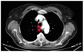
Fig. 2 Aberrant right subclavian artery arising as the fourth branch of the aortic arch on thoracic CT.

DISCUSSION
Dysphagia lusoria is caused by compression of the arteria lusoria, an aberrant right subclavian artery. It is usually asymptomatic but can become symptomatic in the case of a compression of the esophagus or the trachea, in the presence of an aberrant subclavian artery aneurysm and with advanced age. This is possibly due to atherosclerotic hardening or fibromuscular dysplasia of the arteries 1. Mild to moderate symptoms are often treated with a dietary modification 2. In case of severe or persistent symptoms, surgical intervention is required to remove the aberrant vessel and reconstruct the vascular supply. In addition, less invasive techniques are now available, such as vascular endoprosthesis and endovascular occlusion. The initial results of these procedures are promising 3.
