


Mi SciELO
 Servicios personalizados
Servicios Personalizados
Revista
Articulo
 texto en
  Inglés (pdf)
 Articulo en XML
 Referencias del artículo
 Enviar articulo por email
Indicadores
-
 Citado por SciELO
 -
 Accesos
Links relacionados
-
 Citado por Google
 -
 Similares en
SciELO
Similares en
SciELO -
 Similares en Google
Compartir

 Permalink
Revista Española de Enfermedades Digestivas
versión impresa ISSN 1130-0108
Rev. esp. enferm. dig. vol.110 no.9 Madrid sep. 2018
https://dx.doi.org/10.17235/reed.2018.5658/2018
SCIENTIFIC LETTERS
Bacterial endogenous endophthalmitis after colonoscopy
1Sección de Aparato Digestivo. Hospital San Agustín. Avilés, Asturias, Spain
2Servicio de Oftalmología. Hospital San Agustín. Avilés, Asturias, Spain
Key words: Endogenous endophthalmitis; Colonoscopy
Dear Editor,
Endogenous endophthalmitis (EE) or metastatic endophthalmitis, is a severe condition with a poor visual prognosis. It results from hematogenous dissemination of bacteria or fungi from a primary infection site to the eyeball 1.
We report the case of a 69-year-old female with diabetes, hypertension and ischemic heart disease who had undergone colonoscopy due to polyps, three and six months previously. She presented to the Emergency Room due to blurry vision and pain in her left eye (LE) over the last three days and was diagnosed with a vitreous hemorrhage. After 24 hours, the visual acuity (VA) was 0.5 in the right eye (RE), which had a normal fundus. This was lower than 0.05 in the LE, which exhibited chemosis, hypopyon and fibrin in the pupillary area, nuclear cataract, IOP at 45 mmHg and impaired fundus visualization due to the vitreous bleeding (Fig. 1). EE was suspected and the patient received intravenous vancomycin, ceftazidime and fluconazole, plus intravitreal vancomycin, which was repeated after 72 hours. Blood cultures were negative. She had an untoward outcome and required evisceration after ten days. Streptococcus bovis was isolated from the eviscerated material.
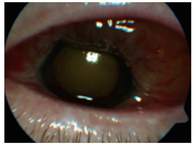
Fig. 1 Large chemosis with fibrin in the pupillary area and vitritis that prevents focusing on the eye fundus.
Discussion
Bacterial EE is an uncommon disease with a very poor visual prognosis 2. It manifests with visual loss, pain, hypopyon, vitritis, Tyndall sign, palpebral edema and ocular hypertension. Some of these manifestations are non-specific, which may result in a misdiagnosis 3. When suspected, empiric therapy should be initiated with intravenous wide-spectrum antibiotics in association with intravitreal doses. A definite diagnosis requires cultures for bacterial identification. Blood cultures are positive in over 50% of cases, whereas positivity ranges from 36% to 73% for intraocular samples 3,4. S. bovis grew in our cultured sample of eviscerated material. This bacterium is typically found in the gastrointestinal system, which is consistent with the patient having undergone a colonoscopy, a procedure that may give rise to transient bacteremia 5).
Bibliografía
1. Díaz-Llopis M, Calonge M, Sainz-de-la-Maza MT, et al. Uveítis y escleritis. Diagnóstico y tratamiento. 90ª Ponencia de la Sociedad Española de Oftalmología. Madrid: Mc Line SL; 2014. [ Links ]
2. Jackson TL, Paraskevopoulos T, Georgalas I. Systematic review of 342 cases of endogenous bacterial endophthalmitis. Surv Ophthalmol 2014;59: 627-35. DOI: 10.1016/j.survophthal.2014.06.002 [ Links ]
3. Sociedad Española de Retina y Vítreo. Endoftalmitis infecciosa. Primera revisión. Guías de práctica clínica de la SERV. 2014. Consultado: 15 septiembre 2017. Disponible en: http://www.serv.es. [ Links ]
4. Lee S, Um T, Joe SG, et al. Changes in the clinical features and prognostic factors of endogenous endophthalmitis. Retina 2012;32:977-84. DOI: 10.1097/IAE.0b013e318228e312 [ Links ]
5. Wu AY, Oestreicher JH. Endogenous bacterial endophthalmitis after routine colonoscopy. Can J Ophthalmol 2011;46:555-6. DOI: 10.1016/j.jcjo. 2011.10.002 [ Links ]
 Este es un artículo publicado en acceso abierto bajo una licencia Creative Commons
