Mi SciELO
Servicios Personalizados
Revista
Articulo
Indicadores
-
 Citado por SciELO
 -
 Accesos
Links relacionados
-
 Citado por Google
 -
 Similares en
SciELO
 -
 Similares en Google
Compartir
Medicina Oral, Patología Oral y Cirugía Bucal (Internet)
versión On-line ISSN 1698-6946
Med. oral patol. oral cir.bucal (Internet) vol.11 no.1 ene./feb. 2006
ORAL MEDICINE AND PATHOLOGY
Acid-etching effects in hypomineralized amelogenesis imperfecta.
A microscopic and microanalytical study
Carmen Sánchez-Quevedo 1, Gregorio Ceballos 2, Ismael Ángel Rodríguez 3, José Manuel García 1, Miguel Alaminos 4 (1) Profesor Titular de Histología y Embriología Bucodental, Departamento de Histología, ABSTRACT
Objectives: The purpose of this study was to use quantitative x-ray
microprobe analysis with scanning electron microscopy to define the
morphostructural and calcification patterns in the enamel of teeth with the
hypomineralized variant of amelogenesis imperfecta. Key words: Acid-etching, microanalysis, quantitative method, scanning
electron microscopy. RESUMEN Objetivos: El objetivo del presente trabajo consistió en definir los
patrones morfológicos y de calcificación en el esmalte de dientes afectados
con amelogénesis imperfecta hipomineralizada, tras la aplicación del grabado
ácido, mediante la utilización de la microscopía electrónica analítica
cuantitativa. Palabras clave: Amelogénesis imperfecta, patrones prismáticos,
microscopía electrónica de barrido, microscopía electrónica analítica
cuantitativa.
Facultad de Medicina y Odontología, Universidad de Granada, España
(2) Jefe de Servicio, Hospital Clínico Universitario San Cecilio, Universidad de Granada, España
(3) Profesor de Histología, Cátedra B de Histología y Embriología, Facultad de Odontología,
Universidad Nacional de Córdoba, República Argentina
(4) Investigador contratado. Unidad Mixta de Investigación. Hospital Clínico Universitario San Cecilio.
Departamento de Histología. Universidad de Granada, España
Study design: We compared 5 fragments of permanent human canines from
patients with clinically diagnosed hypomineralized amelogenesis imperfecta and 5
normal permanent canines from subjects without amelogenesis imperfecta. All
specimens were etched with phosphoric acid for morphological and microanalytical
examination.
Results: Two types of etching patterns were found; in addition, islets of
pattern I were seen within areas of pattern II. Microanalysis detected no
significant differences in calcium concentration between specimens with
amelogenesis imperfecta and normal control specimens after acid etching. Pattern
III was not observed.
Conclusions: The changes and their distribution in the enamel structure
after 30 s of acid etching are described in teeth with this rare disorder.
Although these data seem to coincide with alterations in prism development, no
alterations in calcium concentration were found.
Diseño del estudio: Se estudian 5 fragmentos de caninos humanos permanentes
de pacientes con amelogénesis imperfecta hipomineralizada y 5 caninos
controles. Todas las muestras fueron tratadas con ácido fosfórico y
posteriormente procesadas para su estudio morfológico y microanalítico.
Resultados: Se observan dos tipos de patrones de grabados, I y II, así como
áreas o islotes de patrón I insertados en extensas áreas homogéneas en las
que predomina el patrón II. No se aprecian diferencias significativas en la
concentración de calcio entre las muestras con amelogénesis imperfecta
hipomineralizada y control después del grabado ácido tras el estudio microanalítico
cuantitativo. El patrón tipo III no fue observado.
Conclusiones: Se describen los cambios y distribución en la estructura del
esmalte después del grabado ácido durante 30 segundos en dientes con amelogénesis
imperfecta hipomineralizada. Aunque los datos obtenidos coinciden con
alteraciones en el desarrollo de los prismas, no se detectan alteraciones en la
concentración de calcio.
Introduction
Acid etching is important in odontology because it can serve as a model for the acid destruction caused by caries (1) and help in the taxonomic and evolutionary classification of mammals on the basis of structural differences in enamel (2). In addition, this technique is important in odontologic treatment because it facilitates the bonding of restorative materials to excavations and fissures it creates (3). Amelogenesis imperfecta is a term that identifies a group of hereditary alterations that affect the formation of the enamel extracellular matrix (4) by altering prism formation (5). Although odontological treatment for amelogenesis imperfecta varies depending on the patients clinical status and the extent of the process itself, there are indications that acid etching can be used as a preliminary step in restorative treatment (6, 7).
The recent application of new electron microscopy quantitative analysis techniques to study mineralized tissues, allows us to establish a very closed relationship between the morphological and chemical patterns in those tissues (8-10). The application of analytical electron microscopy to odontology is an important advance to define the normal and pathological patterns of the mineralized dental tissues and the response of these structures to dental therapy (11, 12).
This study was designed to use quantitative x-ray microprobe analysis with scanning electron microscopy to characterize the structural and calcification patterns in the hypomineralized variant of amelogenesis imperfecta. The findings make it possible to analyze the effects of this technique on enamel prisms, and suggest how these effects might influence restorative treatments that use acid etching as a preliminary conditioning step.
Material and Method
The material consisted of fragments of 5 permanent human canines from the coronal part of permanent teeth obtained in the course of preparation for crown replacement from patients with autosomal dominant clinically and genetically diagnosed hypomineralized amelogenesis imperfecta. All patients were members of the same family (13). As the control material we used 5 coronal fragments from normal included canines surgically extracted.
The tooth fragments were plunge-frozen in liquid nitrogen. The samples were transferred to Polaron E 5350 the freeze-drying apparatus and dried at 80ºC for 24 h. The amelogenesis imperfecta fragments were subjected to acid etching with 35% phosphoric acid for 30 seconds, then washed in double-distilled water and air-dried for 1 h. Each fragment was mounted on an SEM stub and coated with carbon for microanalysis, and later with gold for morphological study with a Philips XL30 SEM and an EDAX DX-4 detector (14, 15)
Specimens prepared as described above were studied in a Phillips XL- 30 scanning electron microscope (SEM) (operating voltage = 15 kV; spot size = 500 nm; tilt angle = 35º; take-off angle = 61.34º). An energy dispersive spectrometer (EDAX DX-4) was used for quantitative analyses (count rate = 1200 cps; live time = 50 s). Spectra were collected by pin-point electron beam at x 40 000, and ten analyses were run with each specimen, so that all 50 separate analyses were done in both amelogenesis imperfecta and control teeth.
The peak-to-background (P/B) ratio method (16, 17) was used to measure the concentration of calcium. Microscrystalline salt standards were used to quantify calcium (10, 15, 18, 19). Microscrystalline salts standards were methodologically treated in similar way to control and amelogenesis imperfecta specimens (20, 21). For statistical analyses we use the Student´s t test.
Results
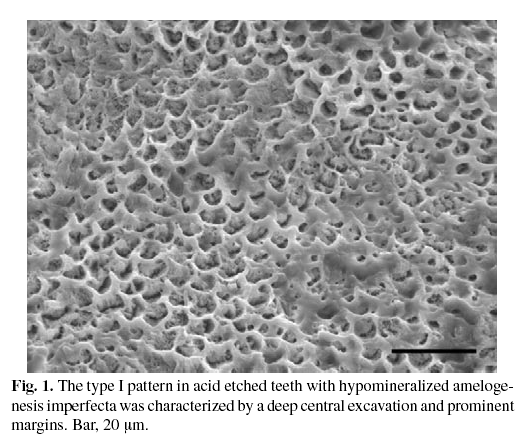
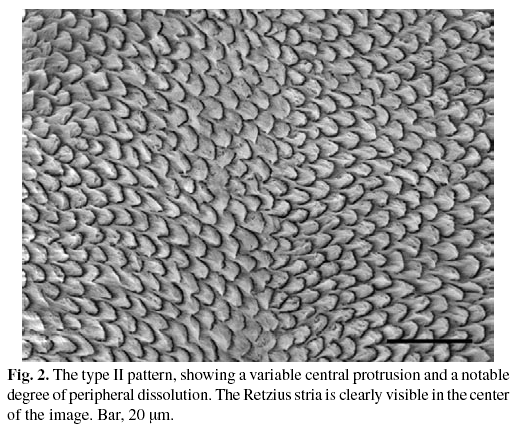
Two types of etched patterns were seen in fragments of teeth with amelogenesis imperfecta. The type I pattern was characterized by deep central excavations and prominent margins (Fig. 1). The type II pattern showed a variable central protrusion and a notable degree of peripheral dissolution. The type II pattern was more widespread in the enamel structure than type I (Fig. 2). In areas where type I prisms were predominant, the size and appearance of the prisms were more variable than in type II. Acid etching also revealed prisms of variable size and appearance in the Retzius stria, but not in the areas between the Retzius lines, which were much more homogeneous in appearance. However, the effect of acid etching appeared to be similar in all prisms regardless of whether they were located within the Retzius stria or in the surrounding areas.
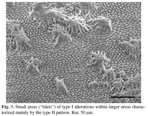
One of the most characteristic findings in specimens from amelogenesis imperfecta teeth was that acid etching revealed small areas ("islets") of type I alterations within large areas characterized mainly by the type II pattern (Fig. 3). The type I islets appeared to arise from mounds of amorphous material.
Percent weight fractions and mean weight fractions of calcium in the acid-etched enamel were 22.90 ± 2.52 in the type I pattern, 22.64 ± 1.95 in the type I pattern islets, and 21.95 ± 2.25 in the type II pattern of amelogenesis imperfecta samples. The values after acid etching in control specimens were 22.50 ± 2.41 in the type I pattern, and 21.61± 2.25 in the type II pattern. There were no statistically significant differences between amelogenesis imperfecta and normal teeth in calcium concentrations after acid etching.
Discussion
Acid etching and examination with scanning electron microscopy have revealed, in several species including humans, three main types of etching patterns (designated I, II and III) as described by Silverstone et al (22). The topographical distribution of the different patterns in the etched enamel has also been reported (23). In teeth fragments with clinically diagnosed hypomineralized amelogenesis imperfecta, we found type I and type II patterns, but not the type III pattern. This contrasts with the report by Seow and Amaratunge (7) who found all three patterns in teeth with amelogenesis imperfecta. In this connection it is important to note that these authors treated their specimens with acid for 1 min, a period that surpasses the maximum therapeutic exposure of 30 s.
Our finding of islets displaying the type I pattern within areas where the type II pattern predominated (as previously reported) suggests that alterations had occurred in prism development. This notion contrasts with the conclusions of Seow and Amaratunge (7) and Wright et al (11) who suggested that the presence of three different patterns in amelogenesis imperfecta was evidence of normal prism structure. The presence of islets meant that an alteration in the normal distribution of patterns established in controls and in previous studies (7-10) was present. In such studies our group has previously demonstrated that enamel prisms use to show parallel and decussated patterns, these latters being less frequent that the formers, and that independently from the spacial distribution, some prisms show filamentous patterns. Microanalytical study with quantitative electron microscopy of the calcium concentrations showed that there were no significant differences in the effects of acid etching between amelogenesis imperfecta and healthy control teeth. Our observations of the morphology and structure of the prisms suggest that acid etching has a more marked effect on the morphostructural features of the tooth enamel in hypomineralized amelogenesis imperfecta than on the mineral concentration as measured by microanalysis. Our studies with quantitative electron microscopy in hypomineralized amelogenesis imperfecta do not confirm the results obtained by Wright et al (24) using qualitative electron microscopy. Our microanalytic results should be considered in the context of the reviewing that is currently being held about the mineralization process related to enamel and dentine in the different clinical modalities of amelogenesis imperfecta. These results should contribute to the current reviewing process of the amelogenesis classical clinical classification (19,25). Dentists should be aware that these effects may influence the outcome of restorative treatments that involve acid etching as a preliminary conditioning step. The knowledge of the acid-etching patterns of the different clinical types of amelogenesis imperfecta could help to prevent the high incidence of failure in the restorative treatments applied to these types of teeth (7).
Acknowledgements
This study was supported in part by the Spanish Ministry of Education and Culture through Project no. PB97-0804, and by the Agencia Española de Cooperación Internacional through Project AECI/98/01. We thank M. Ángeles Robles for her competent technical assistance.
![]() Correspondence
Correspondence
Profª. Carmen Sánchez-Quevedo
Departamento de Histología
Facultad de Medicina y Odontología
Universidad de Granada
E-18071 GRANADA, España
Tel.: +34-958-243514
Fax:+34-958-244034
E-mail: mcsanchez@histolii.ugr.es
Received: 12-03-2004
Accepted: 18-05-2005
References
1. Boyde A, Jones SJ, Reynolds PS. Quantitative and qualitative studies of enamel etching with acid and EDTA. Scanning Electron Microsc 1978;II:991-1002. [ Links ]
2. Martin LB, Boyde A, Grine FE. Enamel structure in primates: A review of scanning electron microscope studies. Scann Microsc 1988;2:1503-26. [ Links ]
3. Retief DH. Effect of conditioning the enamel surface with phosphoric acid. J Dent Res 1973;52:333-41. [ Links ]
4. Dong J, Gu TT, Simmons D, Mac Dougall M. Enamelin maps to human chromosome 4q21 within the autosomal dominant amelogenesis imperfecta locus. Eur J Oral Sci 2000;108:353-8. [ Links ]
5. Wright JT, Butler WT. Alteration of enamel proteins in hypomaturation amelogenesis imperfecta. J Dent Res 1989;68:1328-30. [ Links ]
6. Seow WK. Clinical diagnosis and management strategies of amelogenesis imperfecta variants. Pediat Dent 1993;15:384-93. [ Links ]
7. Seow WK, Amaratunge FA. The effects of acid-etching on enamel from different clinical variants of amelogenesis imperfecta: an SEM study. Pediatr Dent 1998;20:37-42. [ Links ]
8. Sánchez-Quevedo MC, Ceballos G, García JM, Gómez de Ferraris MEG, Rodriguez IA, Campos A. Quantitative analytical electron microscopy of calcium in hypomineralized amelogenesis imperfecta. Scanning 23:2001;152-3. [ Links ]
9. Sánchez-Quevedo MC, Ceballos G, Rodriguez IA, García JM, Campos A. Quantitative X-ray microanalytical and histochemical patterns of calcium and phosphorus in enamel in human amelogenesis imperfecta. International Journal of Developmental Biology 2001;45:115-6. [ Links ]
10. Sánchez-Quevedo MC, Ceballos G, García JM, Rodriguez IA, Gómez de Ferraris MEG, Campos A. Scanning electrón microscopy and calcification in amelogenesis imperfecta in anterior and posterior human teeth. Histol Histopathol 2001;16:827-32. [ Links ]
11. Wright JT, Duggal MS, Robinson C, Kirkham J, Shore A. The mineral composition and enamel ultrastructure of hypocalcified amelogenesis imperfecta. J Craniofac Genet Dev Biol 1993;13:117-26. [ Links ]
12. Zambrano M, Nikitakis NG, Sánchez-Quevedo MC, Rivera H. Oral and dental manifestations of vitamin D-dependent rickets type I: Report of a pediatric case. Oral Surg Oral Med Oral Pathol Oral Radiol and Endodontics 2003;95:705-9. [ Links ]
13. Ceballos A, Ceballos G. Estudio de una familia con amelogenesis imperfecta. Avances en Odontoestomatología 1988;4 (3):125-8. [ Links ]
14. Campos A, Rodriguez IA, Sánchez-Quevedo MC, García JM, Nieto-Albano OH, Gómez de Ferraris ME. Mineralization of human premolar occlusal fissures A quantitative histochemical microanalysis. Histol Histopathol 2000;15:499-502. [ Links ]
15. Sánchez-Quevedo MC, Nieto-Albano OH, García JM, Gómez de Ferraris ME, Campos A. Electron probe microanalysis of permanent human enamel and dentine. A methodological and quantitative study. Histol Histopathol 1998;13: 109-13. [ Links ]
16. Statham PJ, Pawley JB. A new method for particle X-ray microanalysis on peak to background measurements. Scanning Electron Microsc 1978;1:469-78. [ Links ]
17. Small JA, Heinrich KFJ, Newbury DE, Myklebust RL. Progress in the development of the peak-to-background method for the quantitative analysis of single particles with the electron probe. Scanning Electron Microsc 1979;II:807-16. [ Links ]
18. López-Escámez JA, Campos A. Standards for X-ray microanalysis of calcified structures. Scanning Electron Microsc 1994; 8:171-85. [ Links ]
19. Sanchez-Quevedo MC, Ceballos G, Garcia JM, Luna JD, Rodriguez IA, Campos A.Dentine structure and mineralization in hypocalcified amelogenesis imperfecta: a quantitative X-ray histochemical study. Oral Diseases 2004;10: 94-8. [ Links ]
20. Warley A. Quantitative X-ray microanalysis of thin sections in biology: appraisal and interpretation of results. En: Sigee DC, Morgan AJ, Sumner AT, Warley A, eds .X-ray microanalysis in biology. Experimental techniques and applications. Cambridge: Cambridge University Press; 1993. p. 47-57. [ Links ]
21. Warley A . X-ray microanalysis for biologists. London: Portland Press. 1997 [ Links ]
22. Silverstone LM, Saxton CA, Dogon IL, Fejerskov O. Variation in the pattern of acid etching oh human dental enamel examined by scanning electron microscopy. Caries Res 1975;9:373-87. [ Links ]
23. Risnes S. Multiplane sectioning and scanning electron microscopy as a method for studying the three-dimensional structure of mature dental enamel. Scann Microsc 1987;1:1893-902. [ Links ]
24. Wright JT, Deaton TG, Hall KI, Yamauchi M. The mineral and protein content of enamel in amelogenesis imperfecta. Conn Tiss Res 1995;32:247-52. [ Links ]
25. Aldred M, Savarirayan R, Crawford P. Amelogenesis imperfecta: a classification and catalogue for the 21st century. Oral Diseases 2003;9:19-23. [ Links ]


 texto en


