


Mi SciELO
 Servicios personalizados
Servicios Personalizados
Revista
Articulo
 texto en
  Inglés (pdf)
 Articulo en XML
 Referencias del artículo
 Enviar articulo por email
Indicadores
-
 Citado por SciELO
 -
 Accesos
Links relacionados
-
 Citado por Google
 -
 Similares en
SciELO
Similares en
SciELO -
 Similares en Google
Compartir

 Permalink
Revista de Osteoporosis y Metabolismo Mineral
versión On-line ISSN 2173-2345versión impresa ISSN 1889-836X
Rev Osteoporos Metab Miner vol.5 no.2 Madrid abr./jun. 2013
https://dx.doi.org/10.4321/S1889-836X2013000200006
Vertebral fractures as a debut to Cushing's syndrome diagnosed after a pregnancy
Fracturas vertebrales como debut de síndrome de Cushing diagnosticado tras un embarazo
Gutiérrez Medina S.1, Medrano Izquierdo P.2, Díaz Curiel M.3
1 Servicio de Endocrinología y Nutrición - Hospital Universitario Fundación Jiménez Díaz - Madrid
2 Servicio de Urgencias - Hospital Severo Ochoa - Leganés - Madrid
3 Servicio de Medicina Interna/Enfermedades Metabólicas Óseas - Hospital Universitario Fundación Jiménez Díaz - Madrid
SUMMARY
The case is described of a 34 year old patient with Cushing's disease diagnosed as a result of having multiple pathological vertebral fractures after giving birth. The sudden appearance of acute fractures in five vertebral bodies, the phenotype characteristics of the patient and her medical history pointed to a diagnosis of Cushing's syndrome, a condition which rarely coincides with pregnancy. After a resection of the hypophysary adenoma and the start of treatment with teriparatide, the patient experienced notable clinical and densitometric improvement. This case demonstrates the importance of suspecting a bone metabolism disorder in the presence of pathological fractures in young patients, even more in certain states, such as pregnancy or lactation.
Key words: Cushing's syndrome, osteoporosis, pregnancy, lactation.
RESUMEN
Se describe el caso de una paciente de 34 años con enfermedad de Cushing diagnosticada a raíz de presentar múltiples fracturas vertebrales patológicas tras el parto. El debut fulminante con fracturas agudas en cinco cuerpos vertebrales, las características fenotípicas de la paciente y sus antecedentes médicos orientaron hacia el diagnóstico de síndrome de Cushing, entidad poco habitual coincidente con un embarazo. Tras la resección del adenoma hipofisario y el inicio de tratamiento con teriparatida la paciente experimentó una notable mejoría clínica y densitométrica. Este caso demuestra la importancia de sospechar un trastorno del metabolismo óseo ante la presencia de fracturas patológicas en pacientes jóvenes, más aún en ciertas etapas, como embarazo o lactancia.
Palabras clave: síndrome de Cushing, osteoporosis, embarazo, lactancia.
Introduction
Cushing's syndrome comprises symptoms and signs associated with prolonged exposure to inappropriately high levels of glucocorticoids. Excluding those cases due to chronic use of glucocorticoids, the great majority (60-70%) are due a hypophysary adenoma; this is what is known as Cushing's disease. Other causes are: tumours and anomalies in the suprarenal glands and ectopic secretion of ACTH.
Osteoporosis is very common in patients with Cushing's syndrome. The functional and structural deterioration of bone is a significant cause of morbidity and incapacity in these patients, who have a higher risk of, fundamentally vertebral, fractures.
On the other hand, pregnancy and lactation result in multiple changes in women, many of which affect the bone.
However, the combination of Cushing's syndrome and pregnancy is highly infrequent due to that fact that hypercortisolism usually coincides with amenorrhea and infertility.
Clinical case
The patient was a Spanish woman of 34 years of age, referred to the bone metabolism disease clinic due to having had several vertebral fractures after having given birth.
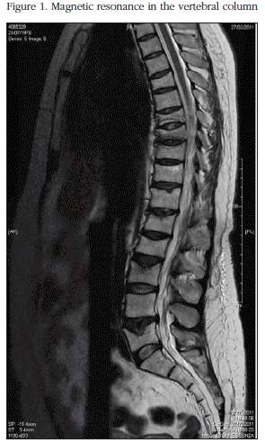
The patient had no family history of fractures. She confirmed having a sufficient daily intake of milk derivatives and low levels of regular physical activity. Of interest in her medical history were having had a transverse traumatic fracture of the patella three years earlier, with complications of pseudarthrosis. The patient had also been diagnosed with persistent polycystic ovarian syndrome due to amenorrhea 14 years previously, and had received continuous treatment with cyproterone acetate and ethenylestradiol, suspending treatment when trying for a pregnancy. After suspending the anovulatories the patient continued with her earlier amenorrhea without achieving gestation, which finally required techniques of assisted reproduction (in vitro fertilisation). The pregnancy was without complications (no gestational diabetes, and no hypertension or preeclampsia). Normal lactation was maintained for 5 months. The patient confirmed having gained weight in recent years, and significantly so after giving birth. A month after the birth persistent pain in the lumbar region started. After two months of lumbalgia resistant to treatment a magnetic resonance scan of the spine was performed, which showed evidence of fractures in various vertebral bodies, both dorsal and lumbar, with a reduction in height of the vertebral bodies being observed in T6, T8, T9, T11, T12, Li, L2, L3 and L5, with acute signs in T8, T9, T10, L1 and L3 (Figure 1). A physical examination showed facial rubicundity, accumulation of supraclavicular and retrocervical fat, with some cutaneous atrophy. The weight of the patient was 75.8 kg, height 1.61 metres, with a body mass index of 29.2 kg/m2. Blood pressure was 150/95 mm Hg, confirmed on various occasions.
Notable from the general analysis were the following values: haemoglobin, 13.4 g/dl; sedimentation velocity in the first hour, 10 mm/h (normal, <25); 25-OH-vitamin D, 8.39 ng/ml (20-50); blood calcium 8.6 mg/dl; intact parathormone (PTH), 26 pg/ml (normal, 10-65); alkaline phosphatase 93 UI/l (normal, 38-126); carboxy-terminal telopeptide of collagen (CTX), 0.595 ng/ml (normal 0.064-0.548), amino-terminal propeptide of procollagen (P1NP), 38.7 ng/ml (normal, 10.4-62); urinary cortisol in 24 hours, 616.00 and 779.9 µg (normal, <200); creatinine in urine, 41.6 mg/dl; ACTH, 81.97 pg/ml (normal, 4.7-48.8); TSH, 0.45 µUI/ml (normal 0.465-468); and electrophoretic spectrum, unchanged.
The bone densitometry (double X-ray absorptiometry, DXA) was compatible with osteoporosis in the lumbar spine and osteopenia in the femoral neck: lumbar spine (L2-L4), 0,709 g/cm2 (T-score = -3.26) and femoral neck, 0.683 g/cm2 (T-score = -1.44).
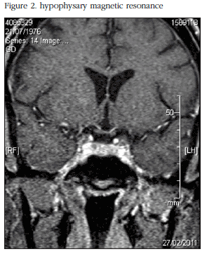
On the evidence of ACTH-dependent hypercortisolism, and after a more than 50% decrease in blood cortisol after a strong dexamentasone suppression test, Cushing's disease was diagnosed. A cerebral magnetic resonance (MR) scan showed up a nodular image of4mm in the right adenophysis, on the superior and anterior margin (Figure 2). Treatment was initiated with fluconazol before surgery which improved pressure levels and provoked a small amount of the spontaneous menstrual bleeding. The patient underwent transsphenoidal surgery for resection of the microadenoma at 8 months postpartum.
Subsequent treatment with calcium and vitamin D supplements (1,500 mg a day of calcium carbonate and 400 mg a day of colecalciferol), daily subcutaneous teriparatide and hydroaltesone at subsitutive doses of 30 mg a day was recommended, as well as recommendations of a diet rich in dairy products and regular physical exercise. After surgery on the hypophysary adenoma the patient started to have spontaneous menstruations without treatment, with a normalisation of blood pressure, progressive disappearance of the Cushingoid phenotype which she had exhibited, and experienced a notable reduction in weight.
A year after the initiation of treatment with teriparatide she showed a notable improvement in DXA in both locations: lumbar spine (L2-L4), 0.837 g/cm2 (T-Score = -2.03) and femoral neck 0.714 g/cm2 (T-score = -1.14) as well as favourable changes in the analyses: alkaline phosphatase, 69 UI/l; 25-OH-vitamin D, 21.9 ng/ml; PTH, 31.4 pg/ml; CTX, 1.830 ng/ml; and P1NP, 286.9 ng/ml.
Discussion
Cushing's syndrome is defined as a combination of signs and symptoms derived from the prolonged exposure to inappropriately high levels of glucocorticoids.
Harvey Cushing was, in 1932, the first to postulate that the syndrome characterised by obesity, plethora, diabetes, hypertension, hirsutism, amenorrhea and osteoporosis, could be caused by hypophysary adenomas [1].
Cushing's syndrome is uncommon during pregnancy because the excess of corticoids and androgens suppress the secretion of gonadotropins, which brings with it ovarian and endometrial dysfunction. The most common cause of Cushing's syndrome during pregnancy is adrenal adenoma; however, there have been cases published in which the cause is hypophysary adenoma [2]. In addition, Cushing's syndrome during pregnancy carries a higher level of maternal morbidity, in up to 70% of cases [3], the most common complications being arterial hypertension and diabetes mellitus. Less frequent are osteoporosis, fractures, psychiatric disease and cardiac insufficiency. Therefore, this pathology is commonly underdiagnosed during gestation, it being confused with other conditions such as preeclampsia or gestational diabetes [4].
The physiopathology of steroid-induced osteoporosis (SIO) is complex, given that there are multiple factors involved, many of which have not been completely clarified [5]. There is a decrease in bone formation due to osteoblast and osteocyte apoptosis [6], as well as an increase in bone resorption due to the activation of the osteoclasts [7], which have a more prolonged useful life. The formation of new collagen is an inhibitor and the degradation of pre-existing collagen an accelerator [8]. The steroids also have effects on bone remodelling at the level of the basic remodelling unit, resulting in a reduction in average trabecular thickness and a lower degree of bone apposition. Furthermore, the steroids reduce the levels of insulin growth factor (IGF-1), growth hormone (GH) and sex hormones. With respect to calcium metabolism, they reduce intestinal absorption and increase the renal excretion of calcium [8].
Patients with Cushing's syndrome may have a decrease in levels of alkaline phosphatase and osteocalcin, which indicates the inhibitory effect of osteoblast function [9], as well as an increase in the parameters for bone resorption.
Bone fractures are present in 19-50% of patients with Cushing's syndrome, including Cushing's disease [10], and specifically, vertebral fractures in 16-20% [11]. In SIO the zones of greatest affectation are those with high trabecular bone content. This implies an increase in vertebral, rib and pelvic fractures.
SIO happens in two phases, an early rapid phase in which bone mineral density (BMD) is reduced by excessive bone resorption, and a slower progressive phase in which BMD decreases due to the damage to bone formation. However, SIO is reversible, the bone recuperation being gradual. It has been reported that the complete recuperation in BMD in patients cured of Cushing's disease may take more than 10 years [10]. Treatment with bisphosphonates and the active fragment of human parathyroid hormone (PTH 1-34) or teriparatide, may accelerate the recuperation of BMD in these patients. The drugs approved in Spain for the treatment of SIO are risedronate, zoledronate, and in cases where there is high risk of fracture, teriparatide. The bisphosphonates have an antiresorptive effect by inhibiting the activation or recruitment of the osteoclasts. Risedronate, in being taken orally, is the drug of first choice in the treatment of this disease. Zoledronate is administered intravenously, once a year, which is more comfortable for patients and results in greater adherence to treatment. PTH 1-34 or teriparatide, obtained through recombinant DNA technology, has an anabolic action, given that it stimulates the formation of bone due to a direct effect on the osteoblasts, indirectly increasing intestinal absorption of calcium and increasing in the kidney the tubular resorption of calcium and the excretion of phosphate. It is also related with an improvement in bone mineral density and in bone quality [12]. It is approved for the treatment of established osteoporosis in postmenopausal women (capillary fragility fracture and BMD with a T-score value of less than -2.5) and in the treatment of steriodal osteoporosis, demonstrating an increase in BMD and a reduction in risk of fracture in these patients [13]. Its use is limited to a maximum of 18 month, and can then be replaced by an antiresorptive drug.
On the other hand, pregnancy is a state of hyperestrogenism which usually inhibits bone resorption [14]. However, on occasion, loss of bone mass and fractures have been reported. Osteoporotic fractures associated with pregnancy are characterised by the presence of pain in the lumbar region and the hip in the third quarter of gestation. Pubic and subcapital femoral fractures have been reported in pregnant women with osteoporosis [14]. However, rapid clinical and radiological resolution post partum is common [15]. In this case, the patient suffered multiple vertebral fractures several months after giving birth, which is not compatible with the transitory osteoporosis of pregnancy.
Maternal lactation may also affect a mother's bones. Some studies have shown that women may lose between 1 and 3% of their bone mass during lactation, due to the growing necessity of the new-born for calcium, the reduction in the production of estrogens and the increase in levels of proteins related with parathyroid hormones (PTHrP) [16]. However, the loss of bone mass which takes place during lactation is usually recovered in the first six months postpartum.
It is the combination of certain characteristics with makes this case interesting. Firstly, the appearance of a pregnancy without complications in a patient with Cushing's syndrome is unusual. Although this point may be arguable due to the absence of cortisol levels prior to the pregnancy, the appearance of prior weight-gain, defects in the consolidation of the facture of the patella and the recovery from sterility or amenorrhea after surgery, makes us think that Cushing's syndrome was present before the pregnancy. Secondly, the presence of a fulminant debut with multiple acute vertebral fractures is also uncommon in young people. Perhaps this point may be explained by the demineralising effect pregnancy and breastfeeding [16] has on bone previously affected by SIO. Thirdly, and lastly, what is interesting is the good response to teriparatide in a case of SIO.
There are multiple causes of secondary osteoporosis in young patients, many related to endocrinopathies. Therefore it is essential to suspect and include in the differential diagnosis Cushing's syndrome when faced with the presence of multiple pathological fractures, even in certain stages such as pregnancy and lactation.
The authors declare that they have no conflict of interest.
![]() Correspondence:
Correspondence:
Sonsoles Gutiérrez Medina
Hospital Universitario Fundación Jiménez Díaz
Servicio de Endocrinología y Nutrición
Avda. Reyes Católicos, 2
28040 Madrid (España)
Correo electrónico:
sgutierrezme@fjd.es
Date of receipt: 22/03/2012
Date of acceptance: 25/06/2012
Bibliography
1. Arnaldo G, Angeli A, Atkinson AB, Bertagna X, Cavagnini F, Chrousos GP, et al. Diagnosis and complications of Cushing's syndrome: A consensus statement. J Clin Endocrinol Metab 2003;88:5593-602. [ Links ]
2. Yoshihara A, Okubo Y, Tanabe A, Sata A, Nishimaki M, Kawamata T, et al. A juvenile case of Cushing's Disease incidentally discovered with multiple bone fractures. Intern Med 2007;46:583-7. [ Links ]
3. Goñi Iriarte MJ. Síndrome de Cushing: situaciones especiales. Endocrinol Nutr 2009;56:251-61. [ Links ]
4. Choi WJ, Sik Jung T, Young Paik W. Cushing's syndrome in pregnancy with a severe maternal complication: A case report. J Obstet Gynecol Res 2011;37:163-7. [ Links ]
5. Adachi JD, Papaioannou A. Corticosteroid-induced osteoporosis. Drug Safety 2001;24:607-24. [ Links ]
6. Manolagas SC, Weinstein RS. New developments in the pathogenesis and treatment of steroid-induced osteoporosis. J Bone Miner Res 1999;14:1061-6. [ Links ]
7. Bressot C, Meunier PJ, Chapuy MC, Lejeune E, Edouard C, Darby AJ. Histomorphometric profile pathophysiology, and reversibility of glucocorticoid-induced osteoporosis. Metab Bone Dis Relat Res 1979;1:303-11. [ Links ]
8. Canalis E. Mechanisms of glucocorticoid action in bone: implications to glucocorticoid induced osteoporosis. J Clin Endocrinol Metab 1996;81:3441-6. [ Links ]
9. Canalis E, Delany AM. Mechanisms of glucocorticoid action in bone. Ann NY Acad Sci 2002;966:73-81. [ Links ]
10. Mancini T, Doga M, Mazziotti G, Giustina A. Cushing's syndrome and bone. Pituitary 2004;7:243-6. [ Links ]
11. Khanine V, Fournier JJ, Requeda E, Luton JP, Simon F, Crouzet J. Osteoporotic fractures at presentation of Cushing's disease: two case reports and a literature review. Joint Bone Spine 2000:67;341-5. [ Links ]
12. Weinstein RS, Jilka RL, Almeida M, Roberson PK, Manolagas SC. Intermittent parathyroid hormone administration counteracts the adverse effects of glucocorticoids on osteoblast and osteocyte viability, bone formation, and strength in mice. Endocrinology 2010;151:2641-9. [ Links ]
13. Carpinteri R, Porcelli T, Mejia C, Patelli I, Bilezikian JD, Canalis E, et al. Glucocorticoid-induced osteoporosis and parathyroid hormone. J Endocrinol Invest 2010;33:16-21. [ Links ]
14. Smith R, Athanasou NA, Ostlere SJ, Vipond SE. Pregnancy-associated osteoporosis. Q J Med 1995;88:865-78. [ Links ]
15. Tajika T, Shinozaki T, Watanabe H, Yangawa T, Takagishi K. Case report of a Cushing's syndrome patient with multiple pathologic fractures during pregnancy. J Orthop Sci 2002;7:498-500. [ Links ]
16. Hirata G, Chaki O. Bone loss in lactating women and post-pregnancy osteoporosis. Clin Calcium 2011;21:1347-52. [ Links ]

