


 Servicios personalizados
 Inglés (pdf)
 Articulo en XML
 Referencias del artículo
 Enviar articulo por email
 Citado por SciELO
  Citado por Google
  Similares en
SciELO
Similares en
SciELO  Similares en Google
 Permalink
CASE REPORT
A 7-year-5-month-old girl, Caucasian, previously healthy, who consulted because for three days she had presented an asymptomatic eruption of multiple small, nonconfluent dark-brown macules on the neck, trunk (Figure 1) and proximal extremities. No history of inflammatory skin lesions or drug exposure. Rest of mucocutaneous examination, systemic assessment, and routine laboratory investigation, were normal. Given the clinical suspicion of acquired macular pigmentation of unknown etiology, a general term that encompasses a group of diseases with similar clinical and histological characteristics, a skin biopsy was performed. Histopathological examination findings were: melanic hyperpigmentation of keratinocytes of the basal layer of the epidermis; pigmentary incontinence with a lack of numerous melanophages in the papillary dermis and no presence of interface changes; slight superficial perivascular lymphocytic infiltrate; normal mast cell count. Histopathological examination confirmed the diagnosis of idiopathic eruptive macular pigmentation (IEMP). The evolutionary prognosis of the dermatosis was explained to the patient and parents and no treatment was prescribed. IEMP remained stable for 26 months, at which point it began to decline until its total disappearance after 37 months.
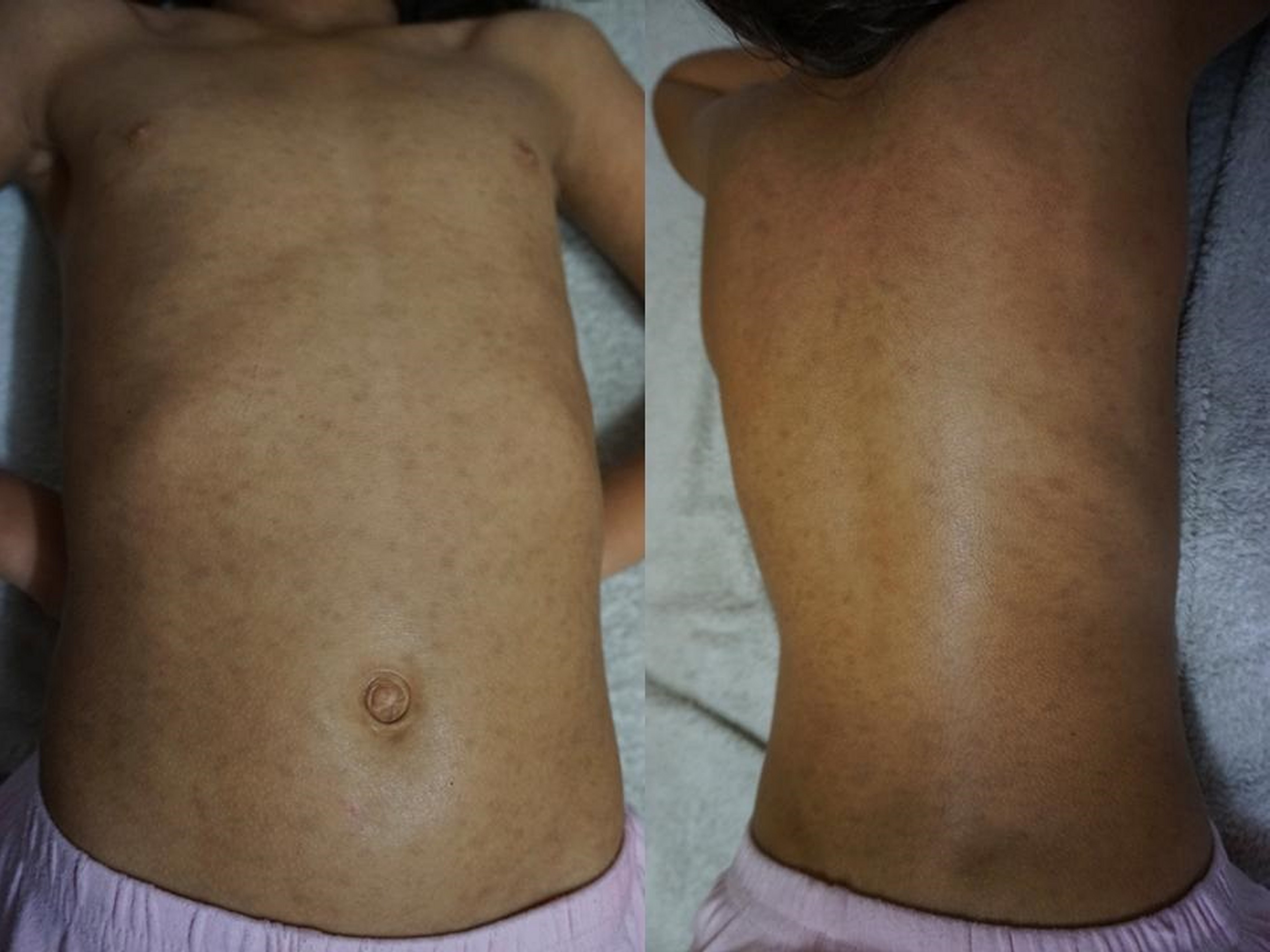
Figure 1. Multiple discrete, dark-brown, round-to-oval, small (< 1 cm in diameter), well-defined, nonconfluent macules over the front and back of the patient´s trunk.
IEMP is a rare dermatosis first described in 1978 by Degos et al. but it was in 1996 when Sanz de Galdeano et al. [1] established the following diagnostic criteria: 1) Eruption of asymptomatic, nonconfluent, brownish-black macules involving the trunk, neck, and proximal regions of the arms and legs in children or adolescents; 2) Absence of preceding inflammatory lesions; 3) No prior exposure to drugs; 4) Hyperpigmentation of the basal cell layer of the epidermis and prominent dermal melanophages without visible damage to the basal layer or lichenoid inflammatory infiltrate; 5) Normal mast cell count.
Since the publication of these criteria, they have been used regularly in case reports. In 2015, Joshi et al. [2] reviewed 24 case reports of IEMP and proposed a revised diagnostic criteria that incorporates a papillomatosis variant and emphasizes the main histological findings. The new criteria are as follows: 1) Eruption of brownish-black, discrete, nonconfluent, asymptomatic macules and/or slightly raised plaques that resemble acanthosis nigricans and involve the face, neck, trunk, and proximal extremities, with complete resolution after months to years; 2) Affects mostly children and adolescents (i.e., those in the first two decades of life); 3) Epidermal hypermelanosis with or without papillomatosis as the main histological finding with an absence of dermal inflammation; 4) A lack of numerous dermal melanophages and no presence of interface changes; 5) Absence of preceding inflammatory lesions; 6) No previous drug exposure; 7) Normal mast cell count.
IEMP is an unusual benign melanosis. Most cases have been reported in Asian populations, and few cases have been reported in white, Hispanic, and black individuals [3]. Its pathogenesis is unknown. It has been hypothesized that it is related to hormonal factors, affecting almost exclusively healthy children and adolescents [4]. Our patient developed thelarchia two months after the beginning of the IEMP and had menarche one month after its total disappearance, facts that would support this pathogenic hypothesis. The diagnosis of IEMP is based on clinical and histopathological criteria, as well as on the evolutionary course [2]. Differential diagnosis should be made with: lichen planus pigmentosum, dyschromic erythema perstans/ashen dermatosis, postinflammatory hyperpigmentation, fixed drug eruption, and urticaria pigmentosa [2, 5]. Knowledge of this rare entity guarantees a correct diagnosis and adequate counseling to avoid unnecessary treatment and to alleviate anxiety for the patient and parents, as the disease is self-limited and resolves without treatment over the course of several months to a few years. So far, there is no documentation of recurrences [4].

