


 Servicios personalizados
 Inglés (pdf)
 Articulo en XML
 Referencias del artículo
 Enviar articulo por email
 Citado por SciELO
  Citado por Google
  Similares en
SciELO
Similares en
SciELO  Similares en Google
 Permalink
Autosomal dominant polycystic kidney disease (ADPKD) is the most common inherited kidney disease, accounting for 10% of cases of chronic kidney disease (CKD) [1]. The two associated gene mutations are PKD1, which accounts for 85% of cases, has an earlier onset and a more severe course, and PKD2, which has a later onset and is less severe [2]. It is characterized by the development and expansion of multiple cysts in the renal parenchyma, with progressive loss of renal function, arterial hypertension, acute and chronic pain, macroscopic hematuria, cyst infections and nephrolithiasis [1, 2, 3]. Genetic diagnosis is not necessary if the clinical and imaging findings are clear; computed tomography (CT) is more sensitive than ultrasound, allowing the detection of cysts as small as 1-2 mm [4].
We present the case of a 42-year-old male with a history of stage V CKD diagnosed 5 years ago, receiving hemodialysis as a replacement therapy, secondary to confirmed ADPKD based on family history (father, paternal uncle, and brother with a history of ADPKD and stage V CKD), imaging, and clinical course. He was admitted to the hospital due to progressive increase in abdominal girth and generalized abdominal pain and in the lumbar region, as well as exertional dyspnea for the past 4 months. Pulse oximetry showed 91% saturation, with no evidence of cardiopulmonary compromise, only palpable renal masses in the abdomen without ascites. Laboratory tests revealed grade II anemia (hemoglobin 10.1g/dl [12 - 14 g/dl]) and blood chemistry showed impaired renal function tests (creatinine 8.7mg/dl [0.4 - 1.2 mg/dl], urea 72.1 mg/dl [12 - 54 mg/dl], blood urea nitrogen 33.7 mg/dl [6 - 20 mg/dl]) without other abnormalities. Abdominopelvic CT scan was performed, revealing massive bilateral polycystic renomegaly (Figure 1). The urology department informed the patient about the indication for bilateral laparoscopic nephrectomy due to symptomatic massive renomegaly, however, the patient declined the surgical procedure for personal reasons. Supportive measures were provided, including supplemental oxygen via nasal cannula, analgesia, and continued hemodialysis sessions following contrast study with clinical improvement. As there was no other indication for hospitalization to avoid nosocomial infections, the patient was discharged and referred to the urology outpatient clinic for follow-up.
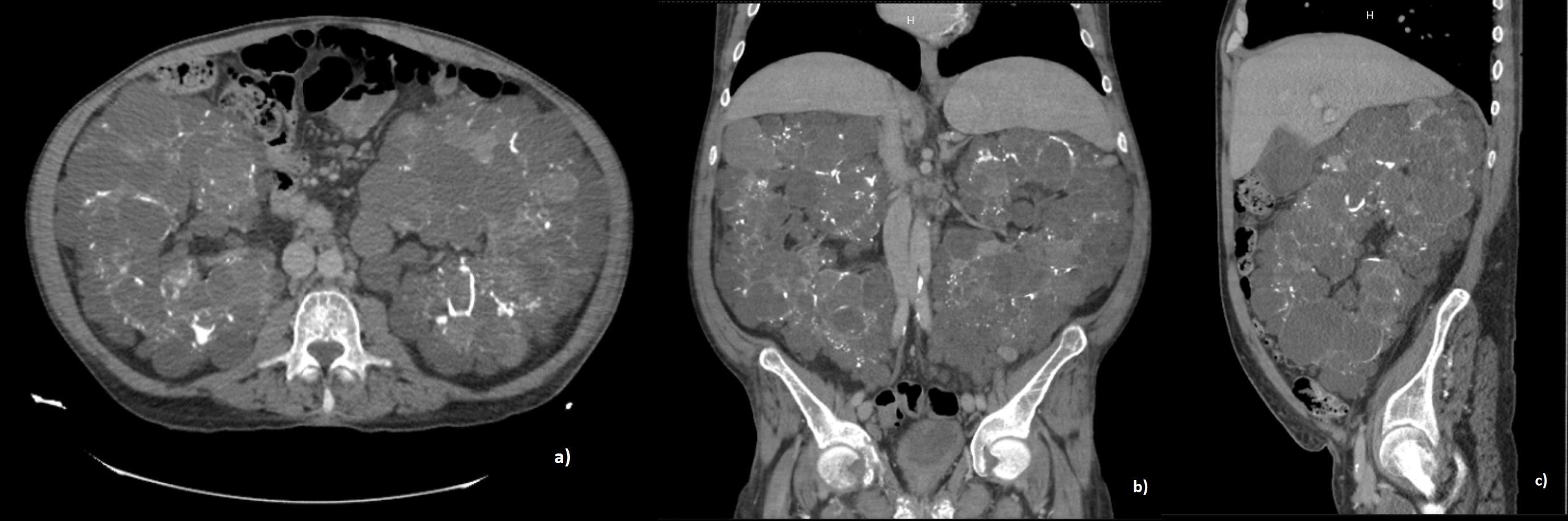
Figure 1: Abdominopelvic contrast - enhanced CT. A) Axial plane. B) Coronal plane. C) Sagittal plane. Both kidneys are enlarged, with loss of the usual morphology and lobulated borders. Multiple rounded images with well-defined hypodense borders are observed, with average attenuation values ranging from -7 to 36 HU. Some of these images have thick and calcified walls, as well as thin septa. There are other hyperdense lesions with attenuation values of up to 60 HU. None of the lesions show enhancement with contrast media. No evidence of pelvicalyceal or ureteral dilatation.
Although nephrectomy is generally avoided in the context of ADPKD due to the complications of removing a partially functional kidney, it may be considered in cases of debilitating abdominal discomfort, marked limitation of daily activities, recurrent cyst infections, recurrent macrohematuria, suspicion of renal cancer, the need for space for future transplantation, and difficult-to-control nephrogenic hypertension [4]. Currently, bilateral laparoscopic nephrectomy is a safe and effective procedure in most cases. However, in kidneys larger than 3500 ml, there is a risk of conversion to open surgery with increased intraoperative and postoperative complications [4]. Therefore, the decision to perform nephrectomy in these patients should be carefully planned, considering the risk-benefit ratio and the personal preferences of each patient.

