


Mi SciELO
 Servicios personalizados
Servicios Personalizados
Revista
Articulo
 texto en
  Inglés (pdf)
 Articulo en XML
 Referencias del artículo
 Enviar articulo por email
Indicadores
-
 Citado por SciELO
 -
 Accesos
Links relacionados
-
 Citado por Google
 -
 Similares en
SciELO
Similares en
SciELO -
 Similares en Google
Compartir

 Permalink
Revista de Osteoporosis y Metabolismo Mineral
versión On-line ISSN 2173-2345versión impresa ISSN 1889-836X
Rev Osteoporos Metab Miner vol.15 no.1 Madrid ene./mar. 2023 Epub 29-Mayo-2023
https://dx.doi.org/10.20960/revosteoporosmetabminer.00009
IMAGE IN OSTEOLOGY
Saber tibia
2Rheumatology Service. Hospital Universitario Clínico San Cecilio. Granada, Spain
CASE REPORT
A 91-year-old woman presented with a 6-month history of pain in the right tibial region, associated with bone deformity and progressive difficulty in walking. Physical examination confirmed these findings, also highlighting an increase in local temperature in the right tibial region.
A basic analytical study with biochemistry and complete blood count was carried out, including phosphocalcic metabolism parameters and bone remodeling markers. Raised levels of alkaline phosphatase (AP) in serum (141 U/L [N = 30-120]) were observed, as well as elevation of bone formation markers (type I collagen amino-terminal propeptide [PINP] 166 ng/mL, [N = 20.2-76.3]) and bone resorption markers (β-CrossLaps [β-CTX] 0.042 ng/mL [N = 0.000-0.028] and C-terminal telopeptide [ICTP] 1.28 ng/mL [N = 0.556-1]).
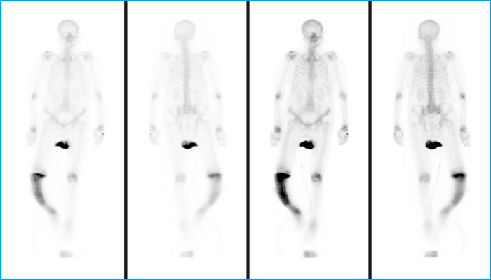
Imaging included X-rays of long bones, pelvis, thoracolumbar spine and skull which revealed a characteristic image of saber shin at the level of the right tibia (Figs. 1-3 3), and bone scan with 99mTc-HDA (Fig. 4). Given these test results, and after only finding alterations (both structural and metabolic) at the level of the right tibia, the patient was finally diagnosed with monostotic Paget’s disease of bone.
DISCUSSION
The case presented is paradigmatic of Paget’s bone disease with a saber tibial deformity. In our case, late diagnosis takes on a special meaning insofar as the observed deformity must have developed over decades without having been diagnosed until then. These highlights the importance of detecting deformities of the musculoskeletal system in any basic examination carried out in a consultation to avoid both its progression and complications derived from the disease itself.
CLINICAL IMAGES
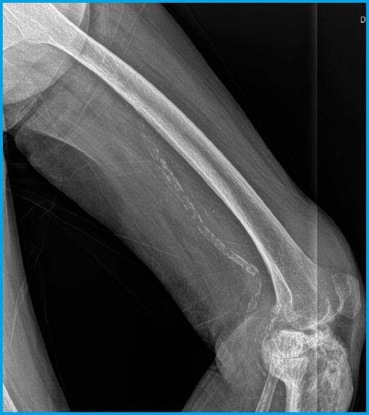
Figure 1. X-ray of the right femur and femorotibial joint: the contrast between the fine reticular trabecular pattern of the femur and the coarse and aberrant trabeculation observed in the tibial plateau stands out. Femorotibial and patellofemoral osteoarthritis. As an incidental finding, calcification of the femoropopliteal artery.
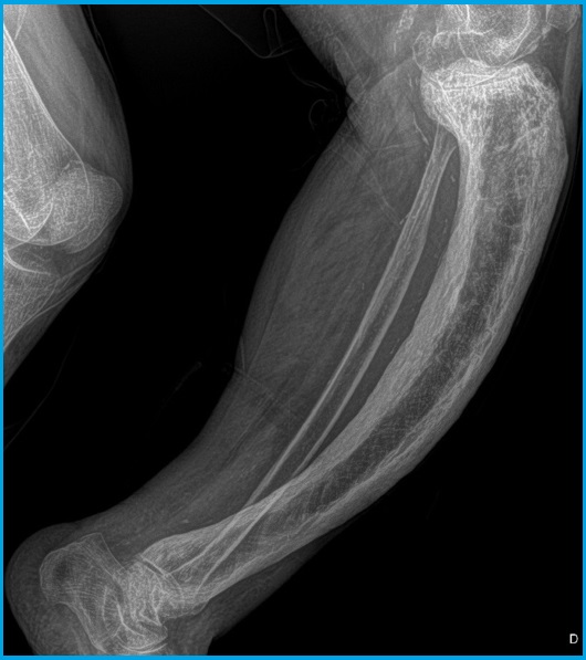
Figure 2. Radiographs of the right tibia. Saber tibia: increased cortical and periosteal thickness, with a coarse and disordered trabecular pattern, as well as a large tibial deformity, which curves laterally with a saber appearance.
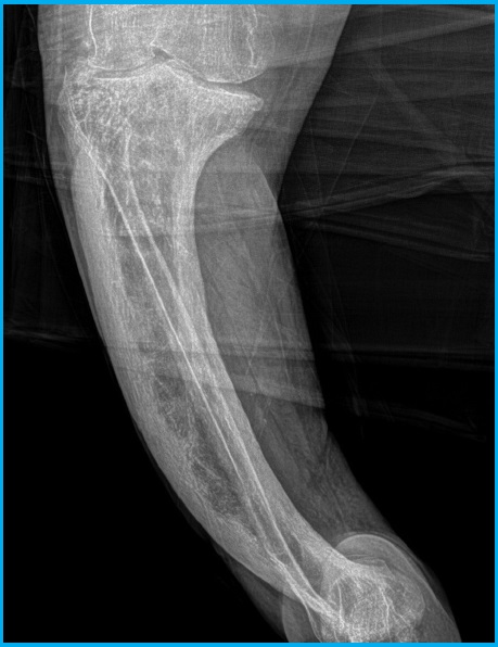
Figure 3. Radiographs of the right tibia. Saber tibia: increased cortical and periosteal thickness, with a coarse and disordered trabecular pattern, as well as a large tibial deformity, which curves laterally with a saber appearance.
Received: June 24, 2022; Accepted: October 10, 2022
 This is an open-access article distributed under the terms of the Creative Commons Attribution License
