


Mi SciELO
 Servicios personalizados
Servicios Personalizados
Revista
Articulo
 texto en
  Inglés (pdf)
 Articulo en XML
 Referencias del artículo
 Enviar articulo por email
Indicadores
-
 Citado por SciELO
 -
 Accesos
Links relacionados
-
 Citado por Google
 -
 Similares en
SciELO
Similares en
SciELO -
 Similares en Google
Compartir

 Permalink
Revista Española de Enfermedades Digestivas
versión impresa ISSN 1130-0108
Rev. esp. enferm. dig. vol.104 no.10 Madrid oct./nov. 2012
https://dx.doi.org/10.4321/S1130-01082012001000008
PICTURES IN DIGESTIVE PATHOLOGY
EUS visualization of the spinal cord from the cervical esophagus: an unusual finding
Visualización por ecoendoscopia de la médula espinal desde el esófago cervical: un hallazgo inusual
Enrique Vázquez-Sequeiros
Unit of Endoscopy. Service of Gastroenterology. Hospital Universitario Ramón y Cajal. Madrid, Spain
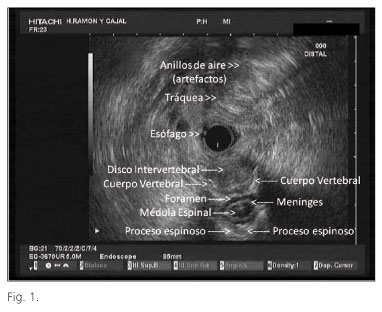
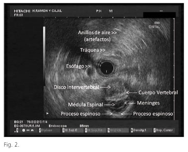
Endoscopic ultrasound (EUS) allows one to efficiently image the posterior mediastinum in search for lesions (1). In patients who present with dysphagia EUS has been shown to be useful, as it may help identify rare entities causing these symptoms (e.g. dysphagia lusoria caused by aberrant right subclavian artery; dysphagia caused by an anterior cervical spine osteophyte) (2-4). These infrequent causes of dysphagia and their EUS appearance were relatively unknown for most endosonographers until its recent publication in endoscopy journals (2-4). The case of a 62-year-old male with unremarkable past medical history who was referred for EUS due to unexplained dysphagia (normal manometry, radiology and endoscopy) is presented. EUS examination of the upper mediastinum showed no lesion or compression in the esophageal wall (vascular or spine) that may be responsible for symptoms. Surprisingly, when searching for causes of these symptoms we identified a "roundish" lesion posterior to the esophagus, with "solid and cystic/anechoic component". The aforementioned lesion was only visible between two spinal vertebral bodies, at the level of the intervertebral disc that allowed ultrasound waves to reach the spinal cord (Figs. 1 and 2). This atypical finding may be difficult to interpret and may misslead the endosonographer towards an incorrect diagnosis like "posterior mediastinum tumor".
References
1. ASGE Standards of Practice Committee, Jue TL, Sharaf RN, Appalaneni V, Anderson MA, Ben-Menachem T, Decker GA, et al. Role of EUS for the evaluation of mediastinal adenopathy. Gastrointest Endosc 2011;74:239-45. [ Links ]
2. Yusuf TE, Levy MJ, Wiersema MJ, Clain JE, Harewood GC, Rajan E, et al. Utility of endoscopic ultrasound in the diagnosis of aberrant right subclavian artery. J Gastroenterol Hepatol 2007;22:1717-21. [ Links ]
3. González-Panizo-Tamargo F, Juzgado-Lucas D, Vázquez-Sequeiros E. Endosonographic diagnosis of aberrant right subclavian artery that leads to disphagia lusoria. Rev Esp Enferm Dig 2011;103:497-8. [ Links ]
4. Peter S, Degen L. A spur sign in the EUS evaluation of dysphagia. Gastrointest Endosc 2008;68:147-8. [ Links ]

