


Meu SciELO
 Serviços customizados
Serviços Personalizados
Journal
Artigo
 texto em
  Inglês (pdf)
 Artigo em XML
 Referências do artigo
 Enviar este artigo por email
Indicadores
-
 Citado por SciELO
 -
 Acessos
Links relacionados
-
 Citado por Google
 -
 Similares em
SciELO
Similares em
SciELO -
 Similares em Google
Compartilhar

 Permalink
Archivos Españoles de Urología (Ed. impresa)
versão impressa ISSN 0004-0614
Arch. Esp. Urol. vol.62 no.1 Jan./Fev. 2009
Giant intrascrotal lipoma
Lipoma intraescrotal gigante
Pastor Casas Agudo, José Manuel Janeiro Pais, Luis Busto Castañón, Daniel López García and Juan González Dacal
Urology Service. Hospital Juan Canalejo. La Coruña. Spain.
We present the case of a 30 year-old man consulting for painless increase of the right scrotum over the last two years. The only antecedent he had was surgical excision of a lipoma of the back two years before. On physical examination n we detected an increase in size of the right hemiscrotum where we could palpate a 15 centimeter mass, independent of the testicle, which displaced it to the inguinal duct (Figure 1).

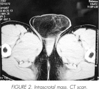
Tumor markers were negative and CT scan was informed as 13 centimetres right intraescrotal mass compatible with lipoma displacing the right testicle towards the inguinal duct (Figure 2).
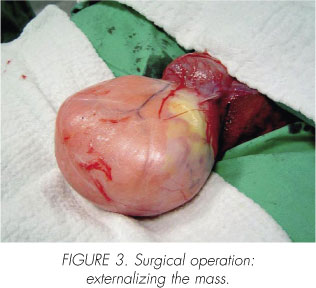
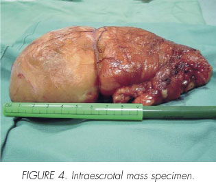
Operattion was carried out removing the mass that extended up to the perineal rafe (Figure 3). Pathology informed lipoma without signs of malignancy (Figure 4).
Paratesticular tumors are rare among intrascrotal neoplasms in comparison with testicular tumors. About 70% are benign, being the most frequent type originating from spermatic cord's fatty tissue.
 Correspondence:
José Manuel Janeiro Pais
Lázaro Cárdenas, 23 - 1ºE
15009 La Coruña (Spain).
janeiropais@canalejo.org

