Meu SciELO
Serviços Personalizados
Journal
Artigo
Indicadores
-
 Citado por SciELO
 -
 Acessos
Links relacionados
-
 Citado por Google
 -
 Similares em
SciELO
 -
 Similares em Google
Compartilhar
Revista Española de Enfermedades Digestivas
versão impressa ISSN 1130-0108
Rev. esp. enferm. dig. vol.101 no.12 Madrid Dez. 2009
LETTERS TO THE EDITOR
An 84-year-old woman who presents with edema in lower limbs
Mujer de 84 años que consulta por edema en miembros inferiores
Key words: Elderly. Malabsorption. Intestinal stenosis.
Palabras clave: Anciano. Malabsorción. Estenosis intestinal.
Dear Editor,
The most frequent causes of edema in lower limbs are heart failure and handle of sodium impairment (renal failure, cirrhosis, etc). Malabsorptive disorders can also be the origin of edema but that only happens infrequently and in advanced stages (1).
Case report
An 84-year-old woman presented with a one-year history of progressive edema in legs with the development, two months before admission, of erythema and cutaneous blisters. The patient's medical history was notable only for type 2 diabetes mellitus in treatment with oral agents, having undergone surgery after a right subcapital hip fracture and also due to a left strangulated inguinal hernia a year before the current admission (without intestinal resection). In the comprehensive history she referred changes in bowel habit, after the surgical repair of the inguinal hernia, with episodes of constipation and watery yellowish diarrhea, borborygmi and increased abdominal girth, without consulting her physician. An 8-kg weight loss was also associated since the beginning of the symptoms. She did not complain of abdominal pain.
On admission blood pressure was 100/57 mmHg, temperature 37.2 oC and heart rate 90 beats per minute. Glycemic capillary index was 80 mg/dl. She had a deficient nutritional state (weight 36 kg, body mass index 16). Jugular venous pressure and cardiac and lung auscultation were normal. The patient's abdomen was slightly distended, with tympany and increased bowel sounds, without organomegaly or masses. There was pitting edema in lower extremities, up to the knees, with inflammatory signs and flaccid blisters, some of them eroded. Initial laboratory data were unremarkable but for a normocytic normochromic anemia -hemoglobin 10.7 g/dl-, the erythrocyte sedimentation rate -52 mm-, hypoproteinemia and hypoalbuminemia -total protein 4.31 g/dl, serum albumin 1.99 g/dl-. Chest radiography, electrocardiogram and echocardiography ruled out the presence of cardiopathy. There was no evidence of proteinuria or nephropathy.
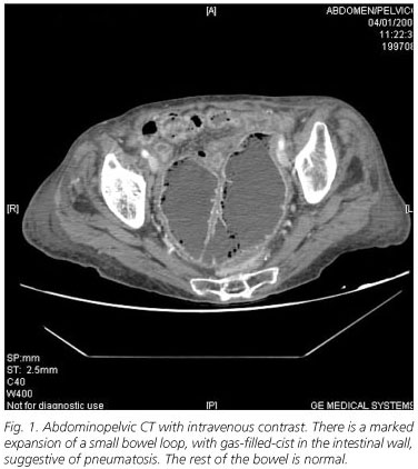
Suspecting intestinal protein loss an abdominal radiography and computed tomography with intravenous contrast were obtained (Fig.1). After these findings were described, decompression with a nasogastric tube was performed. Total parenteral nutrition was also administered. The surgical intervention was diagnostic and therapeutic. A laparotomy showed a 20 cm-stenotic intestinal -jejunal- segment, that was resected with intestinal anastomosis. Pathological examination disclosed no neoformative process or acute focal ischemia, being the final diagnosis mucous and segmental mural infarct with dense chronic inflammatory infiltrate, composed of lymphoplasmocytoid cells, in the theoretical area of the previous abdominal surgery (strangulated hernia). The patient was discharged, without symptoms, nineteen days after her admission. At that moment the serum albumin was 2.52 g/dl and, at a 5-month follow-up visit, 3.83 g/dl.
The patient's final diagnosis was a malabsorptive disorder due to a -jejunal- intestinal stenosis.
Discussion
Malabsorptive causes in the elderly are the same than in other groups of patients, although bacterial overgrowth, after small intestine motor or anatomic abnormalities, has a special importance (2,3). Malabsorption usually manifests insidiously and thus the diagnosis is delayed, giving way to more severe nutritional deficiency states (2,3). It leads to weight loss, hypoproteinemia and hypoalbuminemia, and, if a partial intestinal disorder is present, to specific nutrient deficits (1). Stenoses (ischemic or radiation enteritis, neoplasms, etc.) are clinical situations that favour bacterial overgrowth as the cause of malabsorption (5). Ineffective mechanical cleansing of bacteria and the increased intraluminal intestinal contents lead to steatorrhea and acid watery diarrhea, the production of abundant intestinal gas, distention and borborygmi (1).
Useful tests in the diagnosis of malabsorptive disorders are laboratory studies such as the D-xylose absorption, carbohydrate or hydrogen breath tests or alpha 1-antrypsin clearance, but they are not available in all hospitals. Besides of that, some of them are less useful in elderly patients because of renal impairment (D-xylose test) (2). The diagnosis of intestinal stenoses is made with radiography studies (above all, computed tomography with intravenous contrast) and anatomopathologic examination (5,6).
S. Sanz-Baena, G. Pérez-Martín, E. Berrocal-Valencia, M. J. Moro-Álvarez, V. Delgado-Cirerol,
M. Sarró-Cañizares1, J. Carvajal-Balaguera2 and J. Lacasa-Marzo
Services of Internal Medicine, 1Radiodiagnosis and 2General Surgery.
Hospital Central de la Cruz Roja San José y Santa Adela. Madrid, Spain
References
1. Fernández Castroagudín J, Domínguez Muñoz JE. Síndrome de malabsorción intestinal (I). Medicine 2004; 9(3): 159-71. [ Links ]
2. Holt PR. Diarrhea and malabsorption in the elderly. Gastroenterol Clin North Am. 2001; 30(2): 427-44. [ Links ]
3. Lobo B, Casellas F, Torres I, Chicharro L, Malagelada JR. Utilidad de la biopsia yeyunal en el estudio de la malabsorción intestinal en el anciano. Rev Esp Enferm Dig 2004; 96(4): 259-64. [ Links ]
4. Carvajal J, Gómez-Pavón J, Camuñas J, Martín M, Oliart S, Macho O, et al. Enfermedad obstructiva del intestino delgado en el paciente geriátrico: diagnóstico diferencial. Mapfre Medicina 2006; 17: 81-9. [ Links ]
5. Holt PR. Intestinal malabsorption in the elderly. Dig Dis 2007; 25(2): 144-50. [ Links ]
6. Megibow AJ, Balthazar EJ, Cho KC, et al. Bowel obstruction: evaluation with CT. Radiology 1991; 180: 313. [ Links ]


 texto em


