


Meu SciELO
 Serviços customizados
Serviços Personalizados
Journal
Artigo
 texto em
  Inglês (pdf)
 Artigo em XML
 Referências do artigo
 Enviar este artigo por email
Indicadores
-
 Citado por SciELO
 -
 Acessos
Links relacionados
-
 Citado por Google
 -
 Similares em
SciELO
Similares em
SciELO -
 Similares em Google
Compartilhar

 Permalink
Revista Española de Cirugía Oral y Maxilofacial
versão On-line ISSN 2173-9161versão impressa ISSN 1130-0558
Rev Esp Cirug Oral y Maxilofac vol.31 no.5 Madrid Set./Out. 2009
Report of a case of Fibrous Ameloblastic Odontoma
Fibro-odontoma ameloblástico: a propósito de un caso
P. Reyes Olave1, C. Álvarez Novoa2, C. Muñoz Torres3
1 Cirujano Maxilofacial.
2 Cirujano Dentista.
3 Cirujano Maxilofacial.
Universidad de Talca. Chile
ABSTRACT
Introduction. Fibrous Ameloblastic Odontoma (FAO) is an uncommon benign odontogenic tumor whose characteristics are generally similar to those of an ameloblastic fibroma except that that it has varying amounts of calcified tissue. It is usually found associated with compacted teeth and its incidence is slightly more common in the inferior maxilla.
Materials and Methods. 17-yearold patient goes to the doctor because of a lesion located near the right mandible angle, found in an X-ray. After performing a physical exam, histological tests and taking images it was diagnosed as FAO and then surgically removed. Results. Although the patient fell into the typical age range and they FAO was located in the typical area, the definitive diagnostic could only be decided until after the results of the histological study of the removed tooth were reviewed.
Discussion. FAO diagnosis normally creates confusion from both a histological and a clinical radiographic point of view. It is difficult differentiate between FAO and Complex Immature Odontoma (OIC). Like in this case, the differences are detected later when the histological study is complete.
Conclusion. FAO is independent from OIC and its diagnostic greatly depends on the histological discoveries and the clinical radiographic correlation carried out when the pathology is first dealt with.
Key words: Fibrous Ameloblastic Odontoma; Complex Immature Odontoma.
RESUMEN
Introducción. El fibroodontoma ameloblástico (FOA) es un tumor odontogénico benigno poco frecuente, con características generales de fibroma ameloblástico, pero con cantidades variables de tejido calcificado. Se encuentra habitualmente asociado a piezas dentarias incluidas y su incidencia es ligeramente mayor en el maxilar inferior.
Material y método. Paciente de 17 años que consulta por una lesión ubicada a nivel del ángulo mandibular derecho, encontrada como hallazgo radiológico. Luego de realizar exámenes físicos, radiológicos e histológicos es diagnosticado un FOA, el cual es eliminado quirúrgicamente.
Resultados. A pesar de que el paciente se encontraba dentro del rango de edad y localización típica de un (FOA), el diagnóstico definitivo se pudo alcanzar solo con el estudio histopatológico de la pieza operatoria, posterior a la cirugía.
Discusión. El diagnóstico de FOA frecuentemente puede generar confusión tanto desde el punto de vista clínico-radiológico como histopatológico, este es una entidad difícilmente diferenciable de OCI. Como ocurrió en este caso, las diferencias pueden ser detectadas a partir de un estudio histopatológico.
Conclusión. FOA es una entidad independiente de un OCI y su diagnóstico depende en gran parte de los hallazgos histológicos, y de la correlación clínico- radiológica realizada al momento de enfrentar esta patología.
Palabras clave: Fibroodontoma ameloblástico; Odontoma complejo inmaduro.
Introduction
Fibrous Ameloblastic Odontoma (FAO) is an uncommon benign odontogenic tumor that has characteristics similar to those of Ameloblastic Fibroma. The difference is that it has variable amounts of calcified tissue.1 Its incidence of appearance is from 1-3%. Compared with all odontogenic tumors its incidence is slightly higher in the inferior maxilla and its location is most common in the premolar and molar region of the maxilla. They are normally found in the first twenty years of ones life, they are equally as common in men as in women. It is clinically asymptomatic and caused by change of the dental eruption.4 FAO diagnosis can cause confusion from the clinical radiographic point of view as well as a histological point of view. The similarities between these distinct pathological entities, the distribution and quantity of the odontogenic tissues can cause diagnostic errors. The following article describes a case of FAO in a 17 year old male Chilean patient and its clinical and histological characteristics patient. This study emphasizes the diagnostic analysis, also including clinical, image and histopathological aspects.
Clinical Case
Male patient, 17 years-old, goes o the Maxillofacial Surgery Service at the University of Talca because of a lesion on his right mandible angle. The lesion was discovered during a routine lateral cervical teleradiography after an automobile accident.
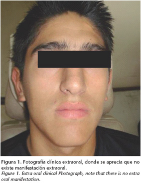
The patient did not report any prior personal or family diseases. He did not smoke, drink alcohol or take drugs, nor medications of any kind. The physical exam did not reveal anything suspicious either. The extra oral clinical exam didn't show any signs of aesthetic or functional changes. Upon palpation there was a small augment in the volume of the zone posterior to the mandible vestibule.
There was an extensive unilocular image was seen in the X-ray. It had mixed density, was radiopaque, surrounded by a radio lucid band with cortilcalized nets located near the 4.8 tooth, partially over projecting the roots of the 4.7 tooth (Fig. 3). The boney table balloons and rejects the 4.8 tooth towards the distal cephalic. The mandible canal is found displaced towards the basilar border of the corresponding part of the mandible body and rejecting the distal of the ascending ramus.


The study was complimented with a CAT that found a hyper dense lesion, with net and cortical limits, enlargement of the bone tables. Its dimensions were: from anterior to posterior 25.9 mm, from vestibule to playtime 21 mm and 28.3 mm was the largest cephalous caudal diameter.
Acquiring the images was done using processing software called Dentascan®. It provides axial reconstruction, panorex and transversals of the area that can be used to change the proximity to the mandible canal and also the commitment of both tables (Fig. 5).


The differential diagnostic for this case was complete immature odontoma, ameloblastic fibro odontoma, calcified epithelial odontogenic tumor, and calcified epithelial odontogenic cyst.
After the radiological study a biopsy was carried out. The incision was made under local anesthetic that included both soft and hard tissues. Calcified dental tissue with less calcified areas and loose fibrous connective tissue was observed, it was similar to stellate reticulum of the dental organ. In the peripheral we see fibrous connective tissue and some small areas that show odontogenic epithelium that show signs of ameloblastic cylindrical peripheral cells. The sample was irregular and there were no denticle structures. According to this, the histological diagnosis was a compatible lesion with forming odontoma and ameloblastic changes.
The enucleation of the lesion was carried out under general anesthetic, using an extra oral approach to the right mandible angle to allow for modeling and placement of a rigid osteosynthesis. In this way we accede to the affected area where we find a disappearing vestibular table. We removed it so that the lesion could be seen and completely removed (Fig. 6 and 7), and finally we performed curettage of the surgical floor. The remaining healthy bone tissue was very poor so a 2.4 reconstruction plate was placed near the angle and body of the mandible to grant more resistance and avoid a pathological fracture. The graft wasn't scheduled immediately due to the possibility of relapse.


In the histopathological test the operated tooth had a higher quantity of ameloblastic tissue which leads us to a diagnosis of Ameloblastic Fibro odontoma (Fig. 8 and 9).


There were no complications healing the wound. Although this lesion has a low recurrence rate, the patient is regularly tested and there were no signs of recurrence to date.
Discussion
The FAO is a mixed odontogenic benign tumor. OMS defines it as a lesion that is similar to ameloblastic fibroma with inductive changes that stimulate the formation of enamel and dentine. It is usually found in young patients who have no genetic predisposition.8 The lesions are usually diagnosed during the first 10 or 20 years of life, 12 was the average age in the literature. The main clinical signs are a painless increase in volume in the affected region and changes in dental eruption. There has been no evidence of malignization.6
The X-Ray shows a well defined radio lucid area that has undefined interior quantities of radiopaque materials that are irregular in size and shape. The extension of these radiopaque and radio lucid areas differs from one lesion to the next making it difficult to diagnose. Sometimes the quantity of mineralized material prevails resembling complex odontoma.6 It can create variable expansion of the corticals and become associated with a retained tooth, which can have its eruption interrupted or be displaced.
The FAO is histologically characterized by having cords and nidus of epithelium mixed with a variable amount of calcified tissue in a myxomatus stroma, hence it could be considered to be a combination of ameloblastic fibroma (AF)2 and complex odontoma (CO).6 The etiology is unknown, although many authors suggest that FAO is an immature CO that is found in a histological differentiation period and hence after an undetermined period of time it would transform into a mature or completely differentiated odontoma. In accordance with this, the clinical data for each one of these entities should back up this theory. In other words the FAO should be found in younger patients and CO should be found in older patients. Furthermore the predisposition based on sex and distribution of these lesions should be the same.13
Just like during this case study, because of its clinical and radiological similarity both pathological entities should be considered differential diagnosis. Since when FAO reunites all the clinical characteristics of location, age and X-ray the initial diagnosis is pretty simple.4,6 However when one of the characteristics doesn't coincide it complicates the initial diagnosis (presumptive). In this case, although the patient was within the age range and typical location for FAO, the definitive diagnosis was reached when the histological study was performed of the operated tooth. The tumor mass was found surrounded by a fibrous capsule compounded by a matrix of fibroblastic connective tissue chords and odontogenic epithelium and immature dental structures including enamel and dentine. Finally differentiating itself from a complex odontoma. Dentine deposits, enamel, cement and pulp tissue were totally available at random, but with less quantity of epithelial and connective tissue.
The differential diagnosis also includes lesions that have mixed radiographic patterns like a calcified epithelial odontogenic tumor and a calcified odontogenic cyst.10 These entities are also related to retained teeth and have locations similar to those of FAO, however, its histology is completely different.
The treatment of choice for this entity is conservative surgery, performing a tedious curettage of the surgical floor to decrease the possibility of relapse as much as possible. There are reports of enucleation surgeries without curettage that preserve the included tooth, which were precautions that were taken when treating this case.6,5
Conclusion
After analysis we conclude that the diagnosis of these pathologies depends a lot on the histological discoveries and the clinical/radiographic correlation that we do when faced with one of these entities. Despite the histological similarities between FAO and CO, both are independent lesions that can be histologically differentiated.
![]() Correspondence:
Correspondence:
Carolina Álvarez
Universidad de Talca. Chile
Recibido: 15.07.2008
Aceptado: 25.08.2009
References
1. Arab Oghli A, Scuto I, Ziegler C, Flechtenmacher C, Hofele C. A large ameloblastic fibroodontoma of the rigth mandible. Med Oral Patol Oral cir bucal 2007; 12:E34-37. [ Links ]
2. Azúa-Romeo J, Saura Fillat E, Usón Bouthelier T, Tovar Lázaro M, Azúa BlancoJ. Fibroma ameloblástico versus quiste folicular hiperplásico. Rev Esp Cir Oral Maxilofac 2004;26. [ Links ]
3. Leon Barnes JW. Eveson, Reichart P, Sidransky D. Pathology & Genetics. Head and Neck Tumours. World Health Organization Classification of Tumours. Lyon, 2005. [ Links ]
4. Carvalho G, Correia B, Carvalho E, Rebello M, Pereira S, Santiago R. Case Report: Ameloblastic Fibro-Odontoma. Oral Oncology Extra 2006;42:217-20. [ Links ]
5. Casap N, Zeltser R, Abu-Tair J, Shteyer A. Removal of a Large Odontoma by Sagittal Split Osteotomy J Oral Maxillofac Surg 2006;64:1833-6. [ Links ]
6. Chang H, Shimizu M, Precious D. Ameloblastic Fibro-Odontoma: a case Report. J Can Dent Assoc 2002;68:243-6. [ Links ]
7. Domínguez Cuadrado L, Martín-Granizo López R. Análisis clínico, radiológico e histológico de los fibromas cemento-osificantes de los maxilares. Rev Esp Cir Oral Maxilofac 2004;26. [ Links ]
8. Furst I, Pharoah M, Phillips J. Recurrence of an ameloblastic fibroodontoma in a 9-year-old boy. J Oral Maxillofac Surg 1999;57:620-3. [ Links ]
9. Huguet P, Castellvi J, Avila M, Alejo M, Autonell F, Basas C, et al. Ameloblastic fibrosarcoma: report of a case. Immunohistochemical study and review of the literature. Med Oral 2001;6:173-9. [ Links ]
10. Morales B, Carvajal L. Quiste odontogénico calcificante (Quiste de Gorlin). Reporte de un caso y su seguimiento. Revisión de la literatura. Revista ADM 1999;2:83-7. [ Links ]
11. Regezi J, Sciubba J. Patología Bucal. Correlaciones Clinicopatológicas. México. McGraw-Hill Interamericana Editores, S.A. 2004. [ Links ]
12. Nikolaos Sakkas N, Schramm A, y cols. The Ameloblastic Fibroodontoma of the Maxilla: Case Report of a Child with Schimmelpenning-Feuerstein-Mims Syndrome/Skin-Eye-Brain-Heart Syndrome. J Oral Maxillofac Surg 2006;64:524-7. [ Links ]
13. Shafer W, Levy B, Maynard H. Tratado de Patología Bucal. México. Nueva Editorial Interamericana 1986. [ Links ]
14. Wood N, Goaz P. Diagnóstico Diferencial de las Lesiones Orales y Maxilofaciales. Madrid 1999. [ Links ]
15. Yagishita H, Taya Y, Kanri Y, Matsuo A, Nonaka H, Fujita H, Aoba T. The secretion of amelogenins is associated with the induction of enamel and dentinoid in an ameloblastic fibro-odontoma. J Oral Pathol Med 2001;30:499-503. [ Links ]

