Meu SciELO
Serviços Personalizados
Journal
Artigo
Indicadores
-
 Citado por SciELO
 -
 Acessos
Links relacionados
-
 Citado por Google
 -
 Similares em
SciELO
 -
 Similares em Google
Compartilhar
Medicina Oral, Patología Oral y Cirugía Bucal (Ed. impresa)
versão impressa ISSN 1698-4447
Med. oral patol. oral cir. bucal (Ed.impr.) vol.9 no.4 Ago./Out. 2004
Odontoameloblastoma: A case report and a review of the literature
MARTÍN-GRANIZO-LÓPEZ R, LÓPEZ-GARCÍA-ASENJO J, DE PEDRO-MARINA M, DOMÍNGUEZ-CUADRADO L.ODONTOAMELOBLASTOMA: A CASE REPORT AND A REVIEW OF THE LITERATURE. MED ORAL 2004;9:340-4.
ABSTRACT
Odontoameloblastoma (OA) is an extremely rare mixed odontogenic tumor appearing within the maxillary bone, with both epithelial and mesenchymal components. The term odontoameloblastoma (OA) was included in the 1971’s WHO classification. Only 23 well-documented cases have been reported in the medical literature. Because of their rarity, controversy exists in the treatment of this tumor. We present a new case of OA involving the mandible mimicking a compound odontoma and a brief review of the related literature.
Key words: Odontoameloblastoma, ameloblastic odontoma, compound odontoma, composite odontoma.
INTRODUCTION
Odontoameloblastoma (OA) is an extremely rare mixed odontogenic tumor appearing within the maxillary bone, with both epithelial and mesenchymal components. Commonly, its clinical presentation mimics an odontoma, and therefore the definite diagnosis is based on the histologic analysis following a simple extirpation and curettage. It is also known as ameloblastic odontoma, although the term odontoameloblas-toma (OA) was included in the 1971’s WHO classification and seems to be more appropriate due to the behavior of the tumor like an ameloblastoma rather than as an odontoma. It is an ameloblastoma in which focal differentiation into an odontoma appears, and since Thoma et al. described in 1944 the first case (1), only 23 well-documented cases have been reported in the medical literature (2-17). Because of their rarity, controversy exists in the treatment of this tumor.
We present a new case of OA involving the mandible mimicking a compound odontoma and a brief review of the related literature.
CLINICAL CASE
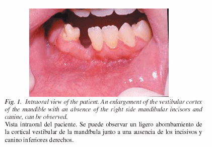
A 12-year-old Caucasian female was referred to the Department of Oral and Maxillofacial Surgery for the complaint of a lack of eruption of the permanent right side inferior incisors and canine during orthodontic treatment. She also referred occasional pain and gingival swelling. Her past medical history included a focal epilepsy and no allergies. On examination, an absence of the right side mandibular incisors and canine was observed with a slight enlargement of the vestibular cortex of the mandible over that site (Fig 1).
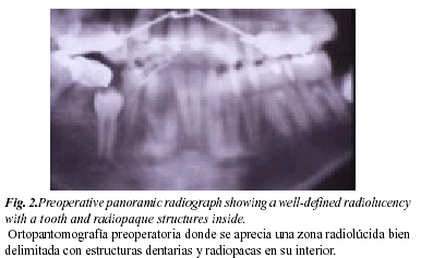
Radiographic test included a panoramic tomography and a periapical radiography, where a cystic-like intraosseous lesion was found, measuring 20 X 30 mm and showing a well-shaped margin with both radiolucid and radiopaque images inside. In addition, some radiopaque structures resembling a tooth could be observed within the cyst along with a lateral displacement of the neighbour dental roots without apical resorption (Fig. 2). With an initial diagnosis of compound odontoma of the inferior jaw, the patient underwent surgery with local anesthesia, although the cyst could not be completely removed due to a lack of patient cooperation. Consequently, a second surgery was carried out with general anesthesia in an outpatient basis, and a complete enucleation of the tumor with curettage of the underlying bone was performed.
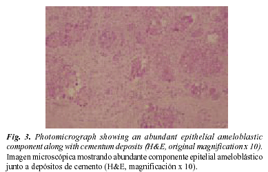
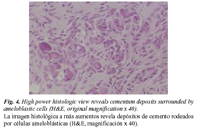
Histologic analysis revealed a compound odontoma with areas of ameloblastic proliferative cells. Microscopically, a proliferation in nests and strands of odontogenic epithelium in a stockade fashion along with stellate cells in the center of the nests, could be observed (Fig 3). The stroma surrounding the epithelial nests showed fibromyxoid areas and collagen zones with abundant deposits of cementum surrounded by ameloblasts and scattered rests of dentine (Fig 4).
Therefore, a periodical control follow-up was carried out including clinical examination and TC, without evidence of recurrence 18 months later.
DISCUSSION
The marked histological polymorphism of odontogenic tumors make the final diagnosis difficult and in some cases it must be made based on clinical, radiologic and histopathologic features. Therefore, confusion exists when describing odontoameloblas-toma (OA), and it has been reported under a variety of names (10). Moreover, Wachter et al. could not histologically differentiate between OA and ameloblastic fibro-odontoma, and herefore suggest that they are the same entity (18). Other authors have found only 19 cases that met the clinicopathological criteria for OA out of 43 reported cases (7,15,17). This tumor is not exclusive of the human race and has also been described in sheep, monkeys, cats and rats.OA affects predominantly to young patients with a median age of 20.12 year-old (2.7-52) in the reported cases, appearing 59% in patients under 15 year-old (Table 1). There is a slight predilection in males and Caucasian and Oriental races (2-17). The incidence of OA is very low; Stypulkowska (14), in a review of 164 odontogenic tumors found one single case (0.6%), and Raubenheimer et al. (13), reported one case out of 108 ameloblastomas (0.9%). This tumor usually occurs in the posterior segments of either jaw, with a slight inclination for the mandible. Only three cases have been reported involving the anterior segment of the mandible, including the present case (8,13). Clinically, OA debut as a slow-growing painless mass that expands the alveolus and vestibular cortex, and courses with an absence of permanent teeth eruption. Radiologic examination usually reveals a multilocular radiolucency with radiopaque areas within resembling mature dental tissue. It commonly exhibits a well defined margin, displacing the surrounding erupted teeth rather than producing root resorption (12,18). Therefore, in many cases OA are often confused with compound or complex odontomas, as in the present case (16,18).
Histopathology of OA reveal islands of odontogenic epithelium resembling an ameloblastoma, surrounded by the mesenchymal component including enamel, dentine and cementum structures in variable degrees of maturity (18). Additionally, dendritic cells containing melanin pigment and fine granules and coarse aggregates of melanin distributed in the epithelial cells of OA have been occasionally described, although its origin and significance is still unknown (9). Furthermore, a squamous differentiation similar to those of the keratocyst and the calcifying odontogenic cyst has been observed (10,18).
Differential diagnosis must be made with mixed radiographic patterns such as ameloblastic fibro-odontoma, calcifying epithelial odontogenic tumor (CEOT), and odontoma. Ameloblastic fibro-odontoma affects adult patients in a slow growing fashion and exhibits a well-defined capsule and a lack of infiltration to the surrounding bone. This permits an easy surgical enucleation with a low recurrence potential. Histologically, it presents immature dental tissue "papila-like" with an absence of ameloblastic differentiation.
REFERENCES
1. Thoma KH, Johnson GJ, Ascario N. Case 43: Adamanto-odontoma. Am J Orthod Oral Surg 1944;30:244. [ Links ]
2. Thoma KH, Goldman HM. Odontogenic tumors: A classification based on observations of the epithelial, mesenchymal, and mixed varieties. Am J Pathol 1946;22:433. [ Links ]
3. Silva CA. Odontoameloblastoma. Oral Surg 1956;9:545. [ Links ]
4. Frissell CT, Shafer WG. Ameloblastic odontoma: Report of a case. Oral Surg Oral Med Oral Pathol 1963;16:582. [ Links ]
5. Choukas NC, Toto PD. Ameloblastic odontoma. Oral Surg Oral Med Oral Pathol 1964;17:10. [ Links ]
6. Jacobsohn PH, Quinn JH. Ameloblastic odontomas: Report of three cases. Oral Surg Oral Med Oral Pathol 1968;26:829-36. [ Links ]
7. LaBriola JD, Steiner M, Bernstein ML, Verdi GD, Stannard PF. Odontoamelo-blastoma. J Oral Surg 1980;38:139-43. [ Links ]
8. Gupta DS, Gupta MK. Odontoameloblastoma. J Oral Maxillofac Surg 1986; 44:146-8. [ Links ]
9. Takeda Y, Kuroda M, Suzuki A. Melanocites in odontoameloblastoma: A case report. Acta Pathol Jpn 1989;39:465-8. [ Links ]
10. Thompson IOC, Phillips VM, Ferreira R, Housego TG: Odontoameloblas-toma: A case report. Br J Oral Maxillofac Surg 1990; 28:347-9. [ Links ]
11. Kaugars GE, Zussmann HW. Ameloblastic odontoma (Odontoameloblas-toma). Oral Surg Oral Med Oral Pathol 1991;71:371-3. [ Links ]
12. Gunbay T, Gunbay S. Odontoameloblastoma: Report of a case. J Clin Pediatr Dent 1993;18:17-20. [ Links ]
13. Raubenheimer EJ, Van Heerden WF, Noffke CE. Infrequent clinicopathologi-cal findings in 108 ameloblastomas. J Oral Pathol Med 1995;24:227-32. [ Links ]
14. Stypulkowska J. Odontogenic tumors and neoplasia-like changes of the jaw bone. Clinical study and evaluation of treatment results. Folia Med Cracov 1998;39:35-141. [ Links ]
15. Aguado A, Valbuena L, Seoane J, Suarez JM, Varela PI. Odontoameloblas-toma. Presentación de un nuevo caso y revisión de la literatura. Rev Esp Cirug Oral y Maxilofac 1998;20:231-5. [ Links ]
16. Goh BT, Teh LY. Odontoameloblastoma: Report of a case. Ann Acad Med Singapore 1999;25:749-52. [ Links ]
17. Mosqueda-Taylor A, Carlos-Bregni R, Ramirez-Amador V, Palma-Guzman JM, Esquivel-Bonilla D, Hernandez-Rojas LA. Odontoameloblastoma. Clinico-pathologic study of three cases and critical review of the literatura. Oral Oncology 2002;38:800-5. [ Links ]
18. Wachter R, Remagen W, Stoll P. Is it possible to differentiate between odontoameloblastoma and fibro-odontoma?. Critical position on basis of 18 cases in DOSAK list. Dtsch Zahnarztl Z 1991;46:74-7. [ Links ]


 texto em


