


Meu SciELO
 Serviços customizados
Serviços Personalizados
Journal
Artigo
 texto em
  Inglês (pdf)
 Artigo em XML
 Referências do artigo
 Enviar este artigo por email
Indicadores
-
 Citado por SciELO
 -
 Acessos
Links relacionados
-
 Citado por Google
 -
 Similares em
SciELO
Similares em
SciELO -
 Similares em Google
Compartilhar

 Permalink
Medicina Oral, Patología Oral y Cirugía Bucal (Internet)
versão On-line ISSN 1698-6946
Med. oral patol. oral cir.bucal (Internet) vol.11 no.3 Mai./Jun. 2006
Resonance frequency analysis after the placement of 133 dental implants
Estudio del análisis de frecuencia de resonancia tras la colocación de 133 implantes dentales
Araceli Boronat López 1, Miguel Peñarrocha Diago 2, Orlando Martínez Cortissoz 1, Ignacio Mínguez Martínez 3
(1) Master of Oral Surgery and Implantology
(2) Associate Professor of Oral Surgery., Director of the
Master of Oral Surgery and Implantology
(3) Assistant Professor of Master of Oral Surgery and
Implantology. University of Valencia
ABSTRACT
Introduction: The primary stability of dental implants is
related to the bone in contact with the latter, and can be evaluated by
resonance frequency analysis.
Material and methods: Measurements were
made in 133 implants (62 in the upper jaw and 71 in the mandible) of resonance
frequency and insertion force to determine implant stability on the day of
surgery, with an evaluation of its relationship to different variables.
Results: The stability quotient of the implants on the day of surgery was
62.1, with an insertion force of 35.7 N. The insertion force was proportional to
the resonance frequency, with an increasing stability quotient with growing
insertion force. The stability quotient was greater in the larger diameter
implants, shorter implants, in mandibular placement and in areas of more compact
bone.
Conclusions: The stability quotient on the day of implant placement
is greater in higher bone density areas.
Key words: Resonance frequency analysis (RFA), insertion force (IF), implant stability quotient (ISQ), dental implants.
RESUMEN
Introducción:
La estabilidad primaria del implante dental
está relacionada con el hueso que se encuentra en contacto con él y se puede
medir mediante el análisis de frecuencia de resonancia.
Material y métodos:
En 133 implantes (62 en maxilar y 71 en mandíbula) se midió la frecuencia de
resonancia y la fuerza de inserción para conocer la estabilidad de los implantes
el día de la cirugía, y estudiar su relación con distintas variables.
Resultados:
El cociente de estabilidad del implante obtenido el día de la
cirugía fue de 621 y el de la fuerza de inserción fue de 357 Nw. La fuerza de
inserción fue proporcional al análisis de la frecuencia de resonancia, a mayor
fuerza de inserción mayor cociente de estabilidad. El cociente de estabilidad
fue mayor en los implantes de diámetro mayor, en longitudes más cortas, en las
fijaciones colocadas en mandíbula y áreas de hueso más compacto.
Conclusiones:
El cociente de estabilidad el día de la colocación de los
implantes es mayor en zonas óseas de mayor densidad.
Palabras clave: Análisis de la frecuencia de resonancia (AFR), valor ISQ, fuerza de inserción (FI), cociente de estabilidad del implante.
Introduction
The most important requirement in oral implantology is to achieve and maintain fixation stability, which can be measured after implant placement and at any time during the healing period by means of resonance frequency analysis (RFA). This analysis yields a value known as the implant stability quotient (ISQ), which ranges from 1 to 100 (the higher the ISQ, the more stable the implant)(1-3).
According to Meredith et al. (1), RFA is a noninvasive clinical measure of dental implant stability the latter being influence by the bone surrounding the implant and by the rigidity of the interface between the implant and bone. The correlation between ISQ and RFA is almost linear (2,3).
It is useful to know the stability of an implant after placement, since such knowledge contributes to define the best moment for implant loading. In this context, it is very interesting to determine the factors that influence primary fixation. The present study involves a series of patients in which the stability of the implants immediately after placement was determined via RFA and the insertion force (IF). An analysis is also made of the influence exerted upon these parameters by the following factors: the length and diameter of the implants; their location in the upper jaw or mandible; placement in the anterior or posterior sector; and the type of bone in which the implant beds are prepared.
Material and methods
In the period between January and June, 2003, a total of 41 patients underwent treatment with Defcon
â implants (Impladent, Sentmenat, Barcelona, Spain) in the Oral Surgery Unit (Valencia University Dental School, Valencia, Spain). The study included the implants placed in mature bone (either with or without fenestrations and dehiscences). In order to ensure sample homogeneity, we excluded one patient with bone regeneration prior to placement of the fixations; two cases of implant placement with direct maxillary sinus lifting; and one patient with implant placement and en bloc bone grafting on the same day; in this context, we excluded those patients in whom important bone regeneration was carried out either previously or at the time of implant placement, since very few cases were involved, and their inclusion could have influenced the statistical analysis.A total of 37 patients without disease antecedents of interest were finally studied, with a mean age of 52.6 years (range 19-75). We placed a total of 133 solid, threaded Defcon
â TSA implants with Avantblast® surface. Sixty-two implants were placed in the upper jaw and 71 in the mandible; 105 corresponded to the posterior sector (in premolars and molar beds) and 28 to the anterior sector (in incisor and canine beds).We positioned 85 implants measuring 4.2 mm in diameter, and 48 measuring 5.5 mm in diameter. The lengths were: 8.5 mm in 7 implants, 10 mm in 17, 11.5 mm in 26, 13 mm in 25, 14.5 mm in 29, and 16 mm in 29 fixations. Based on the classification of Lekholm and Zarb (4), the bone surrounding the implant was: type I in 15 implants, type II in 36, type III in 69, and type IV in 13. In our series there were 18 fenestrations and 9 dehiscences. In 30 implants maxillary sinus penetration was carried out via the indirect approach using osteodilators according to the technique developed by Summers (5). Bone shavings were placed to cover the fenestrations, dehiscences and/or to increase the bone crest, in 60 implants: 56 corresponded to autologous bone harvested from drilling of the bone bed, while one case involved Bio-Oss
â (Geistlich, Wolhusen, Switzerland) and three a mixture of autologous bone and Bio-Ossâ. In 15 implants we used Bio-Guideâ reabsorbable membranes (Geistlich, Wolhusen, Switzerland).Surgery was performed by the same surgeon (MPD) in all cases, in the same operating room and adopting a conventional mechanized approach with abundant sterile saline solution irrigation and the use of osteodilators. The implants were inserted leaving the rough surface in contact with the bone, and the smooth surface in contact with the epithelium, in compliance with the recommendations of the manufacturer. The insertion force was determined with a Oseocare
â physiodispenser allowing perforation and placement of the implants while at the same time determining insertion torque, while implant stability was assessed with the Osstellâ (Integration Diagnostics, Savedalen, Sweden) which measures implant stability via resonance frequency analysis.
Results
The mean ISQ corresponding to all the measurements on the day of surgery was 62.1, with a mean IF of 35.7 N. The statistical analysis showed the insertion force to be proportional to the findings of the resonance frequency analysis, i.e., insertion force increased with increasing ISQ (rxy= 0.284, p
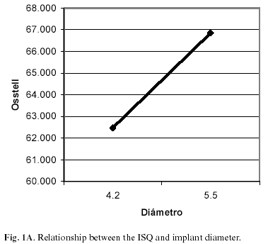
< 0.05).Table 1 shows the statistical data relating IF and ISQ to different variables. No significant differences in relation to patient age or sex were observed for either RFA or the IF. A greater implant diameter was in turn associated with a greater ISQ (t= -2.744, p
<0.05)(Fig. 1A), while increased implant length was correlated to a lower ISQ (F= 3.59, p< 0.05) the most significant differences being between implants with lengths of 11.5 and 16 mm (with mean quotients of 68 and 60.5, respectively)(Fig. 1B).

A significant relation was observed between the type of bone and both RFA and IF: the more compact the bone, the greater ISQ (F=9.53, p
< 0.05) and IF (F=4.58, p< 0.05). Regarding location of the implant, the ISQ was greater in the mandible than in the maxilla, with mean values of 68.9 and 57.8, respectively. There were no significant differences between the anterior and posterior sectors. On relating the location and position of the implants, the ISQ values were found to be higher in the posterior upper jaw than in the anterior sector of the maxilla, while in the case of the mandible no significant differences were recorded between the anterior and posterior sectors (Fig. 2).

RFA was lower in the case of dehiscence (t=3.025, p< 0.05). The ISQ was greater in those cases in which no reabsorbable membranes were placed (mean 65.5) than in the presence of such membranes (mean 57.5)(F=15.134, p<0.05). Finally, the fixations exhibited lower ISQ values when maxillary sinus lifting was carried out with the indirect technique (t=3.642, p<0.05).
Discussion
Park et al. (6) reported a mean ISQ when using Brånemark® MK III implants of 76.6 of the day of surgery this being slightly greater than our own value of 62.1. Meredith et al. (1) in turn observed a near-linear relationship between ISQ and RFA. In coincidence with this finding, we recorded a statistically significant correlation between these two parameters (2,3).
There were no statistically significant differences in terms of age and sex for RFA and IF. Our review of the literature yielded no studies on the way in which RFA and IF vary in relation to patient age and sex. In our series the mean patient age was 52 years, and there were not many young individuals. It was therefore difficult to assess the impact of age upon these variables.
According to Horwitz et al. (7), the greater the implant diameter, the greater RFA and IF. Our own findings coincide with this observation. Regarding implant length, Horwitz et al. (7) reported no correlations for fixation length and RFA, while Balleri et al. (8) recorded greater RFA values with short implants, upon measuring after one year of loading. In our study we obtained similar results on the day of the intervention.
Meredith et al. (1) observed increased resonance frequency values for implants that were placed in mature bone. We in turn recorded a positive relation between bone hardness and IF and RFA. Merdith et al. (1) used RFA to measure implant fixation as it penetrated the bone. They found that with increased fixation exposure, the RFA values tended to decrease a positive correlation being noted between RFA and the amount of implant submerged (r=0.94; p<0.01). Huang et al. (9) obtained greater ISQ in type I bone in an in vitro study. Barewal et al. (10) conducted measurements in 27 implants during 10 weeks; after three weeks they found decreasing values in all bone types though particularly in type IV bone.
According to different authors (7,8,10), there is greater primary stability in the mandible than in the upper jaw, in coincidence with our own observations. According to Nedir et al. (11), most upper maxillary implants presented ISQ <60, versus >60 in the case of the mandible.
Regarding implant position (anterior or posterior), we found no differences in coincidence with the data published by Balleri et al. (8), who studied the same parameter after one year of loading. In our study, on relating the location and the position of the implants, the ISQ values were found to be greater in the upper posterior sector than in the upper anterior zone, while no differences were noted in the case of the lower jaw.
The stability quotient measured immediately after implant placement was greater for the larger diameter and shorter length designs, in mandibular implants, and in implants inserted in more compact bone.
![]() Correspondence:
Correspondence:
Dr. Miguel Peñarrrocha Diago
Clínica Odontológica
Gascó Oliag, 1
46021 Valencia
E-mail: miguel.Penarrrocha@uv.es
Received: 25-04-2004
Accepted: 17-12-2005
References
1. Meredith N, Alleyne D, Cawley P. Quantitative determination of the stability of the implant-tissue interface using resonance frequency analysis. Clin Oral Implants Res 1996;7:261-7. [ Links ]
2. Meredith N, Book K, Friberg B, Jemt T, Sennerby L. Resonance frequency measurements of implant stability in vivo. A cross-sectional and longitudinal study of resonance frequency measurements on implants in the edentulous and partially dentate maxilla. Clin Oral Implants Res 1997;8:234-43. [ Links ]
3. Friberg B, Sennerby L, Linden B, Grondahl K, Lekholm U. Stability measurements of one-stage Branemark implants during healing in mandibles. A clinical resonance frequency analysis study. Int J Oral Maxillofac Surg 1999;28:266-72. [ Links ]
4. Lekholm U, Zarb G. Patient selection and preparation. In: Bränemark P-I, Albrektsson T, eds. Tissue-Integrated Prostheses: Osseointegration in Clinical Dentistry. Chicago: Quintessence; 1985.p.199-210. [ Links ]
5. Chan-Jin Park, Yung-Soo Kim, Chang-Whe Kim, Lee-Ra Cho, Yang-Jin Yi. A study on the change of implant stability using resonance frequency analysis. J Korean Acad Prosthodont 2003;41:271-87. [ Links ]
6. Horwitz J, Zuabi O, Peled M. Resonance frequency análisis in inmediate loading of dental implants. Refuat Hapeh Vehashinayim 2003;20:80-8. [ Links ]
7. Balleri P, Cozzolino A, Ghelli L, Momicchioli G, Varriale A. Stability measurements of osseointegrated implants using Osstell in partially edentulous jaws after 1 year of loading: a pilot study. Clin Implant Dent Relat Res 2002;4:128-32. [ Links ]
8. Huang HM, Lee SY, Y eh CY, Lin CT. Resonance frequency assessment of dental implant stability with various bone qualities: a numerical approach. Clin Oral Implants Res 2002;13:65-74. [ Links ]
9. Barewal RM, Oates TW, Meredith N, Cochran DL. Resonance frequency measurement of implant stability in vivo on implants with a sandblasted and acid-etched surface. Int J Oral Maxillofac Implants 2003;18:641-51. [ Links ]

