


Meu SciELO
 Serviços customizados
Serviços Personalizados
Journal
Artigo
 Inglês (pdf)
 Artigo em XML
 Referências do artigo
 Enviar este artigo por email
Indicadores
-
 Citado por SciELO
 -
 Acessos
Links relacionados
-
 Citado por Google
 -
 Similares em
SciELO
Similares em
SciELO -
 Similares em Google
Compartilhar

 Permalink
Medicina Oral, Patología Oral y Cirugía Bucal (Internet)
versão On-line ISSN 1698-6946
Med. oral patol. oral cir.bucal (Internet) vol.12 no.3 Mai. 2007
Recurrent central giant cell granuloma in the mandible: Surgical treatment and dental implant restoration
Pedro Infante Cossío1, Rafael Martínez de Fuentes2, Andrés Carranza Carranza3, Daniel Torres Lagares4, José Luis Gutiérrez Pérez5
(1) Assistant Professor of Oral Surgery, Faculty of Dentistry, Staff Surgeon, Department of Oral and Maxillofacial Surgery, Virgen del Rocio University Hospital, University of Seville, Spain.
(2) Assistant Professor of Prosthodontic, Faculty of Dentistry, University of Seville, Spain.
(3) Resident, Department of Pathology, Virgen del Rocio University Hospital, Seville, Spain.
(4) Professor of Oral Surgery, Faculty of Dentistry, University of Seville, Spain.
(5) Professor of Oral Surgery, Faculty of Dentistry, Head of the Department of Oral and Maxillofacial Surgery, Virgen del Rocio University Hospital, University of Seville, Spain
ABSTRACT
Central giant cell granuloma is an uncommon benign intraosseus lesion of jaws. Traditional treatment has been local curettage, although aggressive sub-types have a high tendency to recur. This patient report describes a recurrent central giant cell granuloma involving the body of the mandible in a 48-year-old-woman. Initial treatment of lesion consisted of curettage and peripheral ostectomy. When recurrence was detected one year later, an en bloc resection and defect regeneration with a composite bone graft of autogenous bone, xenograft, and autologous platelet-rich plasma was carried out. Adequate new bone formation was observed during follow-up of 24 months. Two dental implants were placed, and implant-supported prosthesis was constructed, providing a satisfactory dental restoration.
Key words: Mandible, giant cell, granuloma, dental implants, dental prosthesis.
RESUMEN
El granuloma central de células gigantes es una rara lesión intraósea benigna de los maxilares. El tratamiento tradicional ha sido el curetaje local, aunque los sub-tipos agresivos tienen una alta tendencia a la recurrencia. Este caso clínico describe un granuloma central de células gigantes recurrente en el cuerpo mandibular de una mujer de 48 años. El tratamiento inicial de la lesión consistió en un curetaje con ostectomía periférica. Cuando se detectó la recurrencia un año más tarde, se realizó una resección ósea en bloque y la regeneración del defecto con un injerto óseo compuesto de hueso autógeno, xenoinjerto y plasma rico en plaquetas autólogo. A los 24 meses de seguimiento se observó una adecuada formación de hueso nuevo. Se insertaron dos implantes dentales y se construyó una prótesis implanto-soportada, proporcionando un restauración dentaria satisfactoria.
Palabras clave: Mandíbula, célula gigante, granuloma, implante dental, prótesis dental.
Introduction
Central giant cell granuloma (CGCG) is an uncommon benign intraosseus lesion that occurs almost exclusively in jaws, introduced for the first time by Jaffé in 1953 (1). Although its aetiology and pathogenesis is even unknown, its histology and clinical behaviour has been studied in detail (2-5). Recently the World Health Organization has defined it as localized benign but sometimes aggressive osteolytic proliferation consisting of fibrous tissue with haemorrhage and haemosiderin deposits, presence of osteoclast-like giant cells and reactive bone formation (6). Many authors have established the differences with other lesions of giant cells (7-9).
The clinical behaviour of CGCG varies from a slowly asymptomatic swelling to an aggressive lesion that manifests with pain, cortical perforation, and root resorption. The traditional treatment of CGCG has been local curettage. However, aggressive sub-types of CGCG show a tendency to recur and necessitate bone resection that may determine extensive defects in jaws (10). The aim of this study is to report the case of a patient with a recurrent CGCG in the mandible, managed by means of local bone resection and defect regeneration with a composite bone graft that consisted of autogenous bone, xenograft, and autologous platelet-rich plasma (PRP), and the dental restoration in a second phase with an implant-supported prosthesis.
Case report
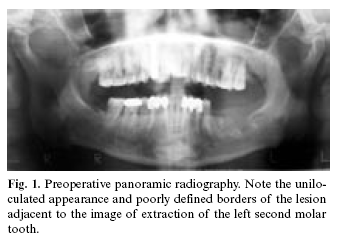
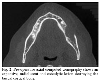
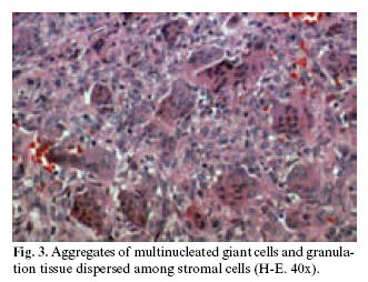
A 48-year-old woman presented to our department in April 2000 with a history of 3 months of swelling and pain in the left body of the mandible. Three months before the patient had felt mobility in the second inferior left molar tooth. She had visited her dentist who carried out the first and second left molar teeth extractions. From then on a swelling involving the left body of the mandible was growing gradually. She reported no pain and no sensory disturbance of the left lower lip and chin. Intraoral examination revealed an expansive bony mass in the left mandibular vestibule from the second cuspid to the angle. Panoramic radiography showed a 2x3 cm radiolucent lesion with poorly defined borders in the left body of the mandible, adjacent to the image of second molar extraction (Fig 1). In computed tomography an expansible lesion was visualized perforating and destroying the buccal cortical bone (Fig 2). Intraoral biopsy performed revealed a large number of multinucleated giant cells, areas of haemorrhage, hemosiderin in deposits, some reactive woven bone and osteoid. The diagnosis was compatible with CGCG (Fig 3). Laboratory values for serum calcium, phosphorous, alkaline phosphate and PTH were within normal limits as were the blood cells count, excluding the brown tumour of the hyperparathyroidism. The patient underwent curettage of the lesion followed by removal of the peripheral bony margins through a retromolar approach. The postoperative course of the patient was uneventful.
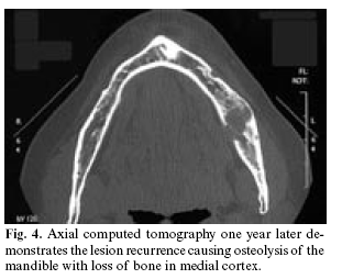
Twelve months later a 1 cm of diameter ill-defined radiolucidency was detected in the panoramic radiograph. The CT scan confirmed the recurrence of the lesion causing erosion of the lingual cortical bone of the left body of the mandible (Fig 4).
In April 2001 the lesion was removed en bloc with a bony security margin of 0.5 cm. The patient received a composite graft of autogenous particulated bone harvested from the mandibular ramus of the left side, and of bioactive glass (Biogram®, 3i Implant Innovations Inc, Florida, USA). 50 cc of venous blood was processed using double centrifugation technique (Platelet Concentrates Collection System PCCS; 3i Implant Innovations Inc, Florida, USA), and separated the two fractions. Before their application, the clot of PRP was activated by means of calcium chloride. The composite graft was mixed with the PRP and the mixture was set into the bony defect. The patient was discharged the same day, and did not refer any symptoms nor there were any postoperative complications. Histological examination of the excised lesion was reported again as CGCG.
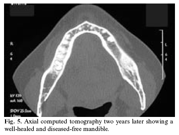
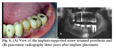
No evidence of clinical and radiological recurrence was observed during follow-up for 24 months. In April of 2003, after ascertainment of graft integration and bone formation by panoramic radiograph and CT (Fig 5), two Osseotite NT® (3i Implant Innovations Inc, Florida, USA) endoosseous implants were placed in the mandible, 5 mm in diameter and 13 and 11.5 mm long. Three months later, second-stage surgery was performed. Partial implant-supported screw retained prosthesis was constructed. After 5 years, no recurrence of lesion has occurred (Fig 6). The patient was seen in maintenance and periodic hygiene checkups. She presented a high level of comfort and satisfaction.
Discussion
The etiopathogenesis of the CGCG of jawbones has not been clearly established but it has been suggested that it is the result of an exacerbated reparative process related to previous trauma and intraosseous haemorrhage that triggers the reactive granulomatous process (9,11). Donoff and Rosenberg (12) discussed a case record of an uncomplicated extraction because of pericoronitis in the area of the lesion and claimed the local changes in the blood flow throughout the bone and local bone dysplasia could be probable etiologic factors. Unal et al (13) presented a 12 year-old girl CGCG in the mandible caused by a molar tooth extraction and explained the pathogenesis by a traumatic aetiology. In our case, we can assume that teeth extractions was the CGCG inciting injury since the lesion was evident and grew rapidly only after molar teeth extractions.
Although CGCGs are benign osseous lesions, some authors separate CGCG into two types, referring to its clinical and radiographic features: (a) Nonaggressive lesion is usually slowgrowing and asymptomatic, does not show cortical resorption by the lesion or root perforation in teeth affected, and it is significantly less likely to recur than the aggressive type (3); and (b) Aggressive lesions, is usually found in younger patients and is painful, grows rapidly, is larger overall, often causes cortical perforation and root resorption and has a tendency to recur (2). Predict the behaviour of CGGCs that will exhibit a higher risk of recurrence after treatment has been problematic. The rate of recurrence varies between 13-49% (14). Whitaker and Waldron (4) reported a mean interval between diagnosis and initial treatment and treatment of a recurrence was 21 months, and stated that very few recurrences were manifested after 2 years of initial treatment. The most reliable factors related to an increased risk of recurrence include clinical activity of lesions (72% of recurrence in the aggressive forms, 3% of recurrence in the nonaggressive forms), younger patients, demonstrated perforation of cortical bone and tumour size (15-17). There has been studies suggesting that the greater functional surface area occupied by giant cells and larger relative size of giant cells may identify tumours with aggressive behaviour (2,8). Recently, Kruse-Loser et al (17) also proved that the aggressive variant of CGCG presented a high number of giant cells, an increased mitotic activity, and a high fractional surface area. However, other studies have not been able to predict the clinical course of CGCGs from known histological or immunohistochemical features (11).
GCGC is considered a non reparative lesion that destroys and grows if it is not treated. Traditionally management has been surgical by means of excision by curettage, which has been associated with a low recurrence rate in the well- located lesions (4,9,10,13). Curettage with peripheral ostectomy and bone resection is reserved for recurrences. Eisebund et al (3) used the technique of curettage or curettage plus peripheral ostectomy. Unal et al (13) recommended microdrill with diamond burr to obtain margins of security after removing the lesion and filled the cavity with iliac crest chips. The most aggressive or recurrent lesions can require en bloc bone resection and reconstruction, since it can determine a bone defect and teeth loss. Becelli et al (18) described a case treated by means of excision of a mandibular CGCG, reconstruction using autogenous iliac crest graft, dental implants and overdenture prosthesis. Non surgical approaches have been introduced, as administration intralesional corticoids (19), or systemic calcitonins that inhibit the osteoclastic activity (20). Other non surgical treatments include interferon-alpha or bifosphonates (16,21). Conservative treatments have shown varying degrees of success and, when successful, have reduced the necessity for reconstructive surgery.
We initially used curettage and peripheral ostectomy to obtain clear surgical margins. When we detected the CGCG recurrence one year later, we carried out an en bloc resection. However, there are no published articles in the literature which refer to the employment of a bone graft combined of autogenous bone and xenograft mixed with PRP for regeneration of the defect after resection of a CGCG recurrence in the mandible. The ascending ramus of mandible represents an alternative source for obtaining particulated autogenous bone for small reconstructions in oral surgery, relatively simple to carry out, with a low complications rate (22). It can be mixed easily with xenografts and with PRP. It seems to be that the addiction of PRP accelerates soft tissues healing and favours bone regeneration especially when using composite bone graft, acting as a scaffold to support new bone formation (23,24).
In our patient, CT scan showed an adequate new bone formation 24 months later. Bone density assessed during insertion of dental implants demonstrated that the newly formed bone had even higher density than bone commonly found in posterior mandible. Two dental implants were successfully placed and loaded, and an implant-supported prosthesis was constructed, providing a satisfactory dental restoration. A check up was carried out every 3 months from then on. At each visit a check on the restoration was made and the prosthesis was unscrewed to examine oral mucosa and bone.
References
1. Jaffe HL. Giant cell reparative granuloma, traumatic bone cyst, and fibrous (fibroosseous) dysplasia of the jaw bones. Oral Surg 1953;6:159-75. [ Links ]
2. Chuong R, Kaban LB, Kozakewich H, Perez-Atayde A. Central giant cell lesions of the jaws: a clinicopathologic study. J Oral Maxillofac Surg 1986;44:708-13. [ Links ]
3. Eisenbud L, Stern M, Rothberg M, Sachs SA. Central giant cell granuloma of the jaws: experiences in the management of 37 cases. J Oral Maxillofac Surg 1988;46:376-84. [ Links ]
4. Whitaker SB, Waldron CA. Central giant cell granulomas of the jaw. A clinical radiologic and histologic study. Oral Surg Oral Med Oral Pathol 1993;75:199-208. [ Links ]
5. de Lange J, van den Akker HP. Clinical and radiological features of central giant-cell lesions of the jaw. Oral Surg Oral Med Oral Pathol Oral Radiol Endod 2005;99:464-70. [ Links ]
6. Barnes L, Eveson JW, Reichart P, Sidransky D, eds. Pathology and Genetics of Head and Neck Tumours. Kleihues P, Sobin LH, series eds. World Health Organization Classification of Tumours. Lyon, France: IARC Press, 2005. p. 324. [ Links ]
7. Murphey MD, Nomikos GC, Flemming DJ, Gannon FH, Temple HT, Kransdorf MJ. From the archives of AFIP. Imaging of giant cell tumor and giant cell reparative granuloma of bone: radiologic-pathologic correlation. Radiographics 2001;21:1283-309. [ Links ]
8. Yamaguchi T, Dorfman HD. Giant cell reparative granuloma: a comparative clinicopathologic study of lesions in gnathic and extragnathic sites. Int J Surg Pathol 2001;9:189-200. [ Links ]
9. Ustundag E, Iseri M, Keskin G, Muezzinoglu B. Central giant cell granuloma. Int J Pediatr Otorhinolaryngol 2002;65:143-6. [ Links ]
10. Stern M, Eisenbud L. Management of giant cell lesions of the jaws. Oral Maxillofacial Surg Clin North Am 1991;3:165-77. [ Links ]
11. Kauzman A, Li SQ, Bradley G, Bells RS, Wunder JS, Kandel R. Central giant cell granuloma of the jaws: assessment of cell cycle proteins. J Oral Pathol Med 2004;33:170-6. [ Links ]
12. Donoff RB, Rosenberg AE. Case records of the Massachusetts General Hospital. Weekly clinicopathological exercises. Case 20-1993. A 23-year-old woman with a rapidly enlarging intraoral mass after a tooth extraction. N Engl J Med 1993;328:1478-83. [ Links ]
13. Unal M, Karabacak T, Vayisoglu Y, Bagis HE, Pata YS, Akbas Y. Central giant cell reparative granuloma of the mandible caused by a molar tooth extraction: Special reference to the manuever of drilling the surgical field. Int J Pediatr Otorhinolaryngol 2006;70:745-8. [ Links ]
14. de Lange J, van den Akker HP, Klip H. Incidence and disease-free survival after surgical therapy of central giant cell granulomas of the jaw in the Netherlands. Head Neck 2004;26:792-5. [ Links ]
15. Minic A, Stajcic Z. Prognostic significance of cortical perforation in the recurrence of central giant cell granulomas of the jaws. J Craniomaxillofac Surg 1996;24:104-8. [ Links ]
16. Bataineh AB, Al-Khateeb T, Rawashdeh MA. The surgical treatment of central giant cell granuloma of the mandible. J Oral Maxillofac Surg 2002;60:756-61. [ Links ]
17. Kruse-Losler B, Diallo R, Gaertner C, Mischke KL, Joos U, Kleinheinz J. Central giant cell granuloma of the jaws: A clinical, radiologic, and histopathologic study of 26 cases. Oral Surg Oral Med Oral Pathol Oral Radiol Endod 2006;101:346-54. [ Links ]
18. Becelli R, Cerulli G, Gasparini G. Surgical and implantation reconstruction in a patient with giant-cell central reparative granuloma. J Craniofac Surg 1998;9:45-7. [ Links ]
19. Khafif A, Krempl G, Medina JE. Treatment of giant cell granuloma of the maxilla with intralesional injection of steroids. Head Neck 2000;22:822-5. [ Links ]
20. Pogrel MA. Calcitonin therapy for central giant cell granuloma. J Oral Maxillofac Surg 2003;61:649-53. [ Links ]
21. Curtis NJ, Walker DM. A case of aggressive multiple metachronous central giant cell granulomas of the jaws: differential diagnosis and management options. Int J Oral Maxillofac Surg 2005;34:806-8. [ Links ]
22. Capelli M. Autogenous bone graft from the mandibular ramus: a technique for bone augmentation. Int J Periodontics Restorative Dent 2003;23:277-85. [ Links ]
23. Sanchez A, Sheridan P, Kupp L. Is platelet-rich plasma the perfect enhancement factor? A current review. Int J Oral Maxillofac Surg 2003;18:93-103. [ Links ]
24. Kanno T, Takahashi T, Tsujisawa T, Ariyoshi W, Nishihara T. Platelet-Rich Plasma enhances human osteoblast-like cell proliferation and differentiation. J Oral Maxillofac Surg 2005;63:362-9. [ Links ]
![]() Correspondence:
Correspondence:
Dr. Pedro Infante-Cossio
Servicio de Cirugía Oral y Maxillofacial
Hospital Universitario Virgen del Rocío
Av. Manuel Siurot
41013 Sevilla, Spain.
E-mail: pinfante@us.es
Received: 4-10-2006
Accepted: 30-01-2007

