SciELO - Scientific Electronic Library Online

 
vol.5 número2Leishmaniasis cutánea en un niño albino: una presentación peculiar índice de autoresíndice de assuntospesquisa de artigos
Home Pagelista alfabética de periódicos  

Serviços Personalizados

Journal

Artigo

Indicadores

Links relacionados

  • Em processo de indexaçãoCitado por Google
  • Não possue artigos similaresSimilares em SciELO
  • Em processo de indexaçãoSimilares em Google

Compartilhar


Iberoamerican Journal of Medicine

versão On-line ISSN 2695-5075versão impressa ISSN 2695-5075

Iberoam J Med vol.5 no.2 Logroño Mai. 2023  Epub 30-Out-2023

https://dx.doi.org/10.53986/ibjm.2023.0008 

Clinical Image

Beaver tail liver: an incidental but crucial anatomical variation in case of acute abdomen

Hígado de cola de castor: una variación anatómica incidental pero crucial en caso de abdomen agudo

Serghei Covantsev (orcid: 0000-0001-7236-5699)*  a  , Ivan Lebedinskyb  , Diana Slepuhkovac  , Maria Kovalevac  , Karina Mulaevac 

aDepartment of Research and Clinical Development, Botkin Hospital, 2nd Botkinsky drive, 5, Moscow, Russia

bDepartment of General Oncology, Botkin Hospital, 2nd Botkinsky drive, 5, Moscow, Russia

cDepartment of Surgery, Russian Medical Academy of Continuous Professional Education, Barrikadnaya st., 2/1, building 1, Moscow, Russia

Keywords: Beaver tail liver; Liver; Anatomy; Spleen

Palabras clave: Hígado de cola de castor; Hígado; Anatomía; Bazo

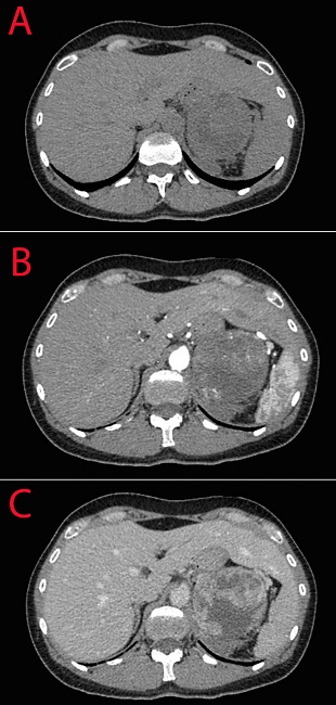
Anatomical variations of the liver are relatively understudied, while they deserve special attention in the day-to-day clinical practice [1, 2]. Beaver tail liver (also known as sliver of liver, sabre-shaped liver or falx-like liver) is a normal anatomical variation of liver development when an elongated left liver lobe extends laterally contacts and/or surrounds the spleen. The true incidence of this developmental variation is not known. It is seen more predominantly in females and usually encountered incidentally during routine ultrasound or computed tomography (CT) evaluation of the abdomen. However, it is a difficult imaging picture in setting of an acute abdomen or trauma. Due to similar density and echogenicity, the two organs may be hard to differentiate during examination. We present a clinical image of a 42-year old female admitted to the emergency department with abdominal pain irradiating to the back that began abruptly with no apparent reason. Her laboratory examination revealed moderate leukocytosis and anemia (leukocytes 12*109/l [normal range 4-9*109/l], erythrocytes 3,2*1012/l [normal range 3,8-5,2*1012/l], hemoglobin 101 g/l [normal range 120-150 g/l]). Her abdominal CT revealed adrenal mass with signs of intraparenchymal hemorrhage and beaver tail liver. Figure 1 demonstrates the difficulties in differentiation of the two organs that seem similar in the native and portal phases of examination and are different only in the arterial phase of the study. The patient undergone supraselective angiography of the adrenal artery, which demonstrated extravasation of contrast material. We performed selective catheterization of adrenal arteries and embolization of her adrenal mass. The postoperative period was uneventful and she was discharged 10 days after the procedure. Further evaluation revealed normetanephrine value 1180 mcg/d (normal value <600 mcg/d), she was consulted by an endocrinologist and scheduled for a planned adrenalectomy, which confirmed adrenocortical carcinoma. Beaver-like liver is an important anatomical variation as it can be mistaken for perisplenitis, subcapsular hematoma, and splenic mass [3, 4]. Abdominal trauma in the left hypochondrum region that usually lead to splenic injury can in turn affect the left lobe of the liver [5].

Figure 1. Abdominal CT scan of the patient (the liver can be differentiated from the spleen during the arterial phase). A: Native phase; B: Arterial phase; C: Venous phase. 

REFERENCES

1. Glenisson M, Salloum C, Lim C, Lacaze L, Malek A, Enriquez A, et al. Accessory liver lobes: anatomical description and clinical implications. J Visc Surg. 2014;151(6):451-5. doi: 10.1016/j.jviscsurg.2014.09.013.Links ]

2. Covanțev S, Mazuruc N, Catereniuc I, Belic O. Accessory liver fissures: a report of two cases and review of their clinical significance. Russ. Open Medical J. 2018; 7(3):e0303.Links ]

3. Atalar MH, Karakus K. Beaver tail liver. Abdom Radiol (NY). 2018 Jul;43(7):1851-1852. doi: 10.1007/s00261-017-1395-x.Links ]

4. Xiang H, Han J, Ridley WE, Ridley LJ. Beaver tail liver: Anatomic variant. J Med Imaging Radiat Oncol. 2018;62 Suppl 1:57. doi: 10.1111/1754-9485.05_12784.Links ]

5. Md Noh MSF, Muhammad SJSS. Beaver in the liver. Pan Afr Med J. 2017;27:138. doi: 10.11604/pamj.2017.27.138.12227.Links ]

The authors declared that this study has received no financial support.

HOW TO CITE THIS ARTICLE:Covantsev S, Lebedinsky I, Slepukhova D, Kovaleva M, Mulaeva K. Beaver tail liver: an incidental but crucial anatomical variation in case of acute abdomen. Iberoam J Med. 2023(2):93-95. doi: 10.53986/ibjm.2023.0008.

Received: December 31, 2022; Revised: February 01, 2023; Accepted: February 06, 2023

*Corresponding author. E-mail address: kovantsev.s.d@gmail.com

CONFLICT OF INTERESTS

The authors have no conflict of interest to declare.

Creative Commons License This is an open access article under the CC BY license