




 Servicios personalizados
 Inglés (pdf)
 Articulo en XML
 Referencias del artículo
 Traducción automática
 Enviar articulo por email
 Citado por SciELO
  Citado por Google
  Similares en
SciELO
Similares en
SciELO  Similares en Google
 Permalink
INTRODUCTION
One of the most prevalent tumors, renal cancer, also known as Renal Cell Carcinoma (RCC), mostly affects people over the age of 50 (1). On a global level, the frequency of this has increased. According to GLOBOCAN, renal cancer cases were 431,288 in 2020 or 2.2 %, and RCC fatalities were 179,368 in total or 1.8 % (2). In the United States, the tenth-largest cause of cancer death in adult men is RCC. Based on GLOBOCAN data from 2012, the death rate for men with renal cancer in Indonesia was 1.6 per 100,000 and 0.8 per 100,000 for women (3). While the rate of RCC increased, there was also a 20-30 % increase in metastases. The use of computed tomography imaging for other illnesses, which improves the detection of renal tumors detected by accident, is to blame for the rise in RCC instances.
Initially used to treat untreatable renal cancer that was accompanied by symptoms, renal artery embolization is now more frequently utilized to support surgical resection in patients with renal tumors. Since resection of renal tumors was linked to a high risk of perioperative morbidity related to bleeding, surgeons started using renal embolization in the 1970s. In some individuals, transarterial embolization is a procedure that makes nephrectomy easier. Embolization reduces the need for transfusions, intraoperative blood loss, and operation time in patients with big or advanced renal tumors, which is advantageous in surgical resection. This implies that there are advantages to renal tumor embolization, such as lower tumor vascularity that enables early renal vessel ligation. The lymphoproliferative condition is increased by post-embolization tu- mor necrosis, and Natural Killer cells respond to the tumor specifically (4).
EVIDENCE ACQUISITION
A literature search was conducted using the PubMed, Embase, and Science Direct databases published between 2013-2023. Keywords used for searches included renal cancer, transarterial embolization, Amplatzer Vascular Plug and Gelatin Sponge Particles. This literature review examines the effectiveness of AVP and GSP in Transarterial Embolization of Kidney Tumors. Studies were assessed for quality and risk of bias, and data were extracted and analyzed.
EVIDENCE SYNTHESIS
The goal of vascular embolization is to interrupt blood flow or cause an inflammatory response in the vessel wall. Renal artery embolization has been used to treat a variety of renal conditions, ranging from symptomatic hematuria to palliative measures in malignant renal tumors. It is also commonly used in preoperative preparation for renal tumors, arteriovenous fistulas and other renal vascular malformations, post-biopsy complications, and traumatic renal hemorrhage (5). Indications for embolization in the therapeutic management of neoplastic renal masses include palliative care for mass loss and bleeding risk in patients requiring total or partial nephrectomy or treatment with radiotherapy, as well as management hematuria and/or bleeding during and before and after trauma (6). Endovascular transcatheter embolization is considered the first-line therapy because of high success rate and shorter hospital stay. Compared to surgery, it has lower morbidity and mortality, which surgery is more invasive and has risk of intraoperative anesthesia. However, percutaneous embolization carries a significant migration risk of embolic material into the venous and pulmonary circulation (7,8).
Classification of embolic agents can be classified based on physical form (solid or liquid), mechanism of action (mechanical or chemical), and primary ingredients (biosynthesis, synthesis, etc.). However, most experts divide them based on the duration of the occlusion into temporary and permanent. Examples of temporary embolic agents are gelatin sponge, oxidized cellulose, and microfibrillar collagen. Meanwhile, permanent embolic agents include polyvinyl alcohol, tris-acryl gelatin microspheres, N-butyl cyanoacrylate, and plugs. Permanent embolic agents are considered modern embolic agents and are non-absorbable. The choice of embolic agent is based on the case to be treated, such as characteristic of renal fistula by size, location and flow rate, the desired duration of occlusion, the required tissue viability, and the patient's clinical condition. In the case of large blood vessels, the embolization type required is permanent embolization (8,9).
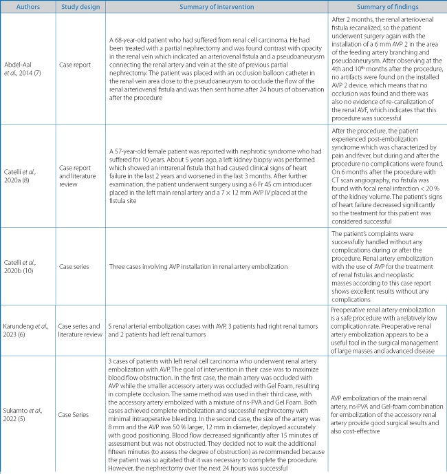
Coils are used most often because of their easy availability, low cost, and ease of use. However, they have a risk of migration into the venous and pulmonary circulation and generally require several spirals to completely occlude the vessel. Coils have been demonstrated to be less effective in embolization as the current preferred technique uses n-butyl cyanoacrylate glue which allows rapid and definitive distal occlusion of a voluminous vascular bed and causes necrosis in perivascular tissue (4). Plugs have entered clinical practice in recent decades, first for the treatment of heart diseases and then for the treatment of peripheral vessels. The Amplatzer Vascular Plug (AVP) is a recent innovation by St. Jude Medical. Its use as a peripheral embolic agent was approved by the United States Food and Drug Administration (FDA) in 2004, and was declared effective in a study published with year. The structure consists of nitinol-based braids that allow self-expansion across the target vascular bed. It can be repositioned and restored at any time after being released. The use of a 30 to 50 % larger device is crucial for achieving optimal blood vessel seal and preventing unintended peripheral embolism. AVP is a very effective, especially in cases related to the structure of the peripheral renal arteries. The structural characteristics of AVP IV allow for irregular and high-flow delivery into blood vessels and rapid targeted occlusion (5,8).
The use of AVP 2 has been widely reported and many have reported the success of use this device. AVP 2 is described in the embolization of renal AVFs in certain vascular territories, more specifically in high-flow renal AVFs. In several reported cases, the use of AVP 2 was found to be effective and safe. AVP 2 is more accurate and stable placement through a detachable system. It has a self-expanding, cylindrical occluding device made out of nitinol mesh wires. It can be safely used in short and high-flow vascular segments, such as renal AVFs, where coils are released with less precision and safety. After placement of the device, an angiogram can be performed to confirm placement and the device can then be deployed from the push wire in a deliberate, controllable and relatively precise by rotation pusher wire. Equipment can be removed and repositioned if the position is unsatisfactory, minimizing migration risks (7).
Several authors in many case reports recommend AVP 2 as the primary choice in cases of renal AVF occlusion with high-flow and short-vascular connection, where the occlusion has to be in an exact location. Its also to be more cost-effective compared to using coils. Compared with coils, the AVP 2 also has minimal metallic artifact features on computed tomography allowing for more accurate interpretation. The AVP 2 device is usually inserted using a sheath or guiding catheter which may be difficult if done in stenotic renal arteries or the distal part of the renal vessels because many fistulas are found in this area. In addition, the AVP 2 delivery cable is relatively stiff compared to the coil, allowing for difficulties in advancing the device in areas of tortuous blood vessel anatomy (7). Futhermore, renal artery embolization in the preoperative setting can decrease perioperative blood loss, creation of a tissue plane of edema facilitating dissection, and reduction in tumor bulk including extent of vascular thrombus, when present. Wide variation in reporting markers such as reduction in intraoperative blood loss, transfusion requirements, surgical procedure time, surgical complications, and survival outcomes has limited its use to local practice patterns (11).
AVP IV can be placed in tortuous and small vessels resulting in complete occlusion of the target blood vessels in a short time. The use of a single device significantly reduces procedural time when positioning coils, in fact, more coils are often needed to completely occlude the blood vessel. The plug was released at the point of arteriovenous anastomosis without any involvement of non-target arteries. The technical characteristics of the device allow performing very precise preliminary examinations, ensuring selective occlusion of the target blood vessel segment with a very low risk of migration. The device's structural characteristics determine complete vessel occlusion within 10 minutes after release as it gradually adapts to the target vessel causing slowing down of the blood flow until it stops completely (8).
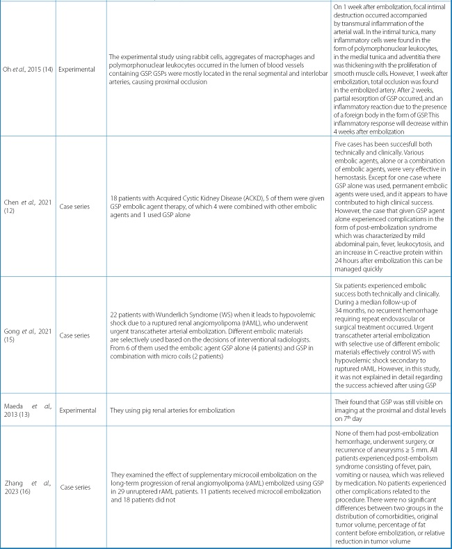
Apart from AVP, renal artery embolization can be carried out with embolic agents such as polyvinyl alcohol (PVA), n-butyl cyanoacrylate (NBCA), micro coils, and gelatin sponge particles (GSP). Embolic agents that are often used are NBCA and PVA as single agents or coils/PVA, GSP/PVA, and coils/GSP as combination agents. GSP is a very effective temporary embolic agent and has a fairly high clinical success rate (12). It is generally used in cases of bleeding, uterine fibroids, and hypervascularization in malignant tumors. It cause an uncontrolled level of occlusion because the instrument is cut by hand, resulting in particles that are difficult to calibrate and very easily deformed. GSP degradation is also said to last quite a long time, around 3 weeks to 4 months, and can be accompanied by a chronic inflammatory response (13).
The usage of GSP in transarterial embolization has increased as a result of its recognition as a potent agent for transitory arterial embolization. Within a few weeks after being embolized with GSP, the embolized arteries are recanalized, preserving normal tissue at the embolization site and enabling additional transarterial embolization to inhibit tumor growth. To achieve partial recanalization and complete thrombus, 4 weeks after embolization with GSP are required. However, the lumen of the recanalized artery has significant stenosis due to thrombus and intimal hyperplasia. This intimal hyperplasia is caused by the migration of smooth muscle cells from the media and collagen deposition. The massive organized thrombi and intimal hyperplasia due to mild embolization with GSP thus lead to arterial stenosis. The massive organized thrombosis appears to be caused by acute inflammatory reactions and foreign bodies, while intimal hyperplasia is due to arterial wall damage due to transmural inflammation and migration of smooth muscle cells after partial loss of continuity of the internal elastic lamina (14).
Several studies report the disadvantages of using GSP. Partial degradation of GSP also causes foreign body inflammation. This finding is to the existing theory that the occlusion caused by GSP is not total or not the same as its size but is smaller due to manual cutting techniques so that the inserted GSP instrument is roughly calibrated and can even change shape. Likewise, the finding by compared study showed that only 88 % of the occlusion was formed after 4 months of embolization, resulting in long-term incomplete recanalization. In this study, some GSPs will be degraded and recanalized around 80 % on day 7. These results may vary due to poor calibration. In another case, 63 % recanalization occurred on day 7 and 100 % on day 14 after embolization. Recanalization is accompanied by partial degradation of GSP and partial destruction and remodeling of the vessel wall with infiltration of inflammatory cells (13).
Tumors that received supplementary microcoil embolization had a significantly greater relative reduction in tumor volume at > 3 years after embolization. The differences in relative tumor reduction only became apparent at > 3 years after embolization suggests that using GSP without microcoil embolization reduced the long-term effects of embolization. Tumor feeders that are not occluded by additional microcoils can be recanalized more easily and quickly than occluded vessels, which may result in tumor regrowth. Indeed, in a representative case, renal arteriography showed revascularization of a tumor feeder that was not occluded by the microcoil during the original embolization (16) (Table I).
CONCLUSION
In several case reports, the use of AVP is effective and safe on transarterial embolization procedure for renal tumors. AVP is very effective in use, especially in cases associated with peripheral renal artery structures. Using GSP requires a long time and recanalization. To achieve partial recanalization and complete thrombus, 4 weeks post-embolization with GSP is required.

