




 Servicios personalizados
 Inglés (pdf)
 Articulo en XML
 Referencias del artículo
 Traducción automática
 Enviar articulo por email
 Citado por SciELO
  Citado por Google
  Similares en
SciELO
Similares en
SciELO  Similares en Google
 Permalink
INTRODUCTION
Latrogenic injury of the thoracic duct or its branches during abdominal, thoracic or cervical neck surgery is a rare condition, that usually it is limited and treated conservatively with drainage, local external compression or diet modification (1-3). However, in patients with lymphatic fistulas that are refractory to conservative therapy the leakage must be surgically treated by ligation or clipping of this lymphatic branches. Several authors have described percutaneous interventions of this disrupted channels by thoracic ductus embolization as a possible alternative (2,4-10).
We report an unusual case of a patient that showed a large neck lymphocele after surgery of a lusoria dysphagia and reimplantation of the aberrant subclavian artery in the common carotid artery, and that after surgical reintervention due to recurrent lymphatic leakage, she underwent a thoracic ductus embolization with glue to be definitively closed.
CASE REPORT
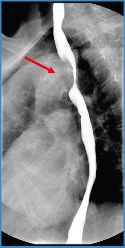
A 68 years old woman, smoker of 30 cigarettes per day, with history of hypertension, helicobacter pylori infection, and liver disease, undergoing digestive follow-up for dysphagia and reflux for approximately six months. She reported weight loss of 4 kg in recent months. A barium shallow study showed normal peristalsis with an indentation at the level of the aortic arch (Fig. 1).
A computed tomography (CT) angiogram showed an aberrant right subclavian artery coursing posterior to the esophagus. The origin from the aortic arch was slightly dilated (12 mm diameter) and located immediately below to the origin of the left subclavian artery. The patient presented a bovine arch with a left carotid artery originating from a common trunk with the right common carotid. Both vertebral arteries were patent. A dysphagia lusoria due to an esophageal extrinsic compression by the aberrant right subclavian artery was diagnosed (Fig. 2), and a hybrid procedure was planned.
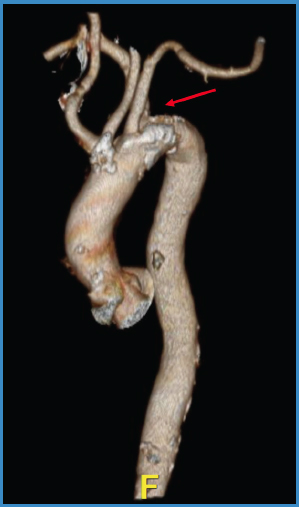
Figure 2. The patient presented a left carotid artery originating from a common trunk with the right common carotid, and an aberrant subclavian artery entrapping the esophagus. A dysphagia lusoria due to an esophageal extrinsic compression was diagnosed.
Under general anesthesia, the right common carotid artery and the right subclavian artery was approached through a right supraclavicular incision. Numerous lymphatic conduits were observed and ligated. The origin of the aberrant right subclavian artery was occluded with a 16 mm Amplatzer Vascular Plug™ (Abbott) through the ipsilateral humeral artery. Later, the stump of the aberrant subclavian artery was sewn with 4/0 suture, and the right subclavian artery was reimplanted in the right common carotid artery with preservation of the vertebral artery (Fig. 3).
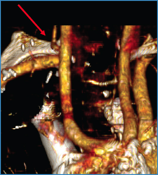
Figure 3. A hybrid procedure was planned by occlusion of the origin of the aberrant right subclavian artery, and reimplantation in the right common carotid artery.
During the postoperative period the patient showed a large right supraclavicular lymphocele (Fig. 4) and she needed of surgical drainage and reintervention. Some lymphatic vessels were clipped, but the milky lymphatic leakage continued despite a free fat diet and local compression. Consequently, an endovascular treatment with embolization of the thoracic ductus (TDE) was scheduled.
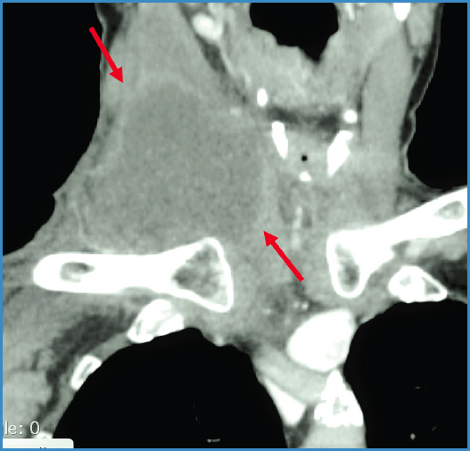
Figure 4. Large postoperative right supraclavicular lymphocele that needed surgical drainage and reintervention.
TDE technique
The patient was positioned on the fluoroscopy table as comfortable as possible since it was a long-time procedure (2-3 hours). An adequate intravenous sedation was employed. An intranodal lymphangiography by ultrasound-guided puncture of inguinal lymph nodes in each groin was undertaken with a 25-G spinal needle with the needle tip positioned in the hilum of the node. Subsequently, an oil-based contrast agent (Lipiodol Ultra Fluide, Guerbet, France) was injected by hand at a rate of about 1-2 ml per 5 min. A total volume of 5 ml of contrast was injected into a lymph node in each groin to opacify the abdominal-pelvic lymphatics, cisterna chyli and thoracic duct with the upper leakage. After the cisterna chyli and its contributing lymphatics were visualized (Fig. 5), a kit for access of 16 cm long and 22-G trocar was used for ductal puncture (Chiba needle, Izasa, Spain). Fluoroscopy was employed to guide a paramedial needle puncture of the cisterna chyli (Fig. 6). A stiff 0.018 wire was gently advanced into the thoracic duct (Fig. 7) (Glidewire Advantage, 300 cm length, Terumo), followed by a 2.6 F microcatheter (Fig. 8) (CXI 0.018, 135 cm, Cook). Contrast was injected through the catheter to demonstrate the thoracic ductus and the lymphatic leakage. Using this access, embolization of the thoracic duct from below the leak point was slowly performed with glue (Glubran2, Italy).
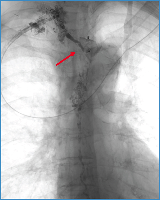
Figure 5. Complete right-sided thoracic duct emptying into the right venous angle, and Amplatzer™ in the origin of the aberrant right subclavian artery.
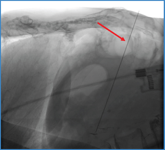
Figure 6. After the cisterna chily was visualized a paramedial needle puncture of the cisterna chyli was used by fluoroscopy to guide the ductal puncture.
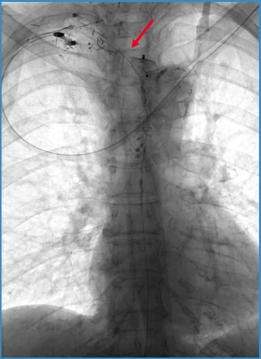
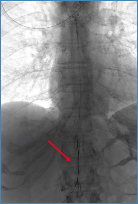
Figure 8. A microcatheter was later introduced for embolization of the thoracic duct from below the leak point.
After successfully TDE, the patient showed a slight abdominal discomfort, but the lymphatic drainage stopped, and the patient was discharged uneventful a few days later.
DISCUSSION
Although the aberrant subclavian artery causing dysphagia lusoria is a congenital anomaly, typically develops symptoms only later in life when the arterial wall becomes more rigid. Symptoms are more likely to occur if there is a bicarotid trunk anteriorly and a retroesophageal aberrant subclavian artery entrapping the esophagus, as seen in the present case.
The surgical dissection to undergo the subclavian transposition to the common carotid artery caused the lymphatic leakage. This was aggravated by the abnormal drainage of the main thoracic ductus into the right subclavian vein. At beginning, it was managed conservatively, but after several days the lymphocele increased, the patient showed pain and neck compression. A surgical drainage and suture of some lymphatic branches was required. Despite reintervention and a fat free diet, the chylous effusion continued, and the embolization of the thoracic ductus was then considered. TDE is a percutaneous minimally invasive technique for treating lymphatic leakage that it has been previously described (2,6,7,10). The procedure has been reported to be successful in over 70 % patients without appreciate morbidity or mortality (11).
Most postoperative lymphoceles are small and resolve without treatment. However, in 4-7 % of patients these lymphoceles persist resulting in pain, infection, or compression of vital structures, which require an intervention (2,6). If the lymphatic leakage persists, it could be associated with a serious depletion of elements (fluid, electrolytes, T-lymphocytes, protein, and fat), and the patient can become further debilitated.
Aortic aneurysm repair is the main vascular surgical cause, but the overall incidence is less than 1 % (2,6,10). Others common causes reported have been retroperitoneal lymphadenectomy or abdominal surgeries with chylous ascites, and thoracic surgeries, particularly esophagectomy, with chylothorax (9-11).
We performed a bilateral intranodal lymphangiography to opacify the lymphatic system, particularly the cisterna chyli and the thoracic ductus, because this approach, contrary to the classical pedal lymphangiography, reduces the procedure time due to a faster transit to visualize the lymphatic system (11-13).
Given the technical difficulty of the transabdominal catheterization of the cisterna chyli and cannulation of the thoracic ductus, fluoroscopic images in different positions is recommended to guide needle puncture. Some authors employ the combination of CT images (2). The technical success of catheterizing the thoracic duct has been reported as 67 % (2,6,11). However, in cases which the catheterization is technically unsuccessful, thoracic duct disruption have been described using twiddling motion with the needle, but we don´t have experience with this technique (2,6,11). The embolization of this traumatic leaks should start from the leak point down into the thoracic ductus. Success is higher when glue is used either by itself or in conjunction with coils (91 %).
Various adverse events have been reported with thoracic duct interventions (2,6,7,10,11), such as hypersensitive to oil contrast, local abdominal pain or perihepatic hematoma that usually resolved without treatment, limited lower extremity swelling or chronic diarrhea. There have also been aortic punctures without observed sequelae.
In conclusion, lymphangiography and TDE is a relatively safe, effective, and a reliable interventional method that provides complete occlusion of the lymphatic leakage, which results in complete resolution of one of the most dreadful and disturbing complications of a postoperative surgery.

