




 Servicios personalizados
 Inglés (pdf)
 Articulo en XML
 Referencias del artículo
 Traducción automática
 Enviar articulo por email
 Citado por SciELO
  Citado por Google
  Similares en
SciELO
Similares en
SciELO  Similares en Google
 Permalink
INTRODUCTION
The liver is the principal metabolic organ in the human body, responsible for numerous complex biochemical processes involving the metabolism of carbohydrates, proteins, and lipids; storage and activation of vitamins; detoxification and excretion of endogenous and exogenous products, among others. As liver function declines, systemic overload increases, leading to a depletion in nutritional status, which is evident even in the early stages of liver disease (1).
Sarcopenia, recognized as a muscular disease characterized by a reduction in both the quality and quantity of muscle mass, has an estimated prevalence of 37.5 % in patients with cirrhosis. When present, it increases the mortality risk of this population by 2.6 times (2).
Aiming at screening for sarcopenia and identifying the risk of poor functional outcomes, the European Working Group on Sarcopenia in Older People 2 (EWGSOP2) in 2019 suggested an algorithm involving the following steps: (i) screening, using the SARC-F questionnaire which subjectively assesses strength, assistance with walking, getting up from a chair, climbing stairs, and falls; (ii) assessment of muscle strength through methods such as handgrip strength (HGS) and the chair stand test; (iii) evaluation of muscle quantity and quality using body composition methods (3). As an alternative to SARC-F, Barbosa-Silva et al. (2016) proposed the SARC-Calf tool, which adds calf circumference to the subjective criteria of SARC-F (4).
Due to the symptomatic characteristics of patients with cirrhosis, such as ascites and edema, the step of assessing muscle quality and quantity becomes challenging, as it hinders the use of bioelectrical impedance analysis, increasing the reliance on imaging methods such as dual-energy X-ray absorptiometry (DXA) and computed tomography (CT), which are costly and difficult to access in clinical practice (5).
Given the challenges and uncertainties in assessing the presence of sarcopenia in patients with cirrhosis, as well as the impact of its development on the quality of life and survival of these individuals, early identification is essential to establish effective clinical and nutritional treatment. In this context, the objective of this study is to identify the best method for diagnosing sarcopenia in patients with cirrhosis, as well as to evaluate the concordance of subjective and anthropometric methods - classically used in the assessment of these patients - with sarcopenia diagnosed by DXA.
PATIENTS AND METHODS
STUDY DESIGN
This is a cross-sectional study conducted in the Infectious and Parasitic Diseases Department of Professor Alberto Antunes University Hospital, Maceió/Alagoas, Brazil, from October 2022 to November 2023.
STUDY GROUPS
Patients aged ≥ 18 years and less than 70 years, of both sexes, diagnosed with liver cirrhosis, were eligible for participation and divided into two groups. One group had score Model for End-Stage Liver Disease-sodium (MELD-Na) ≥ 15, eligible for Liver Transplant (LT), while the other group had MELD-Na ≤ 14, with portal hypertension. Eligible criteria for portal hypertension were ascites presence, splenomegaly, esophagogastric varices, or the presence of portosystemic collaterals (patent paraumbilical vein, splenorenal collaterals, dilated left gastric veins, and short veins). Exclusion criteria included: (a) neoplasia; (b) acute liver failure; (c) pregnant and lactating women; (d) human immunodeficiency virus infection; (e) patients listed for liver transplantation due to special conditions (intractable pruritus, recurrent cholangitis, refractory ascites, persistent hepatic encephalopathy); (f) history of organ failure affecting nutritional status, such as renal replacement therapy, respiratory, and cardiac failure.
SAMPLE SIZE
This is an exploratory study derived from an original research project aiming to identify the prevalence of sarcopenia among LT candidates. A relative risk of 3 for sarcopenia prevalence was expected, considering a baseline prevalence of 25 % in the control group (patients with liver cirrhosis but without LT indication). Assuming 80 % power and a 5 % alpha level, 19 patients were required in each group (Group 1: MELD-Na ≤ 14; Group 2: MELD-Na ≥ 15).
EVALUATION OF LIVER DISEASE SEVERITY
The severity of liver disease in patients was assessed MELD-Na scores and participants were categorized into two groups: ≤ 14, indicating patients not eligible for LT, and ≥ 15, indicating patients eligible for LT. These scores were determined through a clinical evaluation conducted by a specialized medical professional, combined with laboratory test results obtained at the time of consultation.
SOCIODEMOGRAPHIC DATA AND CLINICAL ASSESSMENT
Personal history, current disease history, presence of signs and symptoms, prior hospitalizations related to hepatic disease decompensation, lifestyle habits, etiology, and time of diagnosis were collected using a standard form.
NUTRITIONAL/FUNCTIONAL ASSESSMENT
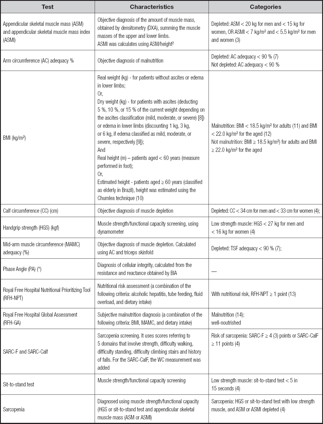
Nutritional and functional tests applied to individuals can be visualized in table I.
EQUIPMENT AND TECHNIQUES
Weight and height measurements followed the technique recommended by Lohman (1988), using a Filizola® digital scale and a metal anthropometer (6). Arm circumference (AC) and calf circumference (CC) were measured with a non-extensible tape measure, while triceps skinfold (TSF) was assessed with a Lange® caliper (6). Tetrapolar bioelectrical impedance analysis (BIA) by Sanny® was used to determine the phase angle (PA). Appendicular Skeletal Muscle Mass (ASM) was obtained through Dual-Energy X-ray Absorptiometry (DXA) analysis using the Lunar Prodigy Primo system from GE HealthCare, with a full-body anteroposterior incidence, and the patient lying supine with extended legs, feet together, arms extended alongside the body, without adornments. Muscle strength was identified through the Individual performance in handgrip strength (HGS) using the Jamar® dynamometer, measured three times on the dominant hand by a trained professional.
ETHICAL CONSIDERATIONS
All patients provided written informed consent. The study was conducted following the ethical guidelines of the 1975 Helsinki Declaration. The protocol was approved by the Ethics Committee on May 26, 2022 (Opinion Number 5432777).
STATISTICAL ANALYSIS
We utilized the Statistical Package for Social Science (SPSS®), version 26.0, for all analyses. Descriptive statistics included frequencies, absolute and relative values (n/percentage), with continuous variables reported as mean and standard deviation. The kappa concordance test (κ) was used to evaluate the agreement between methods, interpreted as poor (< 0.0), slight (0.01-0.2), fair (0.21-0.4), moderate (0.41-0.6), substantial (0.61-0.8), and almost perfect (0.81-1.00) (15). We initially compared individual diagnostic methods with muscle mass as assessed by DXA. Subsequently, we combined techniques for assessing muscle strength and mass to determine whether any of these combinations showed concordance with the sarcopenia diagnosis obtained via DXA. The alpha value was set at 5 %.
RESULTS
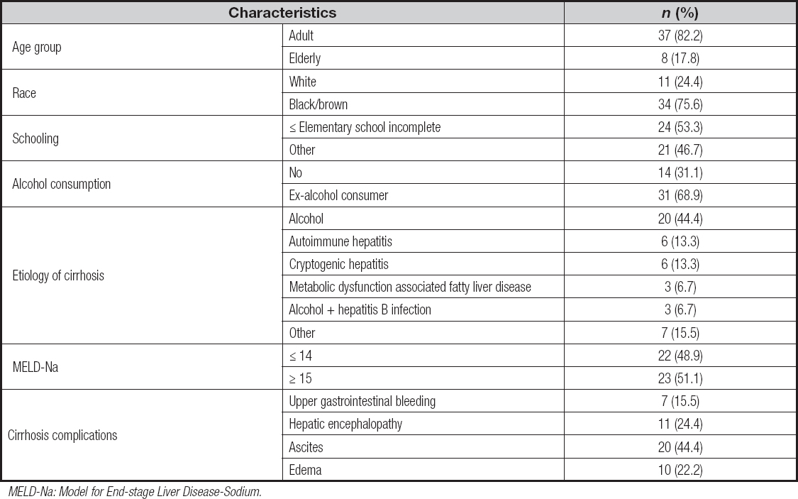
We analyzed 45 patients, with the majority being male (68.9 %), and a mean age of 47.5 ± 14.2 years. Most resided in rural areas (60 %), and 57.8 % reported being married or in a stable relationship.
The most common etiology of liver disease was alcoholic (44.4 %). Among the individuals analyzed, approximately 37.8 % had comorbidities such as systemic arterial hypertension (SAH), diabetes mellitus (DM), obesity, dyslipidemia, and hypothyroidism. Additionally, 46.7 % reported episodes of hepatic decompensation in the last 6 months, including upper gastrointestinal bleeding (UGIB), ascites, and hepatic encephalopathy (HE) (Table II). Among these, 28.6 % reported a combination of ascites and HE, and 19 % had UGIB, ascites, and HE in the last 6 months. The presence of ascites and/or edema at the time of data collection is detailed in table II.
Table II. Sociodemographic and clinical characteristics in outpatients with liver cirrhosis
MELD-Na: Model for End-stage Liver Disease-Sodium.
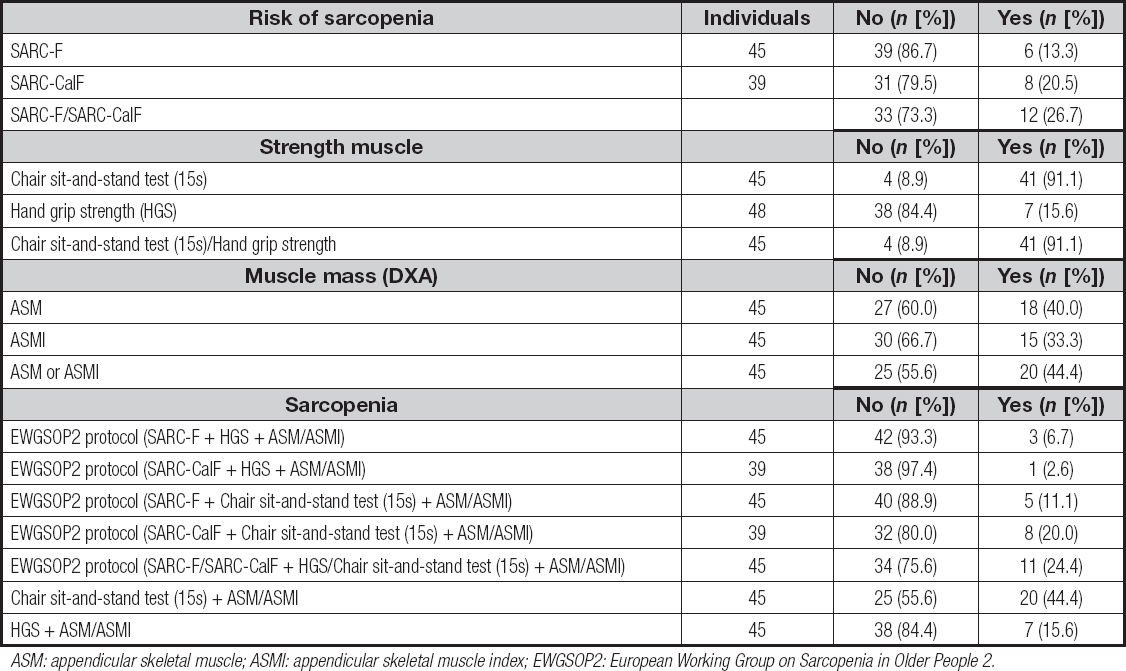
The prevalence rates of sarcopenia risk, low muscle strength, and reduced muscle mass are shown in table III. The SARC-Calf identified more patients at risk for sarcopenia compared to the SARC-F, with rates of 20.5 and 13.3, respectively. It is noteworthy that, due to the presence of lower limb edema and the consequent inability to measure CC, the SARC-Calf was applied to fewer patients than the SARC-F (86.7 of those evaluated).
Table III. Prevalence of sarcopenia risk, low strength muscle, and muscle depletion in outpatients with liver cirrhosis
ASM: appendicular skeletal muscle; ASMI: appendicular skeletal muscle index; EWGSOP2: European Working Group on Sarcopenia in Older People 2.
The prevalence of low muscle strength was identified in 91.1 by the chair stand test compared to 15.6 by handgrip strength. Therefore, the chair stand test proved to be a more efficient screening method for sarcopenia than handgrip strength, identifying nearly six times more patients with reduced strength than dynamometry.
Muscle depletion was highly prevalent, observed in 40 % of the evaluated patients. Interestingly, using the sarcopenia diagnostic protocol suggested by EWGSOP2, the prevalence of sarcopenia varied widely depending on the combination of assessment tools used. It ranged from 2.6 % (SARC-Calf + HGS + ASM/ASMI) to 20.0 % (SARC-Calf + Chair sit-and-stand test (15s) + ASM/ASMI), indicating significant variability in the diagnosis of sarcopenia among outpatients with cirrhosis. When positivity in any of the forms (SARC-F or SARC-Calf) and strength tests (HGS or chair sit-and-stand test) was considered, the prevalence increased to 24.4 %.
Notably, the combination of the chair stand test with reduced muscle mass (ASM/ASMI) identified 44.4 % of patients with cirrhosis as having both low strength and low muscle mass, classifying them as sarcopenic. Furthermore, those identified with muscle depletion by DXA were the same individuals classified as sarcopenic, indicating that reduced muscle mass in this group necessarily reflects low strength. This finding aligns with the sarcopenia screening sequence, where decreased strength precedes muscle mass reduction, and underscores the importance of assessing muscle mass in these individuals.
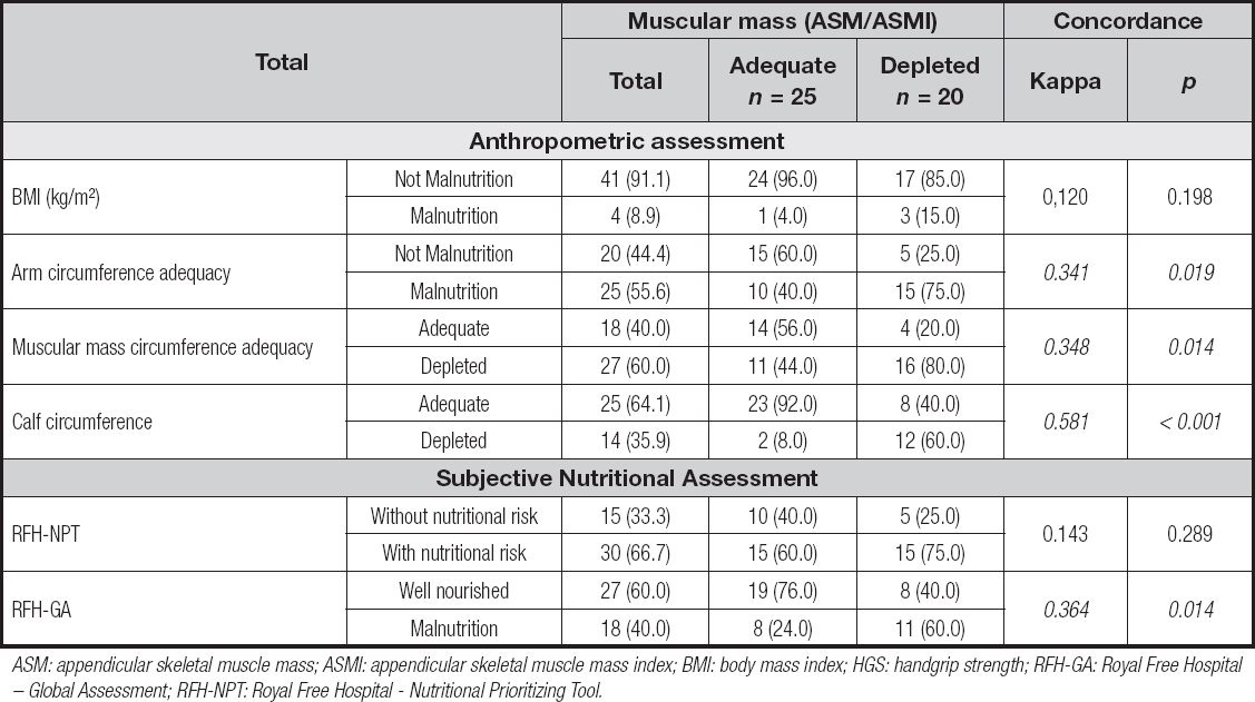
To determine the best method for diagnosing sarcopenia in outpatients with cirrhosis, we compared different muscle and nutritional assessment techniques with muscle depletion diagnosed by DXA. The concordance analysis (Table IV) showed that among anthropometric assessments, CC demonstrated the highest agreement with muscle depletion (κ = 0.581; p < 0.001), successfully identifying 60 % of patients with reduced muscle mass (MMEA/IMMEA). This indicates that CC is the most reliable anthropometric measure for identifying muscle depletion in this population when DXA is unavailable.
Table IV. Concordance between muscular mass by dual-energy X-ray absorptiometry (DXA) and anthropometrics assessment and subjective instruments used in outpatients with liver cirrhosis
ASM: appendicular skeletal muscle mass; ASMI: appendicular skeletal muscle mass index; BMI: body mass index; HGS: handgrip strength; RFH-GA: Royal Free Hospital - Global Assessment; RFH-NPT: Royal Free Hospital - Nutritional Prioritizing Tool.
Other anthropometric measures, such as AC (κ = 0.341; p = 0.019) and MMAC adequacy (κ = 0.348; p = 0.014), showed fair agreement with DXA being less effective than CC. BMI displayed slight agreement (κ = 0.120; p = 0.198), highlighting its limited utility in detecting sarcopenia in patients with cirrhosis.
Regarding subjective nutritional assessments, the RFH-GA (κ = 0.364; p = 0.014) and RFH-NPT (κ = 0.143; p = 0.289) showed a fair level of agreement with muscle depletion. These findings suggest that while subjective assessments may offer some insights, they cannot replace more objective measures, particularly DXA and CC, in accurately diagnosing sarcopenia.
Therefore, identifying muscle depletion using CC, especially in settings without DXA, appears to be a practical and effective approach for diagnosing sarcopenia in outpatients with cirrhosis.
DISCUSSION
Studies confirm that early identification of nutritional risk, as well as risk of sarcopenia and sarcopenia itself, is crucial for ensuring accurate treatment, potential reversal of the condition, improved prognosis, and quality of life for the affected individual (16-18). When proposing to identify nutritional risk or sarcopenia risk, screening instruments are used to maximize true positives within a sample, with subsequent steps aiming to discard false positives (19,20).
Individuals with cirrhosis experience a progressively worsening condition, often leading to episodes of decompensation that frequently require hospitalization, thereby increasing morbidity and mortality rates. Key complications include ascites, UGIB, and HE, which elevate the mortality risk by 5 to 10 times in this population (21). The average survival for patients experiencing these complications is merely 1 to 2 years, while compensated individuals have a survival expectancy of 10 to 12 years (22). Given its impact on global health (as the 11th leading cause of death, accounting for 2 million fatalities) and its substantial cost (with $32.5 million spent in the US alone in 2016) (21), preventing hepatic decompensation through pharmacological or non-pharmacological interventions is crucial to reduce hospitalizations, healthcare expenses, and improve patients' quality of life.
Nutritional status is heavily impacted by cirrhosis progression. Reduced food intake, energy-protein imbalances, altered macronutrient and micronutrient metabolism, diminished absorptive capacity, as well as muscle dysfunction and sarcopenia, are common nutritional complications seen in individuals with cirrhosis (23).
As cirrhosis negatively impacts nutritional status, the presence of nutritional and functional deficits also adversely affects the clinical progression of patients with cirrhosis. This influence extends to quality of life, with increased risks of infection, HE, ascites, and mortality, making it a prognostic factor for individuals with liver cirrhosis (24). However, the identification of nutritional deficits, especially malnutrition and sarcopenia, remains a challenge in cirrhosis due to the frequent occurrence of fluid retention (edema and ascites) in these patients. This retention hampers the use of more affordable and accessible anthropometric and body composition measures, such as weight, CC, and bioimpedance, across various clinical nutrition monitoring settings for these patients.
The EWGSOP2 recommends using the SARC-F tool for sarcopenia risk screening (3). In a study involving patients with cirrhosis, Singla et al. (2024) demonstrated good sensitivity of the SARC-F score for bedside screening in the Indian population (25). However, a meta-analysis by Voelker et al. (2021) suggested applying sarcopenia diagnostic criteria independently of risk screening due to the SARC-F's low sensitivity, which may lead to the detection of only severe cases (26). Our findings support this, as the SARC-F and SARC-Calf showed low efficacy in identifying individuals with low muscle strength and depletion in outpatients with cirrhosis, indicating that these tools should not be solely relied upon for sarcopenia diagnosis in this population.
Our study is pioneering in that it evaluates sarcopenia prevalence using different methods and assesses the agreement between sarcopenia diagnosis, based on decreased strength (sit-to-stand test) and muscle mass (DXA), and various nutritional and functional assessment methods in outpatients with cirrhosis. Following the diagnostic criteria for sarcopenia, decreased strength precedes skeletal muscle depletion, which is why strength tests, such as the sit-to-stand test and HGS, should precede body composition assessment. In our work, we found that relying on HGS could result in a high number of false negatives, potentially depriving many patients with cirrhosis of timely and appropriate interventions involving physical exercise and nutritional adjustments, which are currently the main treatment options, given the lack of effective pharmacological treatments (27).
Although HGS is widely used to measure strength, its limitations are evident, as it primarily assesses hand and forearm muscles, which are not critical for activities that involve supporting body weight. Despite showing moderate correlation with strength in other body compartments (28), HGS might not be as effective as the sit-to-stand test, which is a more comprehensive tool for assessing functional capacity and muscle power (3). Additionally, several mechanisms contribute to muscle strength impairment in patients with cirrhosis, including muscle quality changes, hormonal alterations, electrolyte imbalances, and systemic complications (8,16,29).
DXA, a recommended method for body composition assessment, accurately evaluates muscle mass and is suitable for individuals with cirrhosis, especially since it can bypass ascites interference when using appendicular skeletal muscle mass (ASM/ASMI) (3,16). Our study confirmed the high prevalence of muscle depletion in outpatients with cirrhosis, reinforcing the importance of incorporating muscle mass measurement for sarcopenia diagnosis.
The sit-to-stand test emerged as a highly effective screening tool for probable sarcopenia, as it identified the largest number of individuals with low muscle strength. Its simplicity, requiring only a chair and timer, makes it more accessible than HGS, and it can be employed across different healthcare settings (30). In contrast CC proved to be the most viable alternative for muscle mass assessment when DXA is unavailable, demonstrating the best agreement with ASM/ASMI. Interestingly, while the SARC-Calf incorporates CC as part of its assessment - adding 10 points to the final score -, our findings showed that CC alone presented a stronger correlation with muscle depletion (ASM/ASMI) compared to SARC-Calf. This suggests that isolating CC as an independent measure may enhance its utility in clinical practice, particularly when the broader SARC-Calf framework shows limitations in identifying low muscle strength and quality.
This association between CC and muscle mass was also identified by Kawakami et al. (2020) in their study of Japanese adults, where CC positively correlated with muscle mass measured by bioimpedance or DXA, regardless of the presence of obesity. Therefore, CC can be considered a useful diagnostic marker for sarcopenia (31).
However, it is important to acknowledge the limitations of using CC in patients with lower limb edema, a common condition in cirrhosis. Given this, the chair sit-and-stand test is recommended as an alternative when edema precludes CC measurement, based on the strong agreement observed in our study regarding sarcopenia diagnosis.
LIMITATIONS AND PERSPECTIVES
The data were collected from outpatients with cirrhosis, meaning that in more severe or decompensated cases, alternative criteria might provide more accurate sarcopenia diagnosis. The inclusion criteria for this study aimed to minimize confounding factors, which may have consequently limited the participation of more compromised individuals, such as those with hepatocellular carcinoma, cardiac, renal, and/or pulmonary complications, or hepatic encephalopathy. Therefore, the findings may not fully represent patients with more advanced disease stages. However, by including patients based on MELD-Na scores, an internationally recognized measure of disease severity, our results can be extrapolated to patients who do not present terminal-stage conditions.
Moreover, although DXA was chosen over computed tomography (CT) — the gold standard for muscle assessment in liver disease - due to greater accessibility, this limitation was mitigated by evaluating appendicular skeletal muscle mass (ASM) and its index (ASMI). We acknowledge that CT imaging, particularly in advanced cirrhosis, where it is often performed for hepatic lesion monitoring or pre-transplant evaluation, could provide more precise muscle assessments and should be considered in future studies.
Finally, the etiology of liver disease may significantly influence nutritional status, especially in alcoholic cirrhosis, as alcohol interferes with nutrient absorption, leading to chronic malnutrition and exacerbating nutritional deficits. However, no specific tools currently exist for nutritional risk or status assessment based on disease etiology. Future assessment tools could consider incorporating alcoholic cirrhosis as a criterion, emphasizing its nutritional impact.
CONCLUSION
Our study demonstrates that the EWGSOP2 algorithm tends to underdiagnose sarcopenia in outpatients with cirrhosis, primarily due to the low sensitivity of the SARC-F tool in this population. Therefore, we suggest that the screening step be excluded or that CC measurement be used as an alternative, provided there is no lower limb edema.
The chair sit-and-stand test emerged as the most reliable method for identifying low muscle strength, effectively capturing a greater number of individuals with probable sarcopenia than HGS. Additionally, CC measurement showed moderate concordance with ASM/ASMI and could serve as a practical alternative in the absence of imaging methods, although it may miss a considerable number of patients with muscle depletion. Hence, CC is not sufficient as a standalone diagnostic measure for sarcopenia in this population.
Our findings reinforce the critical need for incorporating imaging techniques such as DXA or CT in the comprehensive care of patients with cirrhosis to ensure accurate identification and appropriate intervention for sarcopenia. Despite the limited availability of DXA in routine clinical settings, its role in accurately assessing muscle mass highlights the necessity for its inclusion, even if performed with reduced frequency.
In alignment with the Delphi consensus from the Global Leadership Initiative in Sarcopenia (GLIS) (32), which emphasizes the practicality and feasibility of sarcopenia assessment components, we propose that combining CC and the chair sit-and-stand test can serve as feasible alternatives for diagnosing and monitoring sarcopenia in cirrhotic patients, particularly in resource-limited settings where imaging methods are not readily accessible.
AUTHORS' CONTRIBUTION
Conceptualization, data curation and methodology, M. D. R. G., N. B. B., R. M. A. F. W., and F. A. M.; investigation, M. D. R. G., F. L. C. A., A. I. A. N. S., A. J. S. W., and F. A. M.; collection of data and materials, M. D. R. G., A. I. A. N. S., A. J. S. W., and F. A. M.; writing original draft preparation, M. D. R. G., F. L. C. A., N. B. B., J. C. F. S., and F. A. M.; writing, review and editing, M. D.R. G., N. B. B., J. C. F. S., and F. A. M. All authors have read and agreed to the published version of the manuscript.

