




 Servicios personalizados
 Inglés (pdf)
 Articulo en XML
 Referencias del artículo
 Traducción automática
 Enviar articulo por email
 Citado por SciELO
  Citado por Google
  Similares en
SciELO
Similares en
SciELO  Similares en Google
 Permalink
INTRODUCTION
Leukemia is a malignant tumor originating from the hematopoietic system, characterized by the abnormal proliferation of white blood cells in the bone marrow and/or peripheral blood. Based on the degree of cell maturity and the speed of proliferation, leukemia can be classified into two major categories: acute and chronic (1). Acute leukemia includes Acute Myeloid Leukemia (AML) and Acute Lymphoblastic Leukemia (ALL), which are primarily characterized by the rapid proliferation of immature cells. Chronic leukemia includes Chronic Myeloid Leukemia (CML) and Chronic Lymphocytic Leukemia (CLL), which progress more slowly and involve an increase in mature cells. Leukemia poses a significant threat to global public health. According to the World Health Organization (WHO), hundreds of thousands of new cases are diagnosed each year. Treatment for leukemia is not only costly but also has a severe impact on patients' quality of life (2).
Vitamin D, an essential fat-soluble vitamin, plays a critical role beyond maintaining bone health. It is involved in immune regulation, anti-inflammatory processes, and antitumor activity (3,4). Sunlight exposure is an effective way to increase vitamin D levels in the body, and studies have shown that adequate vitamin D status is associated with a reduced risk of cancer (5). In recent years, increasing attention has been given to the relationship between vitamin D and leukemia, with research suggesting that vitamin D may positively influence survival outcomes in children with leukemia (6-8) . However, findings regarding the association between vitamin D levels and leukemia risk have been inconsistent. Some epidemiological studies have found that higher vitamin D levels appear to be associated with a lower risk of leukemia (9), while clinical evidence has also indicated no significant relationship between vitamin D levels and the prognosis of leukemia patients (10). These conflicting results may be due to limitations in study design, such as inadequate control of confounding factors, small sample sizes, or differences in measurement methods.
Mendelian randomization (MR) is a method that utilizes genetic variants as instrumental variables to assess the causal relationship between exposures and diseases. This approach offers a strategy to reduce the influence of confounding factors and reverse causality. By leveraging the fixed and randomly assigned nature of genetic variants throughout an individual's lifetime, MR provides more reliable causal inferences (11). In this study, we applied Mendelian randomization to investigate the causal relationship between vitamin D levels and the risk of various types of leukemia. Using data from large-scale genome-wide association studies (GWAS), this research aims to resolve the conflicting findings in the existing literature regarding the link between vitamin D and leukemia and to provide evidence-based guidance for leukemia prevention and treatment strategies. The findings from this study may help public health policymakers and clinicians better understand the potential role of vitamin D in leukemia prevention, thereby improving patient management and treatment strategies.
MATERIALS AND METHODS
STUDY DESIGN
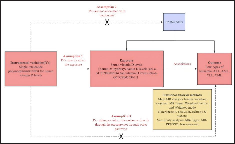
As shown in figure 1, this study utilized a Mendelian randomization (MR) design to systematically investigate the association between vitamin D levels and the risk of leukemia. Since this study involved the reanalysis of publicly available genome-wide association study (GWAS) data, no additional ethical approval was required.
DATA SOURCES
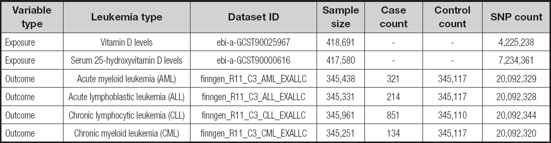
In this study we conducted a comprehensive Mendelian randomization (MR) analysis to explore the causal relationship between vitamin D levels and the risk of leukemia. The patients included in this study met the diagnostic criteria defined by the International Classification of Diseases (ICD). The exposure variables in this study were serum 25-hydroxyvitamin D levels (ebi-a-GCST90000616) and overall vitamin D levels (ebi-a-GCST90025967), both sourced from genome-wide association studies (GWAS). Specifically, the dataset for ebi-a-GCST90025967 included 418,691 samples, analyzing 4,225,238 single nucleotide polymorphisms (SNPs) (12), while the dataset for ebi-a-GCST90000616 included 417,580 samples, analyzing 7,234,361 SNPs (13).
The outcome variables included the following four leukemia phenotypes: acute myeloid leukemia (AML), acute lymphoblastic leukemia (ALL), chronic lymphocytic leukemia (CLL), and chronic myeloid leukemia (CML). AML is characterized by the abnormal proliferation of immature cells in the bone marrow and impaired maturation; ALL primarily affects lymphoid cells, particularly B or T cells; CLL often originates from B lymphocytes and has a slow disease course; and CML is typically associated with the BCR-ABL fusion gene (Philadelphia chromosome), progressing through chronic, accelerated, and blast phases.
The specific data sources for each leukemia subtype are as follows:
- AML: data from a 2024 Finnish dataset (finngen_R11_C3_AML_EXALLC), including 321 cases and 345,117 controls, analyzing a total of 20,092,329 SNPs.
- ALL: data from a 2024 Finnish dataset (finngen_R11_C3_ALL_EXALLC), including 214 cases and 345,117 controls, analyzing a total of 20,092,328 SNPs.
- CLL: data from a 2024 Finnish dataset (finngen_R11_C3_CLL_EXALLC), including 851 cases and 345,110 controls, analyzing a total of 20,092,344 SNPs.
- CML: data from a 2024 Finnish dataset (finngen_R11_C3_CML_EXALLC), including 134 cases and 345,117 controls, analyzing a total of 20,092,320 SNPs.
The specific information on exposure and outcome is shown in table I.
SELECTION OF INSTRUMENTAL VARIABLES
Mendelian randomization (MR) analysis is based on three fundamental assumptions: 1) the genetic variants must be strongly associated with the exposure factor; 2) the genetic variants must be independent of confounding factors; 3) the genetic variants must not be influenced by the outcome factors (11,14). An initial genome-wide significance threshold of 5 × 10⁻⁸ was set to identify single nucleotide polymorphisms (SNPs) significantly associated with lipid metabolism traits (15). To ensure the independence of these SNPs, we applied a linkage disequilibrium (LD) threshold of r2 = 0.001 and required a physical distance of more than 10,000 kb to ensure that no LD occurred between the selected SNPs (16).
Additionally, we calculated the F-statistic for each instrumental variable (IV) to assess the potential for weak instrument bias. The F-statistic was calculated using the formula F = Beta2/SE2, where Beta represents the effect size of the allele and SE is the standard error of Beta. Only IVs with F-values greater than 10 were retained for further analysis to minimize the risk of bias due to weak instruments (17).
Finally, to further ensure the robustness of our analysis, all selected SNPs were cross-checked using the Phenoscanner database (www.phenoscanner.medschl.cam.ac.uk) on June 10, 2024, to exclude any SNPs potentially associated with confounding phenotypes (18). SNPs that were not associated with confounders were retained for the final analysis, ensuring the validity and reliability of our MR study.
MENDELIAN RANDOMIZATION ANALYSIS
In this study, we employed the inverse variance weighted (IVW) method as the primary approach to estimate causal effects. IVW is regarded as a robust tool for detecting causal relationships in two-sample Mendelian randomization (MR) analysis. To ensure the robustness of our findings, we performed supplementary analyses using MR-Egger regression, weighted median, and weighted mode methods (19).
Additionally, we conducted a series of sensitivity analyses to further validate the results. First, Cochran's Q test was applied to assess heterogeneity, which could influence causal estimates. If the p-value was greater than 0.05, heterogeneity was considered negligible, and a fixed-effect model was used. Conversely, if the p-value was less than or equal to 0.05, a random-effects model was adopted to account for heterogeneity (20).
To detect horizontal pleiotropy that might bias the MR results, we performed an MR-Egger intercept test. Furthermore, the MR-PRESSO outlier detection method was used to identify and correct for outliers among the SNPs, addressing any potential horizontal pleiotropy (21). Residual sensitivity analyses were also conducted to assess the robustness of the findings. Statistical analyses were performed using R software (version 4.3.1), with the “TwoSampleMR” and “MRPRESSO” packages facilitating the MR analysis, and the “forest plot” package used for visualization. A p-value of less than 0.05 was considered statistically significant.
RESULTS
INSTRUMENTAL VARIABLES
In this study, we conducted a detailed Mendelian randomization analysis to explore the causal relationship between serum 25-hydroxyvitamin D levels, overall vitamin D levels, and various types of leukemia. SNPs relevant to different types of leukemia were initially selected from large-scale genomic data. For acute lymphoblastic leukemia (ALL), we excluded 5 SNPs from an initial pool of 115, retaining 110 SNPs for the final analysis. The same selection criteria were applied for the analyses of acute myeloid leukemia (AML) and chronic myeloid leukemia (CML). For chronic lymphocytic leukemia (CLL), we excluded 6 SNPs from 115, retaining 109 for further analysis.
In the analysis of overall vitamin D levels, we identified 88 SNPs for ALL, AML, and CML, and excluded 1 SNP in the CLL analysis, retaining 87 SNPs. The selection and exclusion of SNPs were based on the core assumptions of Mendelian randomization to ensure the validity and robustness of the analysis. Detailed information on the selected SNPs and further specifics can be found in supplementary table I (https://www.nutricionhospitalaria.org/files/8598/ADMA1-05541-01.xlsx). These meticulous methodological steps ensured the rigor of our causal inference.
TWO-SAMPLE MENDELIAN RANDOMIZATION ANALYSIS
We employed four methods—inverse variance weighted (IVW), MR-Egger, weighted median, and weighted mode—to assess the causal relationship between vitamin D levels and leukemia. As shown in figure 2, serum 25-hydroxyvitamin D levels did not demonstrate a significant causal relationship with acute lymphoblastic leukemia (ALL) (OR = 0.86, 95 % CI: 0.42-1.74, p = 0.68), acute myeloid leukemia (AML) (OR = 0.95, 95 % CI: 0.54-1.68, p = 0.87), or chronic lymphocytic leukemia (CLL) (OR = 1.05, 95 % CI: 0.73-1.53, p = 0.78). However, a protective relationship was observed with chronic myeloid leukemia (CML) (OR = 0.46, 95 % CI: 0.26-0.81, p = 0.007).
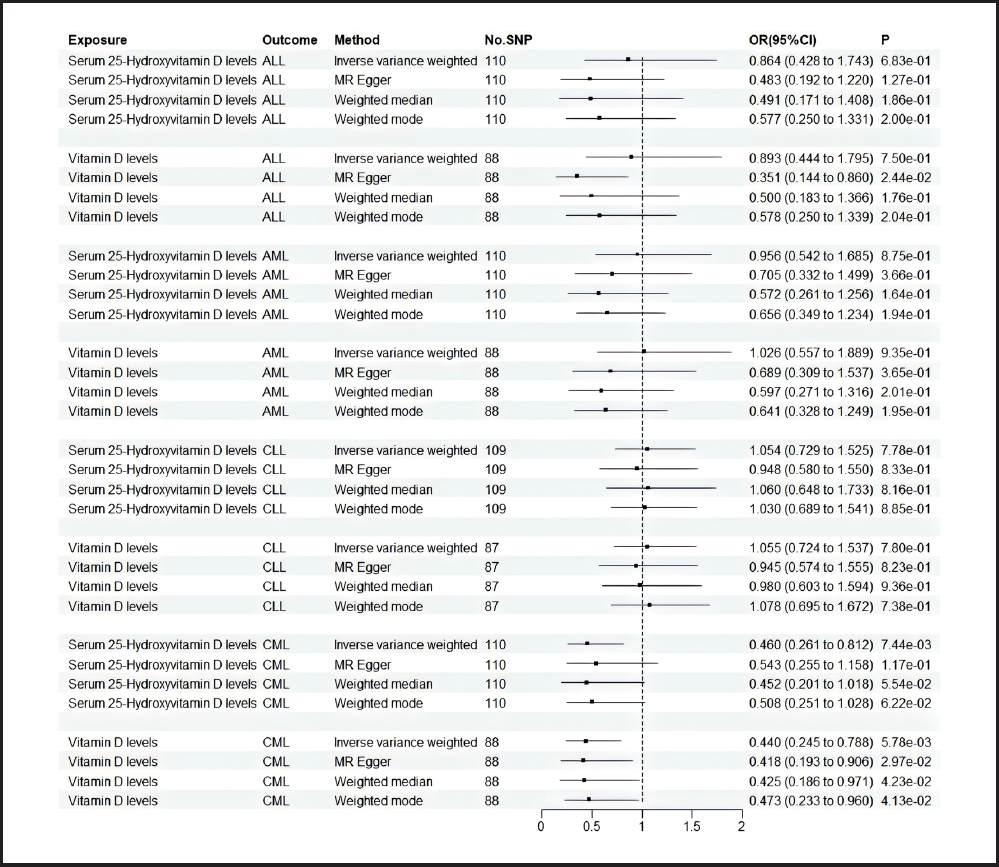
Similarly, overall vitamin D levels showed no significant causal relationship with ALL (OR = 0.89, 95 % CI: 0.44-1.79, p = 0.75), AML (OR = 1.02, 95 % CI: 0.55-1.88, p = 0.93), or CLL (OR = 1.05, 95 % CI: 0.72-1.53, p = 0.78). In contrast, a protective relationship was observed with CML (OR = 0.44, 95 % CI: 0.25-0.78, p = 0.005).
RELIABILITY EVALUATION
Pleiotropy test
As shown in table II, the MR-Egger intercept results for the analysis of serum 25-hydroxyvitamin D levels and overall vitamin D levels with CML indicated that the Egger intercept (p > 0.05) was not significantly different from zero. This suggests that there was no evidence of horizontal pleiotropy in our Mendelian randomization analysis.
Heterogeneity analysis
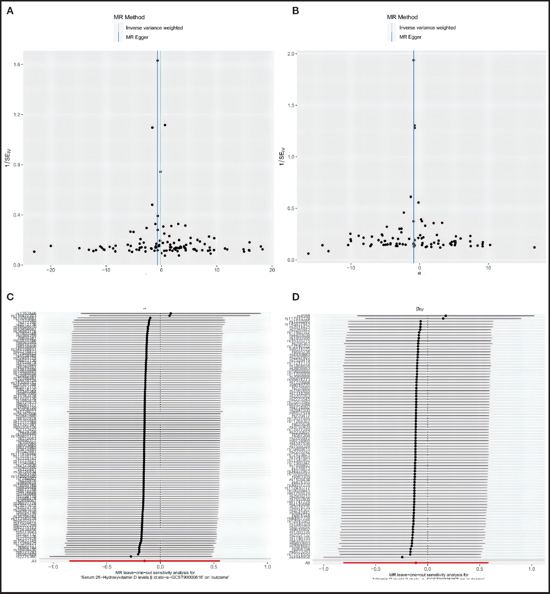
Table II also shows that the Cochran's Q test for the analysis of serum 25-hydroxyvitamin D levels and vitamin D levels with CML revealed p-values greater than 0.05, indicating no heterogeneity in the MR analysis. Furthermore, the MR funnel plots for each group showed a symmetrical distribution of scatter points for the causal association effects, suggesting no potential bias (Fig. 3).

Sensitivity analysis
The leave-one-out test results indicated that excluding individual SNPs one at a time yielded IVW analysis results consistent with those that included all SNPs. In the analysis of serum 25-hydroxyvitamin D levels with CML, SNPs rs1352846 and rs116970203 had a significant influence on the causal association estimates. Similarly, in the analysis of vitamin D levels with CML, SNPs rs4588 and rs117913124 had a significant impact on the causal estimates. Repeating the MR analysis after excluding these influential SNPs showed that the causal effect of serum 25-hydroxyvitamin D levels on CML was not significant (OR = 0.48, 95 % CI: 0.22-1.02, p = 0.06). However, vitamin D levels remained a protective factor for CML (OR = 0.44, 95 % CI: 0.19-0.98, p = 0.04). These results further confirm the stability of the findings (Fig. 3); “a” represents the funnel plot of causal effects between serum 25-hydroxyvitamin D levels and CML, “b” represents the funnel plot of causal effects between vitamin D levels and CML, “c” represents the sensitivity analysis of the leave one method between serum 25-hydroxyvitamin D levels and CML, and “d” represents the sensitivity analysis of the leave one method between vitamin D levels and CML.
DISCUSSION
Vitamin D exerts its antitumor effects through multiple mechanisms, including inhibiting cell proliferation, inducing differentiation and apoptosis, regulating immune responses, and suppressing pro-inflammatory reactions within the tumor microenvironment (22). In this study, we employed various Mendelian randomization (MR) methods to evaluate the relationship between serum 25-hydroxyvitamin D levels and different types of leukemia. The results indicated a significant protective association between serum 25-hydroxyvitamin D levels and chronic myeloid leukemia (CML). Using the inverse variance weighted (IVW) method, we found a significant inverse relationship between serum 25-hydroxyvitamin D levels and the risk of CML (OR = 0.46, 95 % CI: 0.26-0.81, p = 0.007). Similarly, overall vitamin D levels also demonstrated a protective effect (OR = 0.44, 95 % CI: 0.25-0.78, p = 0.005).
However, for acute myeloid leukemia (AML), acute lymphoblastic leukemia (ALL), and chronic lymphocytic leukemia (CLL), our analysis did not reveal significant associations between vitamin D levels and the risk of these leukemia types. This may suggest that the protective effect of vitamin D varies among different leukemia types or that the limited sample size led to insufficient statistical power to detect smaller effects. For example, the study by Sameer A. Parikh et al. (6) found that vitamin D deficiency may be associated with poor prognosis in CLL patients. Similarly, Yasmeen Jramne-Saleem et al. (7) showed that vitamin D derivatives could enhance the differentiation of AML cells by modulating the Nrf2/ARE pathway and its downstream targets, including glutathione and the AP-1 transcription factor. Furthermore, Eliza Turlej et al. (8) reported that vitamin D analogs (VDAs) could directly inhibit the proliferation of ALL-derived B cells and normal B cells, both of which express vitamin D receptors (VDR).
To validate the robustness of our results, we conducted extensive sensitivity analyses. For instance, MR-Egger regression and leave-one-out sensitivity analyses indicated that although individual SNPs may significantly influence the estimates, the overall results consistently supported the protective effect of vitamin D levels on CML. Kazuki Kanno et al. (23) further emphasized the importance of vitamin D supplementation in specific patient groups through the AMATERASU randomized clinical trial. In this trial, vitamin D supplementation significantly improved the 5-year recurrence-free survival in patients within the p53 immune response subgroup but had no significant impact on patients outside this subgroup, suggesting that p53 may be a key factor in vitamin D's anticancer effects.
Additionally, this study highlights the potential application of serum 25-hydroxyvitamin D as a biomarker for leukemia prevention. Given vitamin D's role in cell proliferation, differentiation, and immune regulation, its potential protective effect against CML and other types of leukemia warrants further mechanistic research and clinical validation. Arkapal Bandyopadhyay et al. (24) conducted a randomized controlled clinical trial that found no significant benefit of vitamin D3 supplementation in early treatment responses in CML patients. However, long-term CML patients treated with imatinib mesylate exhibited significant vitamin D deficiency (25), indicating that vitamin D levels and their interaction with imatinib mesylate could play an important role in CML treatment.
It should be noted that this study was based on publicly available genome-wide association study (GWAS) data, which may be subject to potential selection bias or information bias. For instance, GWAS data often derive from specific populations, which may not fully represent leukemia patients from other ethnic groups or regions. Therefore, the generalizability of our findings may be limited, especially in populations with distinct racial or environmental differences. Additionally, vitamin D levels are influenced by various factors, including sun exposure, dietary intake, and lifestyle habits. While the Mendelian Randomization approach helps reduce confounding factors present in traditional epidemiological studies, it cannot entirely eliminate the influence of non-genetic factors on vitamin D levels. Moreover, this study only explored the causal relationship between vitamin D and leukemia without delving into the specific biological mechanisms or the dynamic interactions between vitamin D and leukemia development.
In summary, these findings support the notion that vitamin D may reduce the risk of certain types of leukemia by modulating tumor-related biological pathways. Our study provides a strong biological basis for the development of future leukemia prevention strategies and supports the potential benefits of vitamin D supplementation in clinical settings. Future research should further investigate the mechanisms by which vitamin D impacts different types of leukemia and evaluate its potential applications in leukemia prevention and treatment. These results provide scientific evidence for the development of vitamin D-based preventive measures, which could positively influence public health policy and clinical practice.

