




 Servicios personalizados
 Inglés (pdf)
 Articulo en XML
 Referencias del artículo
 Traducción automática
 Enviar articulo por email
 Citado por SciELO
  Citado por Google
  Similares en
SciELO
Similares en
SciELO  Similares en Google
 Permalink
INTRODUCTION
Ulcerative colitis (UC) is a nonspecific inflammatory bowel disease primarily affecting the rectum and colon, with an unclear pathogenesis (1). It is a significant health burden, and the global incidence of UC has increased in recent years, particularly in Asian countries, including China (2,3). Patients with UC often experience recurrent abdominal pain and bloody stools with mucus associated with disease activity. However, an absence of symptoms does not necessarily indicate complete remission, necessitating methods for monitoring disease activity at the onset of symptoms and during treatment (4). Biologics are among the key therapies used for UC. The United States Food and Drug Administration (FDA) and China’s National Medical Products Administration approved the use of vedolizumab for UC treatment in 2014 and 2020, respectively. Numerous studies have confirmed the efficacy of vedolizumab in inducing and maintaining clinical remission and mucosal healing in UC, although not all patients respond to this treatment (5,6). Thus, given the high cost and potential adverse effects associated with vedolizumab, there is an urgent need to predict and identify patients who are likely to respond to this treatment before initiating treatment as this would reduce the economic burden on patients and optimize the therapeutic efficacy of vedolizumab.
The deposition of adipose tissue, particularly of visceral fat which is known to have a pro-inflammatory role, has been found to be involved in both the pathogenesis and progression of UC. Several studies have demonstrated that visceral adiposity can not only predict the onset of inflammatory bowel disease but is also closely associated with clinical and endoscopic remission in patients undergoing biological therapy (7,8). Visceral fat tissue is traditionally assessed using computed tomography (CT) and magnetic resonance imaging (MRI) (9). However, these imaging techniques are expensive, with long waiting times for appointments, and CT exposes patients to radiation. While endoscopy can reflect disease activity, it is an invasive procedure requiring time-consuming bowel preparation, and is thus impractical for frequent monitoring. Similarly, the use of fecal calprotectin, despite being a non-invasive marker for assessing disease activity, is often associated with difficulties in the prompt collection of samples by patients for analysis. There is thus a long-standing need for a method that is non-invasive, permits repeated assessments, and can monitor disease activity effectively.
Currently, several indicators are used to reflect adiposity deposition. These include body mass index (BMI), waist circumference, visceral adiposity index (VAI), lipid accumulation product index (LAP), and the body roundness index (BRI) (10-13). However, these indices have limitations in that they either do not provide an accurate reflection of visceral adiposity deposition or may not represent the true characteristics of the Chinese population. Xie et al. (14) reported the use of the Chinese visceral adiposity index (CVAI) to diagnose visceral adiposity deposition in the Chinese population, and its predictive ability was validated in a cross-sectional study involving over 450 participants (14). This index is also closely associated with several obesity-related diseases, including diabetes and hypertension, and it is known that obesity can influence the progression of inflammatory bowel disease (IBD) and the response to treatment (15,16).
The CVAI is calculated using age, BMI, waist circumference, and the levels of triglycerides (TG) and high-density lipoprotein cholesterol (HDL-C). These measurements are easy to obtain, can be measured repeatedly, and are non-invasive. This study aimed to evaluate the relationship between the CVAI and disease activity in UC, as well as explore its value in predicting the response to vedolizumab treatment in UC.
MATERIALS AND METHODS
STUDY POPULATION
This retrospective study involved 84 patients with UC who were treated at the IBD Center of Jinhua Hospital, affiliated with Zhejiang University School of Medicine, between January 2021 and December 2023. All patients were aged 18 years or older. The exclusion criteria included the presence of malignant tumors, severe cardiac, pulmonary, hepatic, or renal insufficiency, and recent use of medications that could affect triglyceride or HDL levels. To minimize the influence of steroids on vedolizumab treatment and the predictive value of CVAI, vedolizumab treatment was initiated two weeks after the last steroid treatment. A control group of 72 age- and sex-matched individuals who underwent routine examinations at the hospital during the same period was also included. All patients provided written informed consent. The study was approved by the Ethics Committee of Jinhua Hospital (Approval No. 2024-29).
SCORING OF DISEASE ACTIVITY
The extent of UC was categorized as follows: 1) E1, rectal type; 2) E2, left-sided colitis (with involvement up to the splenic flexure); 3) E3, extensive colitis (with involvement beyond the splenic flexure or including the entire colon). Disease activity was assessed using the modified Mayo score. Clinical remission was defined as a Mayo score ≤ 2 with no individual subscore > 1, while an individual subscore > 1 indicated non-remission, and a Mayo score > 2 represented active disease (with 3-5 points indicating mild activity, 6-10 points indicating moderate activity, and 11-12 points indicating severe activity). During follow-up, clinical response was defined as a ≥ 50 % reduction in patient-reported outcomes (PRO2). The severity of mucosal disease was scored using the Mayo Endoscopic Score (MES) system as follows: 1) 0 points, normal mucosa or no active lesions; 2) 1 point, mucosal erythema, reduced vascular pattern, and mild friability; 3) 2 points, pronounced mucosal erythema, lack of vascular pattern, friability, and mucosal erosion; 4) 3 points, spontaneous bleeding and ulcer formation.
CLINICAL EVALUATION AND LABORATORY TESTS
Data on demographics, laboratory test results, and other relevant information were collected from the electronic medical records system. During the induction phase, 300 mg of vedolizumab was administered intravenously at weeks 0, 2, and 6, while, during the maintenance phase, 300 mg was given intravenously every 8 weeks. Blood tests, including routine blood tests, C-reactive protein (CRP), liver and renal function tests, erythrocyte sedimentation rate (ESR), and lipid profiles, were performed before each infusion. Effectiveness was assessed 12-14 weeks after the initial vedolizumab infusion and was classified into clinical response and primary non-response based on the PRO2 score. Additionally, the CVAI index was calculated before and after treatment. The CVAI was computed as follows: For males: -267.93 + 0.68 × age + 0.03 × BMI + 4 × WC (cm) + 22 × Log10 TG (mmol/L) - 16.32 × HDL-C (mmol/L). For females: -187.32 + 1.71 × age + 4.32 × BMI + 1.12 × WC (cm) + 39.76 × Log10 TG (mmol/L) - 11.66 × HDL-C (mmol/L).
STATISTICAL METHODS
Normally distributed continuous variables are expressed as mean ± standard deviation, with comparisons between groups conducted using t-tests. Non-normally distributed continuous variables are expressed as median and interquartile range, with group comparisons performed using the Mann-Whitney U-test. Categorical data are expressed as percentages, and were compared using chi-square or Fisher’s exact tests. For comparisons among the mild, moderate, and severe UC groups, the Kruskal-Wallis test was used, with pairwise comparisons conducted using Dunn’s test. The ability of the CVAI to differentiate between different groups was assessed using receiver operating characteristic (ROC) curves. The correlation between the CVAI and disease activity was evaluated using Pearson’s correlation coefficient. A p-value of less than 0.05 was considered statistically significant. Statistical analyses were performed using the Prism, version 9.0 software.
RESULTS
BASELINE CLINICAL CHARACTERISTICS
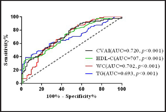
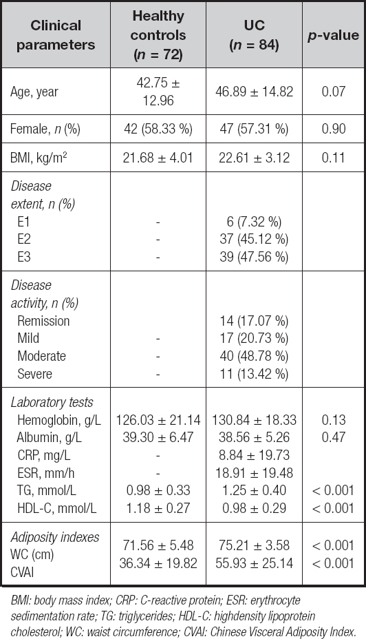
Table I presents the baseline clinical data for healthy controls and patients with UC. There were no significant differences in hemoglobin and albumin levels between the two groups. However, patients with UC had significantly higher TG levels, WC, and CVAI compared to the healthy controls (TG: 0.98 ± 0.33 vs. 1.25 ± 0.40, p < 0.001; WC: 71.56 ± 5.48 vs. 75.21 ± 3.58, p < 0.001; CVAI: 36.34 ± 19.82 vs. 55.93 ± 25.14, p < 0.001). Conversely, HDL-C levels were lower in patients with UC than in healthy controls (1.18 ± 0.27 vs 0.98 ± 0.29, p < 0.001). Notably, the CVAI index in patients with UC was 1.5 times higher than that in the healthy population. The ROC curve analysis revealed that CVAI had the highest area under the curve (AUC = 0.720) value among the four metrics, indicating superior predictive ability compared to TG (AUC = 0.693), WC (AUC = 0.702), and HDL-C (AUC = 0.707) (Fig. 1). Thus, the CVAI may be a practical clinical marker for UC.
CLINICAL AND ENDOSCOPIC ACTIVITY OF UC AS REFLECTED BY THE CHINESE VISCERAL ADIPOSITY INDEX
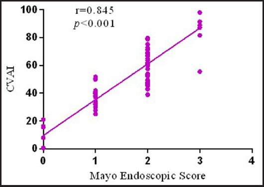
A modified Mayo score was used to classify UC clinical activity into remission and active phases. It was observed that the CVAI index was significantly elevated in patients during the active phase (33.64 ± 21.99 vs. 60.27 ± 22.87, p < 0.001) (Fig. 2A). The ROC curve analysis confirmed that the CVAI index was effective as an indicator to differentiate between the remission and active phases of UC, with an AUC of 0.809, a cut-off value of 45.87, a sensitivity of 0.857, a specificity of 0.764, and a Youden index of 0.622 (Fig. 2B). After the classification of active UC into mild, moderate, and severe stages, it was found that the CVAI index values were significantly higher in moderate and severe UC compared to mild UC (41.27 ± 15.64 vs. 57.19 ± 11.27, p < 0.001; 41.27 ± 15.64 vs. 100.86 ± 12.43, p < 0.001) (Fig. 3A). In addition, ROC curve analysis showed that the CVAI index could distinguish between mild and moderate-to-severe UC activity, with an AUC of 0.843, a cut-off value of 46.88, a sensitivity of 0.765, a specificity of 0.882, and a Youden index of 0.647 (Fig. 3B). These results indicate that the CVAI index can be used to monitor UC disease activity. Apart from symptom control, mucosal healing is also an important aspect of the treatment of UC. Mucosal activity was assessed using the MES and was found to be positively correlated with the CVAI index (Pearson’s r = 0.845, p < 0.001) (Fig. 4). These data demonstrate that the CVAI index is an effective reflection of endoscopic activity in patients with UC and is especially useful in those who require frequent endoscopic examinations to evaluate mucosal healing.
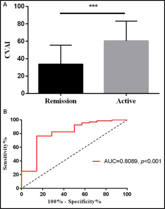
Figure 2 Correlation between the CVAI index and disease activity in patients with UC. A. Association of the CVAI index with disease activity, divided into into remission and active disease according to the modified Mayo score. B. Receiver operating characteristic curve in which the CVAI index was used to distinguish between patients with UC in remission and those with active-phase UC. p < 0.05 was defined as statistically significant. ***p < 0.001.
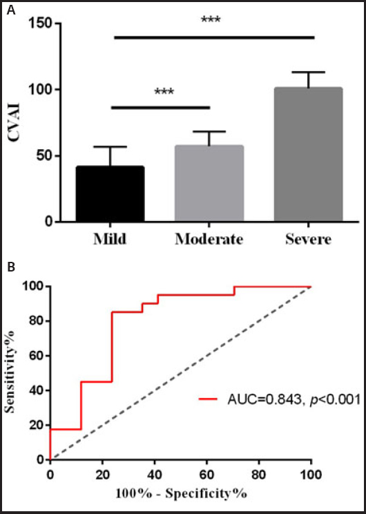
Figure 3 Correlation between the CVAI index and disease severity in patients with UC. A. The CVAI index was associated with disease severity, classified as mild, moderate, and severe. B. Receiver operating characteristic curve in which the CVAI index was used to distinguish patients with mild and moderate to severe UC. p < 0.05 was defined as statistically significant. ***p < 0.001.
PREDICTIVE VALUE OF THE CHINESE VISCERAL ADIPOSITY INDEX INDEX FOR RESPONSE TO VEDOLIZUMAB IN PATIENTS WITH UC
A total of 51 patients with UC were treated with vedolizumab. According to the PRO2 assessment, 17 patients were found to be non-responders, while 34 patients achieved a clinical response. Before each infusion of vedolizumab, patients underwent hematological tests, measurements of fecal calprotectin and WC, and calculation of the CVAI index. It was found that patients who achieved a clinical response had a significantly lower CVAI index before treatment compared to the primary non-responders (54.14 ± 15.45 vs 72.84 ± 21.44, p = 0.002) (Fig. 5A). The ROC curve analysis indicated that the CVAI index was an effective predictor of the response of patients with UC vedolizumab, with an AUC of 0.789, a cut-off value of 53.18, sensitivity of 0.853, specificity of 0.706, and Youden’s index of 0.559 (Fig. 5B). During the 1-year follow-up period, responders to vedolizumab showed reduced CVAI indices (72.84 ± 21.44 vs. 50.07 ± 19.82, p < 0.001) (Fig. 6A), in contrast to non-responders who exhibited no significant change in the CVAI index (54.14 ± 1 vs. 59.73 ± 19.70, p = 0.364) (Fig. 6B).
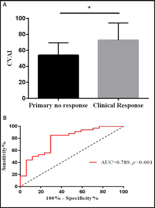
Figure 5 The CVAI index predicts the response to vedolizumab in patients with UC during induction. A. Comparison of the CVAI index of patients with primary non-response and those with clinically responsive UC before treatment. B. Receiver operating characteristic curve analyzing the ability of the pre-treatment CVAI index value in distinguishing between primary nonresponse and clinical response. p < 0.05 was defined as statistically significant. *p < 0.05.
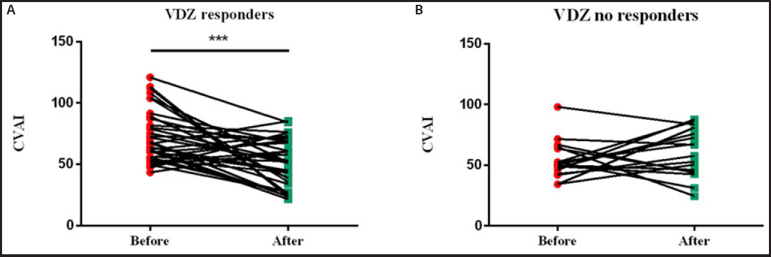
Figure 6 The CVAI index predicted the response of patients with UC to vedolizumab during follow-up. A. Comparison of the values of the CVAI index between the pre-treatment and follow-up periods in patients with UC who achieved clinical response to vedolizumab. B. Comparison of the values of the CVAI index in patients with UC who did not respond to vedolizumab before treatment and during follow-up. p < 0.05 was defined as statistically significant. ***p < 0.001.
DISCUSSION
Many studies, including those on inflammatory bowel disease, have explored the use of simple tools, often based on routine laboratory tests or readily accessible measurements, to assess disease severity and long-term prognosis (17,18). Currently, the methods available for evaluating and monitoring UC remain somewhat limited. This study is the first to evaluate the relationship between the CVAI and UC activity, as well as the response to vedolizumab in patients with UC. It was found that CVAI scores were higher in patients with UC relative to healthy individuals. Further analysis using the modified Mayo score revealed a close relationship between the CVAI and UC activity. Furthermore, the MES results indicated a significant correlation between the CVAI and UC severity as assessed by endoscopy, suggesting that the CVAI represents an effective measure of endoscopic inflammatory activity in UC. Additionally, the study found that patients with UC who responded to vedolizumab had lower CVAI scores prior to treatment compared to non-responders. ROC curve analysis further confirmed that a low CVAI score is a good predictor of response to vedolizumab in patients with UC. It can thus be concluded that the CVAI may be a valuable and promising indicator for predicting both UC disease activity and response to vedolizumab.
The assessment of disease activity in patients with UC, particularly the degree of mucosal inflammation, is crucial for effective treatment. Accurate and objective evaluations, along with timely judgments, are necessary for the implementation of appropriate therapeutic strategies (19). Objective methods for assessment are essential and include endoscopic and imaging evaluations. Imaging methods, such as CT, MRI, and intestinal ultrasound, have limitations. Specifically, CT involves radiation exposure, MRI is relatively expensive, and intestinal ultrasound, while useful, is not universally available in all hospitals in China and its accuracy is dependent on the skill level of the operator. These methods are thus not suitable for frequent use (20-22). Although endoscopy is the gold standard for assessing and monitoring intestinal inflammation in patients with UC, it requires thorough bowel preparation and is an invasive procedure with associated risks such as perforation and bleeding, thus restricting its frequent use (23,24). The measurement of fecal calprotectin is a non-invasive and relatively inexpensive method for evaluating UC activity; however, the collection of samples is often inconvenient, and many patients have difficulty delivering samples promptly to the hospital for testing (25). Therefore, the use of simple hematological indices or easily accessible testing tools for evaluating and monitoring UC activity is highly desirable and could improve patient compliance.
Many recent studies have shown that adipose tissue, particularly visceral adipose tissue, plays a role in the development and progression of IBD (26,27). However, another single-center cross-sectional study found no association between visceral adiposity and the extent of UC lesions (28). Visceral adiposity appears to be more closely related to IBD outcomes, while traditional measures, such as BMI and waist circumference, although indicative of obesity, do not effectively represent abdominal visceral adiposity. A recent retrospective study involving 100 UC patients demonstrated that patients with IBD and a high visceral adiposity index were more likely to experience adverse IBD-related events within a short period; however, similar associations were not found with BMI (29). Traditionally, assessments of visceral adiposity in patients with IBD have relied on CT, MRI, or biopsies, each of which has limitations and results that are highly dependent on the operator’s experience.
The CVAI index is associated with various diseases, including metabolic-associated fatty liver disease, diabetes, and renal injury. A study by Li et al., which included 99.201 individuals enrolled between 2015 and 2017, utilized ROC curve analysis to demonstrate that the CVAI index was more effective in predicting the risk of early hypertension compared to other indices, such as BMI and WC (30). The CVAI index incorporates age, physical measurements (BMI and WC), and biochemical markers (TG and HDL-C). A study from Korea found that in individuals with abdominal obesity, a higher WC increased the likelihood of progression to Crohn’s disease (CD), although this was not observed in patients with UC (31). Similar results were reported in American women, where a WC greater than 84 cm was associated with a 1.56-fold increased risk of progressing to CD compared to a WC value of less than 70 cm, whereas no such risk was noted for UC (32). However, a recent study has suggested that abdominal visceral adiposity is as important for UC as for CD (33). It was previously thought that creeping abdominal fat might be a primary initiator of inflammatory factor production in patients with CD, although this study posits that creeping fat should be considered a result rather than a cause of intestinal inflammation, and is thus significant for both UC and CD. Several researchers have also observed a trend of decreasing WC with disease improvement during UC treatment, although changes in BMI and body weight were not noted, suggesting a possible link between WC and UC (34). These differing conclusions may be partly attributed to differences in study populations, as adiposity distribution can differ by region. Studies have reported that Asians might be more prone to the accumulation of visceral adiposity compared to Caucasians (35,36). Another factor could be variations in disease activity levels among study groups; most studies have included patients with UC who are in remission and have not analyzed the relationship between disease activity and visceral adiposity indices. The relationship may vary between mild and moderate to severe cases, and the present study provides further analysis in this regard. IBD is associated with an increased risk of coronary heart disease, with dyslipidemia being an independent risk factor. A study involving 701 patients with IBD found that compared to healthy controls, patients with IBD (including CD and UC) were more likely to exhibit elevated triglyceride levels, which are associated with IBD surgical events (37). Furthermore, Japanese researchers reported a positive correlation between complete mucosal healing in UC and HDL-C levels, observed in patients with UC not undergoing lipid-lowering treatment (37). This finding aligns with the conclusions of the present study. Therefore, the CVAI index, which includes these indicators, may be more suitable for assessing visceral adiposity levels in the Chinese population.
Vedolizumab is a recombinant humanized IgG monoclonal antibody that binds specifically to integrin α4β7 on the surfaces of targeted memory T-lymphocytes. It inhibits the migration of lymphocytes to sites of inflammation in the gastrointestinal tract, thereby preventing intestinal inflammation. Numerous randomized controlled trials (RCTs) and real-world studies have demonstrated the efficacy of vedolizumab in treating UC. A study by Kim et al. reported a 68.0 % clinical response rate at 14 weeks when treating UC patients with vedolizumab, while a real-world study from China reported a response rate of 73.4 %. This indicates that nearly 30 % of patients may not achieve a clinical response at 14 weeks, representing the primary non-responders (38,39). The early identification of these patients would allow effective treatment adjustments, thus avoiding prolonged damage and helping to alleviate the economic burden. Several earlier studies have explored the relationship between abdominal visceral adiposity and biological therapy patients with IBD. Most of these studies suggested that the presence of abdominal visceral adiposity may influence the efficacy of anti-TNF therapy (33,40). However, there is limited information on the relationship between abdominal visceral adiposity and vedolizumab treatment. The present study found that the value of the CVAI index before treatment can predict whether patients with UC will respond to vedolizumab. Patients with lower CVAI indices may exhibit a better clinical response to vedolizumab.
This study found several advantages in the utilization of the CVAI index. First, the CVAI index is easy to measure, requiring only routine hematological indicators and simple physical data, and is thus non-invasive. Second, the CVAI index can be measured repeatedly and offers objectivity without reliance on human factors. Third, the CVAI index holds potential value for assessing disease activity and treatment outcomes. However, there are limitations to this study. First, it was a single-center retrospective study, and thus prone to bias. Prospective longitudinal studies are needed to verify the current observations. Second, the sample size was relatively small, and some patients with incomplete data were not included. The absence of calprotectin data represents a limitation; this was caused primarily by the high cost of testing in China and the lack of available testing infrastructure at our institution during the study period. Future multicenter collaborations using standardized biomarker protocols may mitigate this issue. Third, there was a lack of long-term follow-up data.
CONCLUSION
In summary, this study demonstrates the potential of the CVAI index in evaluating disease activity and predicting treatment response to vedolizumab in patients with UC. To our knowledge, this is the first clinical study applying the CVAI index to reflect abdominal visceral adiposity in patients with UC. Further large-scale prospective studies are needed to confirm these findings.

