




 Servicios personalizados
 Inglés (pdf)
 Articulo en XML
 Referencias del artículo
 Traducción automática
 Enviar articulo por email
 Citado por SciELO
  Citado por Google
  Similares en
SciELO
Similares en
SciELO  Similares en Google
 Permalink
INTRODUCTION
Hip and vertebral fractures are major public health concerns worldwide, contributing substantially to disability, mortality, and significant socioeconomic burdens among older adults (1). Globally, it is estimated that more than 1.26 million hip fractures occur each year, a number projected to rise to 4.5 million by 2050 (2). Vertebral fractures, particularly vertebral compression fractures, are also highly prevalent in the elderly population and are often underestimated due to their asymptomatic nature and high recurrence rates (3). Both hip and vertebral fractures significantly increase the risk of long-term disability and are strongly associated with depression, cardiovascular diseases, reduced quality of life, and premature mortality, posing serious threats to public health (4-7).
Current strategies for fracture risk reduction mainly include pharmacological interventions (such as bisphosphonates, calcitonin, and RANKL inhibitors), lifestyle modifications (e.g., increasing physical activity, fall prevention), and surgical repair (8-10). However, pharmacological treatments often suffer from issues such as poor adherence and significant side effects, particularly in the context of long-term chronic management. In contrast, nutritional interventions, which are natural, safe, and feasible for long-term use, have gained increasing attention. Adequate nutritional intake not only supports bone health but may also reduce fracture risk through antioxidant, anti-inflammatory, and bone metabolism-regulating mechanisms (11).
Among various nutritional factors, serum vitamins, carotenoids, and retinyl esters, as critical biomarkers of micronutrient status, have demonstrated unique potential for intervention. Previous studies have confirmed the protective roles of vitamin E and carotenoid compounds in cardiovascular diseases, diabetes, chronic inflammatory conditions, and cancers. Nevertheless, research investigating the associations between serum levels of vitamins, carotenoids, retinyl esters, and fracture risk remains limited, often focusing on small, specific populations or relying on traditional dietary intake measures. Particularly within American populations, large-scale epidemiological studies systematically examining these associations based on serum biomarkers are still lacking.
Therefore, this study aimed to systematically investigate the associations between serum levels of vitamins, carotenoids, and retinyl esters and the risks of hip and vertebral fractures using representative data from the NHANES, with the goal of providing new epidemiological evidence to inform nutritional strategies for fracture prevention.
METHODS
STUDY POPULATION
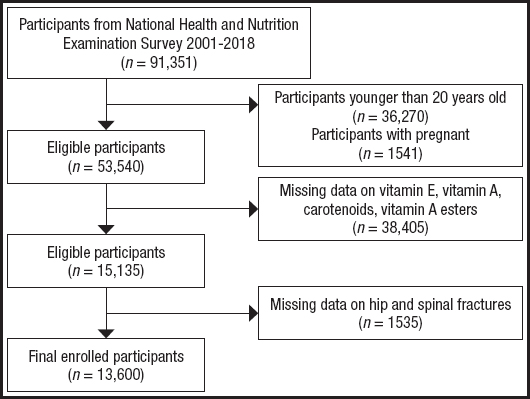
Participants were selected from the NHANES database (https://wwwn.cdc.gov/nchs/nhanes/), covering data collected between 2001 and 2018 by the NCHS. A total of 91,351 individuals were initially identified. We subsequently excluded participants who were under 20 years old or pregnant (n = 37,811), those missing data on serum nutrient biomarkers (n = 34,805), and those without information on hip or vertebral fractures (n = 1,535). Ultimately, 13,600 eligible participants were included in the final analysis (Fig. 1). The NHANES data used in this study are publicly available and de-identified, with no involvement of personal privacy information. The survey protocols were approved by the NCHS Ethics Review Board, and all participants provided written informed consent.
DEFINITION OF STUDY VARIABLES
The serum biomarkers assessed in this study included vitamin E, vitamin A, and its derivatives (carotenoids: α-carotene, β-cryptoxanthin, lutein and zeaxanthin, all-trans β-carotene, cis-β-carotene, and all-trans lycopene; and vitamin A esters: retinyl palmitate and retinyl stearate), all derived from NHANES laboratory datasets. Hip and vertebral fracture events were determined through questionnaire-based interviews conducted by trained interviewers using Computer-Assisted Personal Interviewing (CAPI) systems.
In this study, BMD measurements were obtained at multiple anatomical sites, including the total femur, femoral neck, trochanter, intertrochanteric region, Ward’s triangle, and total spine. All BMD data were acquired using dual-energy X-ray absorptiometry (DXA). Femoral scans were performed with the Hologic QDR-4500A DXA system, while spinal scans were conducted using the Hologic Discovery A DXA device (both manufactured by Hologic Inc., Bedford, MA, USA). The scanning software version was Apex 3.2, and data analysis was carried out using Hologic APEX 4.0 software. All DXA assessments were conducted by certified technicians who had received standardized training. The procedures involved low radiation exposure (< 20 μSv) and strictly adhered to the standardized protocols established by the NHANES project. To ensure the accuracy and consistency of the BMD measurements, rigorous quality control procedures were implemented, including routine device calibration, operator monitoring, and centralized data auditing. This study utilized raw BMD values (expressed in g/cm²) directly obtained from DXA scans. T-scores or Z-scores derived from reference populations were not calculated, and no diagnostic stratification based on scoring systems was applied. Analyses of fracture risk were based on BMD as a continuous variable, and all related conclusions were drawn from the original BMD values.
Covariates were categorized into three main groups: 1) Demographic and socioeconomic variables: age, gender, race/ethnicity, education level, marital status, and the family poverty income ratio (PIR; defined as low income ≤ 1.3, middle income ≤ 3.5, and high income > 3.5); 2) Anthropometric and biochemical variables: BMI, waist circumference, alanine aminotransferase (ALT), and aspartate aminotransferase (AST); and 3) Clinical and lifestyle factors: including hypertension, diabetes, cardiovascular disease (CVD), smoking status, and alcohol consumption. Detailed definitions and diagnostic criteria for diseases and lifestyle factors are provided in the supplementary material (https://www.nutricionhospitalaria.org/files/ 9196/ADMA1-05938-02.pdf).
STATISTICAL ANALYSIS
All statistical analyses were conducted using R software (version 4.2.2) and the EmpowerStats platform (version 4.1.2). Continuous variables were expressed as weighted means with 95 % CIs, and differences between groups were evaluated using weighted linear regression. Categorical variables were presented as weighted percentages and compared using weighted chi-square tests. Multivariable logistic regression models were employed to calculate ORs and 95 % CIs. Three models were constructed: an unadjusted model (Model 1), a minimally adjusted model (Model 2), and a fully adjusted model (Model 3), progressively adjusting for demographic, socioeconomic, lifestyle, and comorbidity confounders.
Smooth curve fitting and two-piecewise linear regression were used to explore potential nonlinear relationships, with the optimal model determined by log-likelihood ratio tests, and inflection points identified via maximum likelihood methods. Subgroup analyses and interaction tests were performed to assess potential effect modifications. Receiver operating characteristic (ROC) curves were generated to evaluate the predictive performance of biomarkers, and the area under the curve (AUC) was calculated. Mediation analysis was conducted to examine the mediating role of bone mineral density indices. A two-sided p-value < 0.05 was considered statistically significant.
RESULTS
BASELINE CHARACTERISTICS OF STUDY PARTICIPANTS
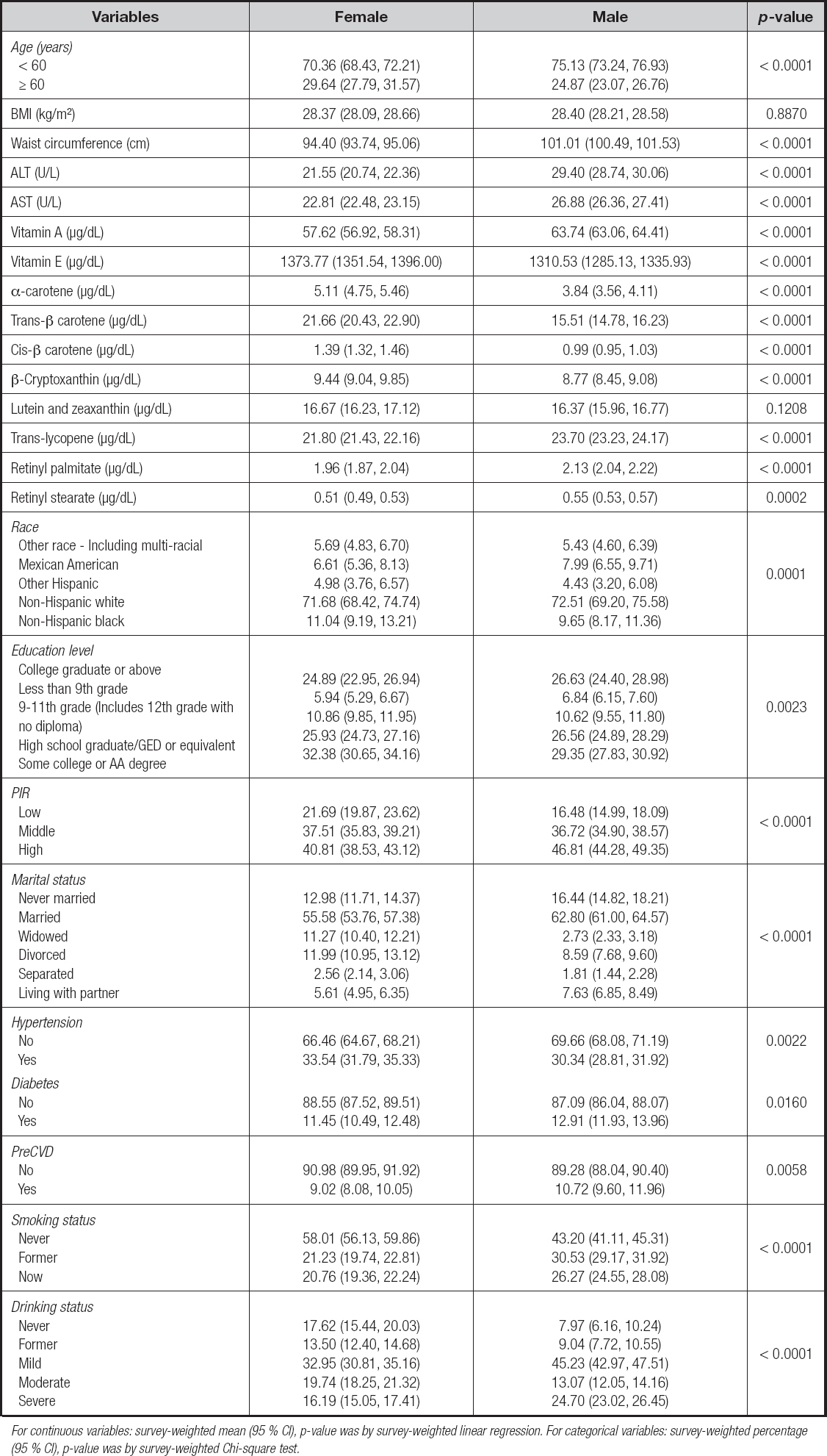
Table I summarizes the baseline characteristics of the study population stratified by gender. Significant differences were observed between males and females across several demographic, anthropometric, and biochemical parameters. Males were generally older, with a higher proportion aged ≥ 60 years (29.64 % vs. 24.87 %), and exhibited a greater mean waist circumference compared to females (101.01 cm vs. 94.40 cm). However, no significant difference in BMI was found between the gender (p = 0.8870). Additionally, males had significantly higher serum levels of ALT (29.40 U/L vs. 21.55 U/L) and AST (26.88 U/L vs. 22.81 U/L). Regarding serum micronutrients, males exhibited higher levels of vitamin A (63.74 μg/dL vs. 57.62 μg/dL), retinyl palmitate (2.13 μg/dL vs. 1.96 μg/dL), and retinyl stearate (0.55 μg/dL vs. 0.51 μg/dL) compared to females. Conversely, females showed significantly higher levels of vitamin E (1373.77 μg/dL vs. 1310.53 μg/dL), α-carotene, trans-β-carotene, cis-β-carotene, and β-cryptoxanthin (all p < 0.0001).
Table I Characteristics of the study population based on gender
For continuous variables: survey-weighted mean (95 % CI), p-value was by survey-weighted linear regression. For categorical variables: survey-weighted percentage (95 % CI), p-value was by survey-weighted Chi-square test.
In terms of sociodemographic characteristics, notable differences were also observed. Males had higher proportions of individuals who were currently married (62.80 % vs. 55.58 %) but lower proportions of widowed individuals compared to females (2.73 % vs. 11.27 %) (p < 0.0001).
Additionally, differences in racial/ethnic composition (p = 0.0001) and educational attainment (p = 0.0023) were noted, with females having a higher proportion of non-Hispanic black individuals and a greater percentage having completed college or associate degrees. Economically, the poverty income ratio (PIR) differed significantly by gender, with a higher proportion of males in the middle-income category (36.72 % vs. 37.51 %, p < 0.0001). Clinically, males had a higher prevalence of hypertension (p = 0.0022), diabetes (p = 0.0160), and PreCVD (p = 0.0058). Lifestyle factors also showed significant gender differences, with males being more likely to be current smokers and former alcohol drinkers compared to females (both p < 0.0001).
ASSOCIATIONS BETWEEN SERUM VITAMINS, CAROTENOIDS, RETINYL ESTERS, AND THE RISKS OF HIP AND VERTEBRAL FRACTURES
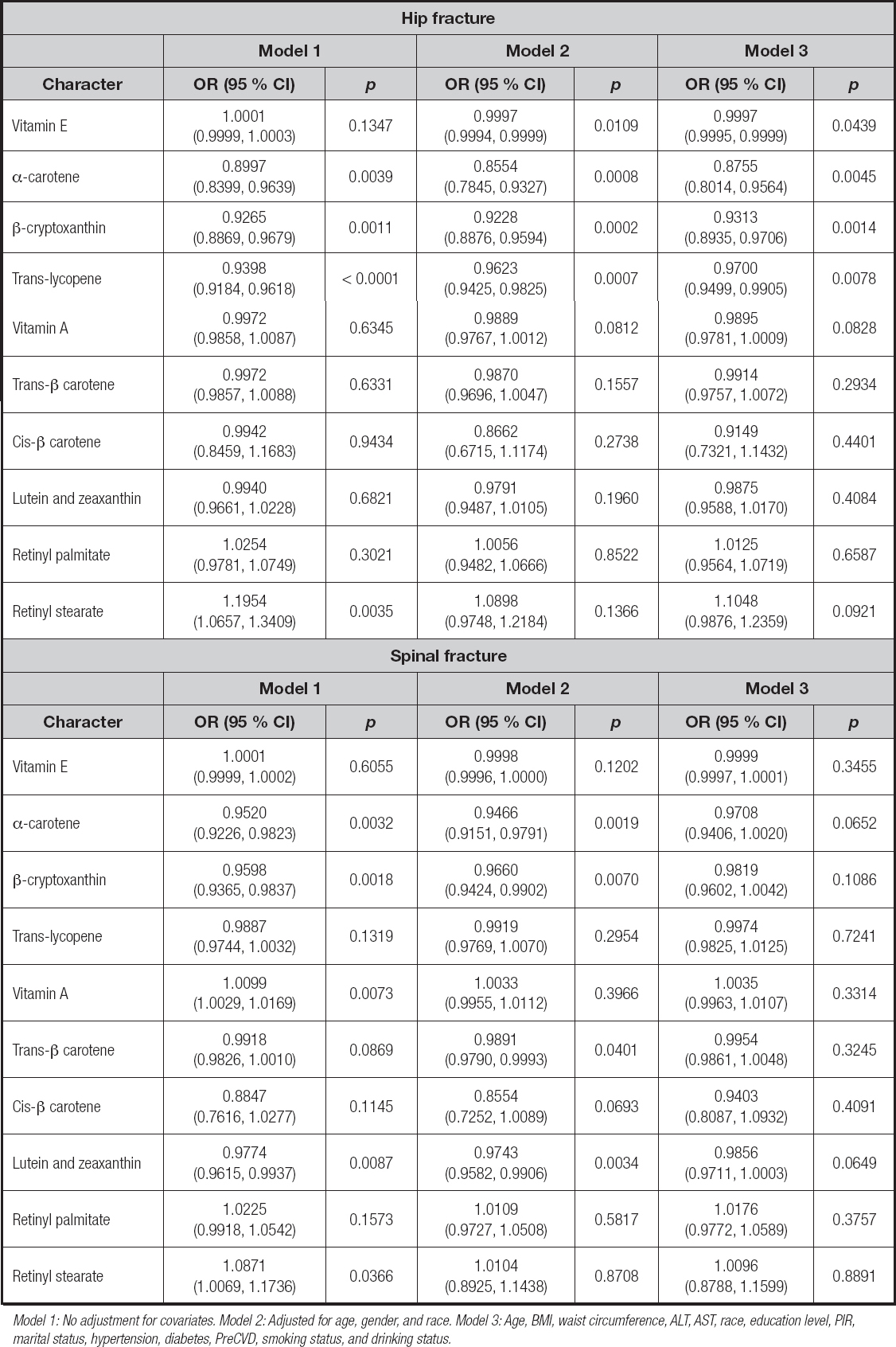
Table II presents the associations between serum nutrient levels and fracture risks. In the analysis of hip fractures, the fully adjusted model (Model 3) showed that serum α-carotene [OR = 0.8755, 95 % CI: (0.8014-0.9564), p = 0.0045], β-cryptoxanthin [0.9313, (0.8935-0.9706), p = 0.0014], and trans-lycopene [0.9700, (0.9499-0.9905), p = 0.0078] were significantly inversely associated with the risk of hip fractures.
Table II Associations between serum vitamins, carotenoids, and retinyl esters and risk of hip and spinal fractures
Model 1: No adjustment for covariates. Model 2: Adjusted for age, gender, and race. Model 3: Age, BMI, waist circumference, ALT, AST, race, education level, PIR, marital status, hypertension, diabetes, PreCVD, smoking status, and drinking status.
Vitamin E also demonstrated a slight inverse association with hip fracture risk [0.9997, (0.9995-0.9999), p = 0.0439]. In the analysis of vertebral fractures, only α-carotene [0.9708, (0.9406-1.0020), p = 0.0652] and lutein and zeaxanthin [0.9856, (0.9711-1.0003), p = 0.0649] approached statistical significance in Model 3. No other serum nutrients showed significant associations with vertebral fracture risk after multivariable adjustment.
NONLINEAR ASSOCIATION ANALYSIS
Figure 2 illustrates potential nonlinear inverse associations between serum levels of vitamins, carotenoids, retinyl esters, and the risks of hip and vertebral fractures. At lower concentration ranges, fracture risk decreased rapidly with increasing serum levels, followed by a plateau phase. The two-piecewise linear regression models presented in table III revealed that the inflection point for α-carotene was 4.15 μg/dL; below this threshold, α-carotene was significantly inversely associated with hip fracture risk [0.79, (0.70-0.88), p < 0.0001], while the association was no longer significant above this concentration. For β-cryptoxanthin, the inflection point was 9.9 μg/dL, with a protective effect observed below the threshold [0.88, (0.84-0.93), p < 0.0001], which diminished at higher concentrations. The log-likelihood ratio tests supported that nonlinear models provided a better fit compared to linear models (p < 0.05). In the analysis of vertebral fractures, the overall nonlinear trends were weaker. Only lutein and zeaxanthin demonstrated a marginally significant inverse association at lower concentration levels (< 15.4 μg/dL) [0.96, (0.93-1.00), p = 0.0362].
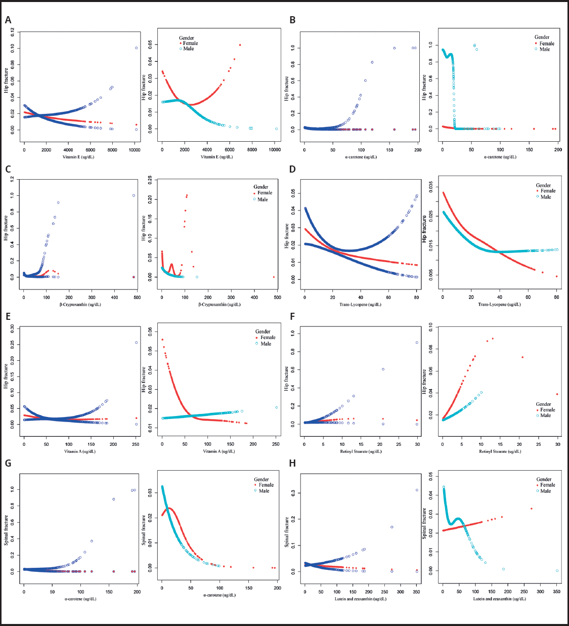
Figure 2 The association between serum vitamins, carotenoids, and retinyl esters in relation to hip and spinal fractures.
Table III Threshold effect analysis of serum vitamins, carotenoids, and retinyl esters in relation to hip and spinal fractures
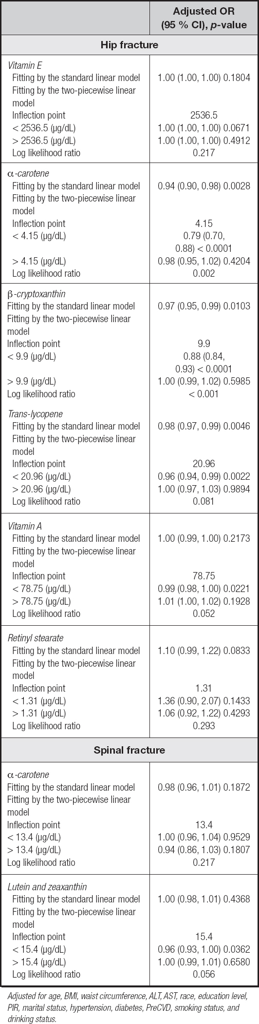
Adjusted for age, BMI, waist circumference, ALT, AST, race, education level, PIR, marital status, hypertension, diabetes, PreCVD, smoking status, and drinking status.
To further explore the potential moderating effect of gender, threshold effect analyses stratified by gender were conducted (Suppl. Table I - https://www.nutricionhospitalaria.org/files/9196/ADMA1-05938-02.pdf). The results indicated that the observed nonlinear associations were primarily evident in females. In women, α-carotene (inflection point = 3.51 μg/dL) showed a significant inverse association with hip fracture risk at lower concentrations [0.75, (0.62-0.92), p = 0.0046], and β-cryptoxanthin (inflection point = 8.2 μg/dL) similarly exhibited a strong protective effect [0.82, (0.75-0.89), p < 0.0001]. Among males, most associations were not statistically significant, except that α-carotene (< 5.5 μg/dL) and trans-lycopene (< 24.8 μg/dL) showed protective trends [0.81, (0.71-0.92), p = 0.0013] and [0.95, (0.92-0.98), p = 0.0030], respectively. Additionally, in females, low serum levels of vitamin A (< 49.6 μg/dL) were also associated with a protective effect against hip fractures [0.96, (0.93-0.99), p = 0.0042].
SUBGROUP ANALYSES
In the analysis of serum trans-lycopene, a consistent inverse association with hip fracture risk was observed across most subgroups (Table IV). Protective effects were noted among heavy drinkers, individuals with hypertension, and participants across different levels of PIR. Notably, a significant interaction was detected with PreCVD status (p for interaction = 0.0069); the inverse association was significant among participants without a history of CVD [0.95, (0.93-0.97), p < 0.0001], but not among those with CVD [1.00, (0.97-1.02), p = 0.7271]. In addition, an interaction trend was observed across age groups (p for interaction = 0.0415), with significant protective effects evident among individuals aged ≥ 60 years [0.96, (0.94-0.97), p < 0.0001], suggesting that age and underlying diseases may modify this association. In contrast, serum vitamin E did not show significant associations with hip fracture risk across any subgroup, and no significant interactions were observed, indicating no evident relationship and no subgroup heterogeneity (Supplementary Table IIA - https://www.nutricionhospitalaria.org/files/9196/ADMA1 -05938-02.pdf).
Table IV Subgroup analysis of the association between serum trans-lycopene and hip fracture risk
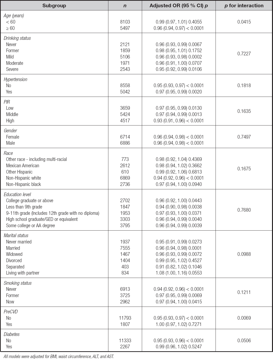
All models were adjusted for BMI, waist circumference, ALT, and AST.
For α-carotene, significant inverse associations with hip fracture risk were observed among participants aged ≥ 60 years [OR = 0.92, p = 0.0009], heavy drinkers [0.77, 0.0228], individuals with high PIR [0.88, 0.0070], females [0.93, 0.0093], never-smokers [0.95, 0.0339], individuals without PreCVD [0.96, 0.0301], and those without diabetes [0.95, 0.0165] (Supplementary Table IIB - https://www.nutricionhospitalaria.org/files/9196/ADMA1-05938-02.pdf). Significant interactions were detected with race/ethnicity (p = 0.0014) and education level (p = 0.0227), with stronger associations observed among Non-Hispanic whites [0.91, (0.87-0.97), p = 0.0015] and those with college education or above [0.86, (0.76-0.98), p = 0.0268].
Similarly, for β-cryptoxanthin, stable protective effects were observed among participants aged ≥ 60 years [0.96, 0.0007], heavy drinkers [0.88, 0.0085], individuals with hypertension, males [0.95, 0.0043], smokers [0.87, 0.0050], and participants without diabetes [0.96, 0.0017] or CVD [0.97, 0.0147] (Supplementary Table IIC - https://www.nutricionhospitalaria.org/files/9196/ADMA1-05938-02.pdf). Significant interactions were identified with PIR (p = 0.0251), race/ethnicity (p = 0.0004), and education level (p = 0.0008). Stronger protective associations were observed among individuals with middle PIR [0.96, 0.0227], high PIR [0.92, 0.0024], non-Hispanic whites [0.92, 0.0001], non-Hispanic blacks [0.89, 0.0143], those who graduated from college or above [0.93, 0.0463], and those with a high school diploma or GED [0.89, 0.0007].
ROC CURVE ANALYSIS
As shown in figure 3A, Model 1—which included α-carotene along with all covariates—demonstrated significantly better predictive performance for hip fracture risk (AUC = 0.773; 95 % CI: 0.7424-0.8031) compared to Model 2, which included only the covariates (AUC = 0.765; 95 % CI: 0.7347-0.7958; p = 0.0247). However, after further adjustment for vitamin E, trans-lycopene, and β-cryptoxanthin, the difference in AUC between the two models was no longer statistically significant (Model 1 AUC = 0.769; 95 % CI: 0.7386-0.8001 vs. Model 2 AUC = 0.775; 95 % CI: 0.7440-0.8053; p = 0.1004) (Fig. 3B). These findings suggest that α-carotene alone may serve as a potential independent predictor of hip fracture risk.
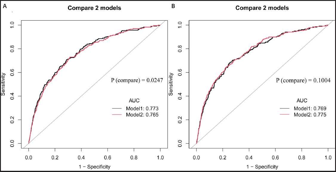
Figure 3 ROC curve analysis evaluating the performance of the models in predicting hip fracture risk.
Supplementary figure 1 (https://www.nutricionhospitalaria.org/files/9196/ADMA1-05938-02.pdf) further evaluated the incremental predictive value of vitamin E, trans-lycopene, and β-cryptoxanthin when added individually to the covariate model. The addition of vitamin E (Model 1 AUC = 0.766; 95 % CI: 0.7359-0.7968 vs. Model 2 AUC = 0.765; 95 % CI: 0.7347-0.7958; p = 0.3371, Supplementary Fig. 1A - https://www.nutricionhospitalaria.org/files/9196/ADMA1-05938-02.pdf), trans-lycopene (Model 1 AUC = 0.769; 95 % CI: 0.7386-0.8001 vs. Model 2 AUC = 0.765; 95 % CI: 0.7347-0.7958; p = 0.2159, Supplementary Fig. 1B - https://www.nutricionhospitalaria.org/files/9196/ADMA1-05938-02.pdf), or β-cryptoxanthin (Model 1 AUC = 0.767; 95 % CI: 0.7357-0.7984 vs. Model 2 AUC = 0.765; 95 % CI: 0.7347-0.7958; p = 0.6668, Supplementary Fig. 1C - https://www.nutricionhospitalaria.org/files/9196/ADMA1-05938-02.pdf) did not significantly improve the model’s predictive performance for hip or spine fracture risk.
MEDIATION ANALYSIS
The results of the mediation analysis are presented in table V. α-carotene exhibited a partial mediating effect on hip fracture risk through intertrochanteric BMD. The estimated total effect of α-carotene on hip fracture risk was [-0.009062 (-0.016754 to -0.004096), p < 0.0001], with an estimated mediated effect through intertrochanteric BMD of [-0.000106 (-0.000273 to -0.000014), p = 0.0140], accounting for a mediation proportion of [1.17 % (0.14 % to 3.62 %), p = 0.0140]. Similarly, β-cryptoxanthin also demonstrated a partial mediating effect on hip fracture risk via trochanteric BMD.
Table V Mediation analysis of the effects of α-carotene and β-cryptoxanthin on hip fracture via intertrochanter and trochanter BMD
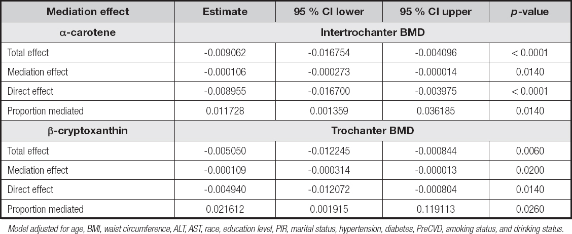
Model adjusted for age, BMI, waist circumference, ALT, AST, race, education level, PIR, marital status, hypertension, diabetes, PreCVD, smoking status, and drinking status.
The estimated total effect of β-cryptoxanthin was [-0.005050 (-0.012245 to -0.000844), p = 0.0060], with an estimated mediated effect of [-0.000109 (-0.000314 to -0.000013), p = 0.0200], corresponding to a mediation proportion of [2.16 % (0.19 % to 11.91 %), p = 0.0260].
As shown in supplementary table III (https://www.nutricionhospitalaria.org/files/9196/ADMA1-05938-02.pdf), α-carotene demonstrated a marginally significant mediating effect through total femur BMD, with an estimated mediated effect of [-0.000085 (-0.000227 to -0.000000), p = 0.0500], accounting for 0.97 % of the total effect. No significant mediation effects were observed for α-carotene through femoral neck BMD, Ward’s triangle BMD, or trochanteric BMD. For β-cryptoxanthin, no significant mediation effects were identified through total femur BMD, femoral neck BMD, Ward’s triangle BMD, or intertrochanteric BMD. Additionally, no significant mediation effects were found for vitamin E or trans-lycopene through BMD measures at any anatomical site (all p > 0.05).
DISCUSSION
This study yielded four major findings. First, we identified significant inverse associations between serum levels of α-carotene, β-cryptoxanthin, vitamin E, and trans-lycopene and the risk of hip fractures. Additionally, α-carotene and lutein-zeaxanthin showed near-significant associations with vertebral fracture risk. Second, a nonlinear inverse relationship was observed between α-carotene, β-cryptoxanthin, and hip fracture risk. Third, ROC analysis demonstrated that α-carotene alone could serve as a potential predictive biomarker for hip fracture risk, without the need for combined modeling with other significant nutrients identified in this study. Finally, mediation analysis revealed that α-carotene partially mediated hip fracture risk through intertrochanteric BMD, while β-cryptoxanthin exerted a partial mediating effect through trochanteric BMD.
In the nonlinear analysis, we observed for the first time that the inflection points for α-carotene and β-cryptoxanthin were 4.15 μg/dL and 9.9 μg/dL, respectively. Protective effects were significant below these thresholds, but not above, suggesting a possible saturation phenomenon in their biological activity. Clinically, these findings imply that supplementation strategies should consider baseline serum levels to optimize dosing and avoid potential plateau effects or diminishing returns (17,18). Moreover, most previous studies exploring the relationships between α-carotene, β-cryptoxanthin, and fracture risk have been based on dietary intake data, with limited evidence from serum biomarker analyses (19). By systematically evaluating these associations using serum concentrations in a nationally representative U.S. cohort, our study provides important supplementary evidence. It is noteworthy that previous findings on the association between α-carotene and fracture risk have been inconsistent. For instance, a study by Wen-Ting Cao et al. in middle-aged and older adults in Guangdong, China, found no significant association between dietary α-carotene intake and hip fracture risk (20). In contrast, Dai Z et al. reported an inverse association among Chinese men in Singapore, but not among women (21). Similarly, Sahni S, et al., using data from the Framingham Osteoporosis Study, found no significant association between α-carotene, β-cryptoxanthin, and fracture risk (22). Conversely, a European study by Hayhoe RPG, et al. suggested that higher α-carotene intake was associated with lower hip fracture risk in men, and dietary carotenoid intake correlated with bone density measures in women, although no association was observed at the plasma level (23). These findings highlight that geographical, racial, gender-based, dietary, and methodological differences (dietary intake vs. serum levels) may contribute to inconsistencies in the observed relationships between α-carotene and fracture risk. In our study, low serum levels of α-carotene (< 3.51 μg/dL) and β-cryptoxanthin (< 8.2 μg/dL) were significantly associated with reduced hip fracture risk among females, whereas among males, only low levels of α-carotene and trans-lycopene showed protective trends. Additionally, low serum vitamin A levels in females were also associated with protective effects. Further subgroup analyses revealed that the inverse association between α-carotene and hip fracture risk was particularly pronounced among non-Hispanic whites and individuals with higher education levels, but not in other racial or socioeconomic groups (p for interaction < 0.05). These findings suggest that gender, race, and socioeconomic factors may modulate the relationship between serum carotenoids and fracture risk, partially explaining discrepancies observed in previous studies.
Furthermore, subgroup analyses revealed that serum trans-lycopene levels were significantly inversely associated with hip fracture risk among individuals aged ≥ 60 years and those without a history of CVD, but not among individuals with CVD or those aged < 60 years. β-cryptoxanthin also demonstrated significant inverse associations in individuals with middle or high PIR, among non-Hispanic white and black populations, and among those with a high school education or higher. Jeffrey B. Blumberg, et al. reported that dietary supplement use improves micronutrient intake across all racial/ethnic groups, with the most pronounced effects observed among non-Hispanic whites (24). These findings further support the notion that gender, race, and socioeconomic status may contribute to the disparities observed in previous studies.
Given that this study utilized quantitative serum nutrient levels, we further employed ROC curve analysis to assess their predictive value for hip fracture risk. The results suggested that α-carotene alone could serve as a potential independent predictor. Previous research has explored potential mechanisms through which carotenoids may influence the skeletal system. In an in vitro model, Michelle Min-Fang Yee, et al. found that mixed carotenoids could promote osteoblast proliferation and inhibit osteoclast differentiation and maturation, thereby improving bone quality (25). Additionally, their systematic review highlighted that vitamin A, via activation of retinoic acid receptor (RAR) signaling, promotes early-stage osteoblast precursor differentiation, although it may inhibit bone mineralization during the later stages of osteogenesis. Provitamin A carotenoids (such as α-carotene and β-cryptoxanthin) are cleaved by β-carotene monooxygenase 1 (BCO1) into retinal, which is subsequently oxidized to all-trans-retinoic acid (ATRA) by retinaldehyde dehydrogenase (RALDH) (26,27). As the natural ligand for RAR, ATRA not only facilitates osteoblast differentiation but also inhibits osteoclastogenesis by suppressing the NF-κB signaling pathway (28). It should be noted, however, that high doses of retinol may exert detrimental effects on bone, particularly cortical bone (29).
Serum α-carotene, β-cryptoxanthin, vitamin E, and trans-lycopene are important micronutrients widely found in everyday diets, particularly in vegetables, fruits, and nuts. α-Carotene is primarily present in deeply colored orange-yellow vegetables such as carrots, pumpkins, sweet potatoes, and winter squash, and serves as a major provitamin A compound (30). β-cryptoxanthin is abundant in tropical fruits, especially citrus fruits (e.g., tangerines, oranges, grapefruits), papayas, and red peppers, and is known for its high bioavailability and potent antioxidant capacity (31). Vitamin E is mainly derived from plant oils (such as wheat germ oil and sunflower oil), nuts (such as almonds and hazelnuts), leafy green vegetables, and some fortified foods, where it functions as a lipid-soluble antioxidant preventing lipid peroxidation in cell membranes (32,33). Trans-lycopene is most abundantly found in tomatoes and tomato-based products (such as tomato paste and juice), with its bioavailability further enhanced through cooking and processing (34). Moderately increasing the intake of foods rich in these nutrients may represent a safe, natural, and feasible strategy for fracture risk prevention, particularly in high-risk populations, with important public health implications. However, nutritional interventions should be tailored to individual baseline levels and overall dietary patterns to avoid potential adverse effects from excessive intake.
It is noteworthy that, although α-carotene and β-cryptoxanthin exhibited statistically significant mediation effects on hip fracture risk through intertrochanteric BMD in this study, the proportion of the effect mediated was relatively small (approximately 1-2 %). This finding suggests that intertrochanteric BMD may play only a partial role in the pathway by which these carotenoids influence fracture risk, indicating the potential involvement of more complex biological mechanisms. Given the pivotal role of BMD in bone metabolism, even a modest mediating effect provides a biologically plausible basis for further investigation into the mechanistic pathways of carotenoids. Future studies may explore the regulatory effects of these nutrients on bone health through pathways related to oxidative stress, inflammatory status, and the balance between osteogenesis and osteoclastogenesis, thereby offering a more comprehensive understanding of their potential mechanisms (35,36).
This study has several notable strengths. First, the study utilized a large sample size with excellent external representativeness, enhancing the generalizability and applicability of the findings. Second, unlike most previous studies that relied on dietary intake questionnaires to estimate carotenoid consumption, this study employed serum concentrations as exposure indicators, providing a more objective and accurate reflection of internal nutritional status and reducing information bias. Third, the study systematically evaluated the associations between multiple serum vitamins, carotenoids, and retinyl esters and the risks of hip and vertebral fractures, incorporating nonlinear analyses, ROC curve assessments, mediation analyses and subgroup analyses. Notably, this study is the first to identify inflection points at lower concentrations of α-carotene and β-cryptoxanthin, below which protective effects against hip fracture begin to emerge. This novel finding enriches the current body of literature and provides a potential quantitative reference for clinical nutritional interventions. The identified threshold values may serve as a basis for guiding dietary intake at both the individual and population levels, particularly in populations with insufficient carotenoid intake or those at high risk of fracture, thereby facilitating precision nutrition strategies. Future studies, including prospective cohort studies and randomized controlled trials, are warranted to further validate the robustness and generalizability of these associations, and to elucidate their practical applications in clinical nutrition. Moreover, these findings may inform the optimization of supplement dosing and the development of targeted fracture prevention strategies, highlighting promising implications for public health translation.
Moreover, the study thoroughly accounted for potential confounding factors such as gender, race/ethnicity, and socioeconomic status, and validated the main findings across multiple subgroups, thus enhancing the reliability and broad applicability of the conclusions. However, several limitations should be acknowledged. As an observational study, this research cannot establish causal relationships between serum nutrient levels and fracture risk; prospective interventional studies are needed to confirm these findings. Additionally, although multiple confounders were adjusted for, residual confounding by unmeasured factors cannot be entirely excluded. Another limitation of this study lies in the age distribution of the included population. Although the overall sample size was relatively large, a substantial proportion of participants were between 20 and 60 years of age. Given that fragility fractures—particularly those involving the hip and lumbar spine—are more prevalent among older adults, the relatively younger age profile of our cohort may have led to an underestimation of the actual fracture incidence. If the study were conducted exclusively in an elderly population, the fracture rate might have been higher and the observed associations potentially more pronounced. Future research focused on older adults is warranted to validate and extend the current findings. Secondly, due to limitations in the NHANES dataset, this study was unable to incorporate serum or urinary bone turnover markers such as C-terminal telopeptide of type I collagen (CTX), procollagen type I N-terminal propeptide (P1NP), osteocalcin, and bone-specific alkaline phosphatase (BSALP). These biomarkers play a critical role in reflecting bone remodeling status and identifying individuals at high risk of fracture. The absence of these data may have limited a more comprehensive assessment of bone metabolic status, underscoring the need for future studies to incorporate bone turnover markers in order to more systematically elucidate the mechanisms by which nutritional factors influence bone health. Finally, several important clinical covariates were not included in this study, such as serum vitamin D levels, parathyroid hormone (PTH) levels, menopausal status, and the use of osteoporosis-related medications (e.g., bisphosphonates, calcitonin, or selective estrogen receptor modulators). These factors play crucial roles in bone metabolism, directly influencing bone mineral density and potentially affecting fracture risk by modulating bone turnover. As such, they are widely recognized as essential confounders to be adjusted for in studies of bone health outcomes. However, due to data availability constraints and substantial missingness within the NHANES survey cycles used in this analysis, we were unable to simultaneously account for all of these variables. Future research should prioritize the inclusion of these key clinical indicators to enhance the comprehensiveness of analytical models and improve the validity and clinical generalizability of the findings. Overall, this study provides important supplementary evidence to the current body of literature and offers a theoretical basis for future interventions targeting serum carotenoids in fracture prevention, although further research is warranted to validate and extend these findings.
CONCLUSION
This study is the first to systematically validate, based on serum levels, the inverse associations of α-carotene and β-cryptoxanthin with fracture risk and to report, for the first time, a significant inverse relationship between serum trans-lycopene and hip fracture risk. These findings expand the research perspective on carotenoids in bone health and partially corroborate and supplement existing evidence regarding serum vitamin E. The analyses revealed that α-carotene and β-cryptoxanthin exert protective effects at lower serum concentrations, partially mediated through bone mineral density indices — specifically, α-carotene through intertrochanteric BMD and β-cryptoxanthin through trochanteric BMD. Moreover, serum α-carotene may serve as a potential independent biomarker for the early identification of individuals at high risk of hip fractures.
ETHICS APPROVAL AND CONSENT TO PARTICIPATE
This study was conducted in accordance with the Declaration of Helsinki and received approval from the Ethics Review Board of the National Center for Health Statistics. All participants provided written informed consent. Detailed methods and protocols for the NHANES study were approved by the CDC/NCHS Research Ethics Review Board. They are publicly available through the CDC.gov website. All methods in this study were performed by the relevant guidelines and regulations. This study was exempt from human subject ethical review as the data is freely available in the public domain.
AVAILABILITY OF DATA AND MATERIAL
The datasets utilized in this study are publicly available through the NHANES database, accessible at https://www.cdc.gov/nchs/nhanes/. All data analyzed are de-identified and freely available for research purposes.
FUNDING
This work was supported by the Natural Science Foundation of Hunan Province (Grant No. 2025JJ70081), the Scientific Research Project of the Hunan Provincial Department of Education (Grant No. 22C0187), the Guiding Project for Scientific and Technological Innovation of Changde City (Grant No. 2024ZD75), the Graduate Innovation Project of Hunan University of Chinese Medicine (Grant No. 2024CX129), and the Science and Technology Innovation Program of Hunan Province (Grant No. 2023RC3167).

