Mi SciELO
Servicios Personalizados
Revista
Articulo
Indicadores
-
 Citado por SciELO
 -
 Accesos
Links relacionados
-
 Citado por Google
 -
 Similares en
SciELO
 -
 Similares en Google
Compartir
Revista Española de Enfermedades Digestivas
versión impresa ISSN 1130-0108
Rev. esp. enferm. dig. vol.108 no.6 Madrid jun. 2016
Accuracy of ASGE criteria for the prediction of choledocholithiasis
Rodrigo M. Narváez-Rivera, José A. González-González, Roberto Monreal-Robles, Diego García-Compean, Jonathan Paz-Delgadillo, Aldo A. Garza-Galindo and Héctor J. Maldonado-Garza
Department of Gastroenterology and Gastrointestinal Endoscopy. Hospital Universitario "Dr. José Eleuterio González". Universidad Autónoma de Nuevo León. Monterrey, México
ABSTRACT
Background/aims: Few studies have validated the performance of guidelines for the prediction of choledocholithiasis (CL). Our objective was to prospectively assess the accuracy of the American Society for Gastrointestinal Endoscopy (ASGE) guidelines for the identification of CL.
Methods: A two-year prospective evaluation of patients with suspected CL was performed. We evaluated the ASGE guidelines and its component variables in predicting CL.
Results: A total of 256 patients with suspected CL were analyzed. Of the 208 patients with high-probability criteria for CL, 124 (59.6%) were found to have a stone/sludge at endoscopic retrograde cholangiopancreatography (ERCP). Among 48 patients with intermediate-probability criteria, 21 (43.8%) had a stone/sludge. The performance of ASGE high- and intermediate-probability criteria in our population had an accuracy of 59.0% (85.5% sensitivity, 24.3% specificity) and 41.0% (14.4% sensitivity, 75.6% specificity), respectively. The mean ERCP delay time was 6.1 days in the CL group and 6.4 days in the group without CL, p = 0.638. The presence of a common bile duct (CBD) > 6 mm (OR 2.21; 95% CI, 1.20-4.10), ascending cholangitis (OR 2.37; 95% CI, 1.01-5.55) and a CBD stone visualized on transabdominal US (OR 3.33; 95% CI, 1.48-7.52) were stronger predictors of CL. The occurrence of biliary pancreatitis was a strong protective factor for the presence of a retained CBD stone (OR 0.30; 95% CI, 0.17-0.55).
Conclusions: Irrespective of a patient's ASGE probability for CL, the application of current guidelines in our population led to unnecessary performance of ERCPs in nearly half of cases.
Key words: Choledocholithiasis. Common bile duct stone. Endoscopic retrograde colangiopancreatography. Cholangitis.
Introduction
Common bile duct (CBD) stones are a frequent complication in symptomatic cholelithiasis (5-10%) (1,2) and biliary pancreatitis (18-33%) (3-5). Endoscopic retrograde cholangiopancreatography (ERCP) is considered as the gold standard for diagnosis and treatment of choledocholithiasis (CL). However, it carries a considerable risk of short-term complications such as post-ERCP pancreatitis, post-endoscopic sphincterotomy bleeding, cholangitis and perforation (6). It has been previously stated that the cost-effectiveness is favorable only when performing ERCP in patients with a high likelihood of CL (7). In order to predict the likelihood of a persistent CBD stone in patients with suspected CL, the American Society for Gastrointestinal Endoscopy (ASGE) published guidelines in 2010 for the prediction of CL with the objective of identifying patients with the highest probability of benefiting from ERCP (8). They classified patients into three risk categories based on the presence of clinical, radiological and biochemical predictors. Patients with high probability of CL (defined as > 50% likelihood) were those with one of the following very strong predictors: CBD stone on transabdominal ultrasound (US), clinical ascending cholangitis or bilirubin > 4 mg/dL, or those with both of the following strong predictors: dilated CBD on US (> 6 mm with gallbladder in situ) and bilirubin level between 1.8-4.0 mg/dL; patients with intermediate probability of CL (10-50% likelihood) were those with the presence of only one strong predictor or any moderate predictor (abnormal liver test, age older than 55 years or gallstone pancreatitis); and low probability of CL (< 10% likelihood) those with no predictors present.
Recently, there has been some evidence from retrospective studies of the lack of accuracy of the ASGE guidelines for predicting choledocholithiasis (9). However, the performance of this risk score has not been prospectively validated. The aim of this prospective study was: a) to assess the performance of the existing guidelines on predicting CL in high- and intermediate-probability patients; and b) to evaluate the strength of association between single clinical and laboratory predictors and documented CL.
Patients and Methods
Approval was obtained from the research and ethics committee of the School of Medicine and Hospital Universitario "Dr. José E. González" of the Universidad Autónoma de Nuevo León. Informed consent was obtained from all participants. The study was performed in compliance with the Declaration of Helsinki for biomedical research.
We prospectively evaluated all patients admitted to our University Hospital with suspected CL that met ASGE high- and intermediate-probability criteria over a 2-year period, from March 1, 2012, to February 28, 2014. Suspected CL was considered when patients presented with upper abdominal pain and clinical manifestations (such as fever, jaundice), cholestatic pattern of liver injury, CBD dilatation (> 6 mm) or CBD stone visualization on transabdominal US.
Study subjects were identified at admission by the gastroenterology residents based on the assessment of clinical, radiological and biochemical variables. Patients were excluded if any of the following conditions that might work as confounding factors were present: history of cholecystectomy (CBD diameter can be changed after cholecystectomy), biliary tract lesion, chronic liver disease (because basal function liver test may be altered) and previous ERCP sphincterotomy. We excluded patients classified by ASGE criteria as "low-probability" because their risk for CL is minimal (< 1%) and no further imaging or endoscopic study is indicated (10).
Patients were classified into high- or intermediate-probability groups for CL according to ASGE guidelines based on initial biochemical liver tests, clinical suspicion of cholangitis, and transabdominal US variables (8). The high-probability group were subjects with the presence of any very strong predictor (CBD stone on abdominal US or clinical ascending cholangitis or bilirubin > 4 mg/dL) or both strong predictors (bilirubin level 1.8-4 mg/dL + CBD on US > 6 mm). The intermediate-probability group was all other patients with the presence of only one strong predictor or any moderate predictor (abnormal liver test, age older than 55 years or gallstone pancreatitis). ERCP (the gold standard for diagnosis and treatment of CL) was performed on all patients in the study by two experienced endoscopists who performed around 250 ERCPs annually (endoscopists knew the ASGE probability of CL in each case). We considered an ERCP as positive when a filling defect was observed on cholangiogram or stones/sludge were photographed within the duodenal lumen after extraction.
Statistical analysis
Baseline characteristics of patients with and without CL as well as in those with and without biliary pancreatitis were compared with the use of Student's t-test or U Mann-Whitney depending on the distribution of continuous variables as appropriate, and Fisher's exact test for categorical variables. We calculated the sensitivity, specificity, positive and negative predictive values, accuracy, and positive and negative likelihood ratios of the ASGE classification on admission as well as the performance of individual predictors. Univariate and multivariate analysis with odds ratio (OR) and 95% confident interval (CI) were performed for all clinical predictors. The SPSS statistical software package (Version 20.0) was used for statistical analysis.
Results
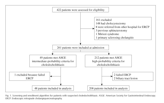
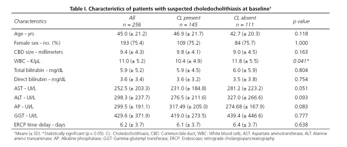
From March 1 2012 to February 28 2014, 422 patients with suspected CL were assessed for eligibility (Fig. 1). After 166 patients were excluded, 256 eligible patients with intermediate- or high-probability for CL were included. The cohort mean age was 45.0 years (± 21.2); 193 (75.4%) were female patients. General characteristics, ultrasonography and biochemical variables among subjects with and without CL are shown in table I. No significant difference was found in the ERCP delay time between the group of patients with and without CL (p = 0.638).
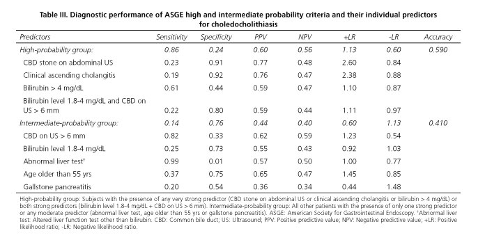
A subset of 208 patients met ASGE high probability-criteria on admission. Of these, 124 (59.6%) had CBD a stone/sludge on ERCP. Of the 48 patients that met ASGE intermediate-probability criteria on admission, 21 (43.8%) had CBD a stone/sludge on ERCP. The performance of ASGE high- and intermediate-probability criteria for CL in our population had an accuracy of 59.0% (85.5% sensitivity, 24.3% specificity) and 41.0% (14.4% sensitivity, 75.6% specificity), respectively. There was no statistically significant difference in the rate of CL detection between ASGE high- and intermediate-probability groups (p = 0.053).
Performance of individual ASGE predictors
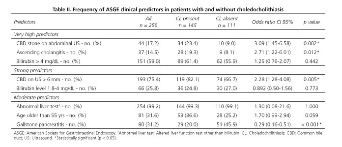
Among the ASGE "very strong predictors" of CL, CBD stone on transabdominal US and clinical ascending cholangitis were the predictors with a statistical significance between subjects with and without CL on ERCP. Among "strong predictors", only CBD size > 6 mm showed a statistical significance between groups (Table II). We also assessed the diagnostic performance of individual predictors for CL in both high- and intermediate-probability groups (Table III).
Significance of biliary pancreatitis
Eighty (31.2%) out of 256 patients in our study presented biliary pancreatitis. The identification of CL on ERCP in the subset of patients with biliary pancreatitis was 36.2% (29/80) compared with 65.9% (116/176) in those without pancreatitis, p < 0.001. Of the 80 patients in our study with biliary pancreatitis, 63 patients (78.8%) met ASGE high-probability criteria for CL. Of these, 25 (39.7%) patients had a stone/sludge on ERCP. Of the 17 patients with biliary pancreatitis that met intermediate-probability criteria, 4 (23.5%) had CBD a stone/sludge on ERCP. The performance of ASGE high- and intermediate-probability criteria for CL in the subset of patients with biliary pancrea-titis had an accuracy of 47.5% (86.2% sensitivity, 25.5% specificity) and 52.5% (13.8% sensitivity, 74.5% specificity), respectively. The comparisons of several variables in patients with and without biliary pancreatitis showed only statistical significance in CBD size (mm), 8.58 (± 3.2) vs. 9.9 (± 4.7), p = 0.044; white blood cell count (K/µL), 13.0 (± 5.3) vs. 10.1 (± 4.9), p < 0.001; and alkaline phosphatase levels (UI/L), 267.9 (± 169.3) vs. 313.7 (± 198.9), p = 0.046, respectively. The ERCP delay time between the group of patients with biliary pancreatitis (6.0 ± 4.01) and without pancreatitis (6.4 ± 3.48) was no significantly different, p = 0.540.
Logistic regression model
Multivariable logistic regression analysis showed that a CBD > 6 mm (OR 2.21; 95% CI, 1.20-4.10; p = 0.011), the presence of ascending cholangitis (OR 2.37; 95% CI, 1.01-5.55; p = 0.046), CBD stone visualized on transabdominal US (OR 3.33; 95% CI, 1.48-7.52; p = 0.004) and the absence of biliary pancreatitis (OR 3.23; 95% CI, 1.81-5.77; p < 0.001) were independently associated with the presence of a stone on ERCP. The occurrence of biliary pancreatitis was a strong protective factor for the presence of a retained CBD stone (OR 0.30; 95% CI, 0.17-0.55; p < 0.001). No additional clinical predictors of CL were identified. When the white blood cell count was introduced in the multivariate logistic regression model, it was not independently associated with the presence of CL on ERCP. However, the white blood cell count was the only studied variable independently associated with the presence of biliary pancreatitis (OR 1.13; 95% CI, 1.06-1.20; p < 0.001).
ERCP-related complications
During the study period, ERCP-related complications were recorded in 11 out of 256 cases (4.3%); we recorded the occurrence of clinically significant post-endoscopic sphincterotomy bleeding (requiring red blood cell transfusion and endoscopic therapy) in only one case and 10 cases of post-ERCP pancreatitis.
Discussion
ASGE guidelines for the prediction of suspected CL lack accuracy. The overall performance for the prediction of CL in our population with high probability was low, 59.0% (85.5% sensitivity, 24.3% specificity). This led to the performance of a significant percentage (> 40%) of unnecessary ERCP procedures. A recent study had similar results, with an accuracy for the ASGE high risk group of 62.1% (47.4% sensitivity, 73% specificity). They evaluated the impact of a second laboratory set, however, it did not increase the performance of ASGE guidelines for CL (9).
Recently, it has been reported that the presence of one very strong criterion or both strong criteria is related to an increased yield of CL (10). In our study, the presence of one very strong predictor or the presence of both strong predictors did not increase the odds ratio for CL in the multivariate analysis. However, in any case the need of noninvasive testing could not be obviated. In general, the usefulness of individual predictors did not increase the diagnostic performance in our population.
Our study, as the one previously reported by Rubin et al. (11), confirms that the ocurrence of biliary pancreatitis is not associated with the presence of a retained CBD stone. The absence of biliary pancreatitis in our study was a major predictor for CL (OR 3.23, 95% CI, 1.81-5.77). This supports the hypothesis that spontaneous biliary stone passage through the CBD to the duodenum occurs due to possible small biliary stone size in the subset of patients with biliary pancreatitis.
With the exception of critically ill patients with suspected ascending cholangitis, the usefulness of performing ERCP early in the course of biliary pancreatitis is controversial (12,13). In this respect, ERCP delay until biliary pancreatitis resolution may be a common practice. This may be a factor that allows the spontaneous biliary stone passage, being biliary pancreatitis probably a negative predictive factor for CL. However, we have demonstrated that ERCP delay time was not different from those with and without biliary pancreatitis, p = 0.540. Our results strongly support the fact that biliary pancreatitis itself may be a protective factor for CL independently of ERCP delay time.
This fact has raised the interest in endoscopic ultrasonography (EUS) as a safe replacement for ERCP in subjects with biliary pancreatitis. First, because it has been demostrated that early EUS has a higher successful examination rate (100 vs. 86%) and a lower morbidity (7% vs. 14%) compared with ERCP (14). In addition, EUS has demonstrated an excellent performance for the detection of small size (median of 4 mm) CBD stones (15), which may be useful in the context of biliary pancreatitis where we showed the risk of CL was significantly lower (36.2%) than without pancreatitis (65.9%) (OR 0.30; 95% CI, 0.17-0.55, p < 0.001).
Due to the morbidity and costs associated with ERCP, it should be limited to those who are most likely to benefit from it. Based on our results, predictive scores have a limited clinical utility for most patients in day-to-day practice. Due to the lack of identification of patients with CL by the current clinical predictive scores, we suggest that all patients with suspected CL (high- or intermediate-probability groups) without a clear indication for ERCP (ascending cholangitis) must have a CL confirmatory image study (magnetic resonance cholangiopancreatography [MRCP] or EUS) before a therapeutic procedure is attempted. Even though 2010 ASGE guidelines already recommended pre-operative EUS or MRCP for those classified as intermediate probability, no formal evidence supporting this recommendation in the ASGE intermediate- or high-probability groups exists.
Prior randomized controlled trials have suggested that intra-operative CBD exploration during cholecystectomy is superior to the two-step pre- or post-cholecystectomy ERCP in terms of CBD stone resolution without additional morbidity or mortality (16). Others have proposed this same approach specifically in the subset of patients with intermediate risk, with a reported shorter length of stay without increased morbidity (17). However, to consider this approach as a definitive recommendation, well designed prospective clinical trials are needed.
Both EUS and MRCP have a high diagnostic accuracy for the detection of CBD stones (18). This strategy can reduce the risk of unnecessary procedures, eliminate complications and may decrease cost. Therefore, we suggest that patients with an EUS or MRCP positive for CL should undergo endoscopic or surgical extraction of CBD stones, and those with negative EUS or MRCP do not need further invasive tests. However, to evaluate the cost-effectiveness of this approach, CL prevalence, center resources and expertise should be considered.
In summary, current ASGE guidelines in our population for the predition of CL lack accuracy, with unnecessary perfomance of negative ERCP in almost half of cases. The optimal cost-effective approach for the management of patients with suspected CL is not known yet. The use of confirmatory imaging studies (MRCP or EUS) in all cases with high and intermediate probability for CL before a therapeutic procedure may be an attractive option to be considered.
Acknowledgements
We thank Sergio Lozano-Rodríguez, MD, for his help in editing the manuscript.
References
1. Hunter JG. Laparoscopic transcystic common bile duct exploration. Am J Surg 1992;163:53-6;discussion 57-8. DOI: 10.1016/0002-9610(92)90252-M. [ Links ]
2. O'Neill CJ, Gillies DM, Gani JS. Choledocholithiasis: Overdiagnosed endoscopically and undertreated laparoscopically. ANZ J Surg 2008;78:487-91. DOI: 10.1111/j.1445-2197.2008.04540.x. [ Links ]
3. Chang L, Lo SK, Stabile BE, et al. Gallstone pancreatitis: A prospective study on the incidence of cholangitis and clinical predictors of retained common bile duct stones. Am J Gastroenterol 1998;93:527-31. DOI: 10.1111/j.1572-0241.1998.159_b.x. [ Links ]
4. Liu CL, Lo CM, Chan JK, et al. Detection of choledocholithiasis by EUS in acute pancreatitis: A prospective evaluation in 100 consecutive patients. Gastrointest Endosc 2001;54:325-30. DOI: 10.1067/mge.2001.117513. [ Links ]
5. Cohen ME, Slezak L, Wells CK, et al. Prediction of bile duct stones and complications in gallstone pancreatitis using early laboratory trends. Am J Gastroenterol 2001;96:3305-11. DOI: 10.1111/j.1572-0241.2001.05330.x. [ Links ]
6. Anderson MA, Fisher L, Jain R, et al. Complications of ERCP. Gastrointest Endosc 2012;75:467-73. DOI: 10.1016/j.gie.2011.07.010. [ Links ]
7. Urbach DR, Khajanchee YS, Jobe BA, et al. Cost-effective management of common bile duct stones: A decision analysis of the use of endoscopic retrograde cholangiopancreatography (ERCP), intraoperative cholangiography, and laparoscopic bile duct exploration. Surg Endosc 2001;15:4-13. DOI: 10.1007/s004640000322. [ Links ]
8. Committee ASoP, Maple JT, Ben-Menachem T, et al. The role of endoscopy in the evaluation of suspected choledocholithiasis. Gastrointest Endosc 2010;71:1-9. DOI: 10.1016/j.gie.2009.09.041. [ Links ]
9. Adams MA, Hosmer AE, Wamsteker EJ, et al. Predicting the likelihood of a persistent bile duct stone in patients with suspected choledocholithiasis: Accuracy of existing guidelines and the impact of laboratory trends. Gastrointest Endosc 2015;82:88-93. DOI: 10.1016/j.gie.2014.12.023. [ Links ]
10. Liu TH, Consorti ET, Kawashima A, et al. Patient evaluation and management with selective use of magnetic resonance cholangiography and endoscopic retrograde cholangiopancreatography before laparoscopic cholecystectomy. Ann Surg 2001;234:33-40. DOI: 10.1097/00000658-200107000-00006. [ Links ]
11. Rubin MI, Thosani NC, Tanikella R, et al. Endoscopic retrograde cholangiopancreatography for suspected choledocholithiasis: Testing the current guidelines. Dig Liver Dis 2013;45:744-9. DOI: 10.1016/j.dld.2013.02.005. [ Links ]
12. Folsch UR, Nitsche R, Ludtke R, et al. Early ERCP and papillotomy compared with conservative treatment for acute biliary pancreatitis. The German Study Group on Acute Biliary Pancreatitis. N Engl J Med 1997;336:237-42. [ Links ]
13. Tarnasky PR, Cotton PB. Early ERCP and papillotomy for acute biliary pancreatitis. N Engl J Med 1997;336:1835;author reply 1836. DOI: 10.1016/S0016-5107(97)80504-4. [ Links ]
14. Liu CL, Fan ST, Lo CM, et al. Comparison of early endoscopic ultrasonography and endoscopic retrograde cholangiopancreatography in the management of acute biliary pancreatitis: A prospective randomized study. Clin Gastroenterol Hepatol 2005;3:1238-44. DOI: 10.1016/S1542-3565(05)00619-1. [ Links ]
15. Vázquez-Sequeiros E, González-Panizo Tamargo F, Boixeda-Miquel D, et al. Diagnostic accuracy and therapeutic impact of endoscopic ultrasonography in patients with intermediate suspicion of choledocholithiasis and absence of findings in magnetic resonance cholangiography. Rev Esp Enferm Dig 2011;103:464-71. DOI: 10.4321/S1130-01082011000900005. [ Links ]
16. Dasari BV, Tan CJ, Gurusamy KS, et al. Surgical versus endoscopic treatment of bile duct stones. Cochrane Database Syst Rev 2013;12:CD003327. [ Links ]
17. Iranmanesh P, Frossard JL, Mugnier-Konrad B, et al. Initial cholecystectomy vs sequential common duct endoscopic assessment and subsequent cholecystectomy for suspected gallstone migration: A randomized clinical trial. JAMA 2014;312:137-44. DOI: 10.1001/jama.2014.7587. [ Links ]
18. Giljaca V, Gurusamy KS, Takwoingi Y, et al. Endoscopic ultrasound versus magnetic resonance cholangiopancreatography for common bile duct stones. Cochrane Database Syst Rev 2015;2:CD011549. [ Links ]
![]() Correspondence:
Correspondence:
José A González-González.
Department of Gastroenterology and Gastrointestinal Endoscopy.
Hospital Universitario "Dr. José Eleuterio González".
Universidad Autónoma de Nuevo León.
Av. Madero y Gonzalitos, s/n Col. Mitras Centro.
64460 Monterrey. Nuevo León, México
e-mail: alberto.gonzalez@uanl.edu.mx
Received: 03-02-2016
Accepted: 04-04-2016


