




 Servicios personalizados
 Inglés (pdf)
 Articulo en XML
 Referencias del artículo
 Traducción automática
 Enviar articulo por email
 Citado por SciELO
  Citado por Google
  Similares en
SciELO
Similares en
SciELO  Similares en Google
 Permalink
INTRODUCTION
Oral squamous cell carcinoma (OSCC) is the most common type of oral malignancy, accounting for over 90 % of all oral cancers. It is one of the top ten cancers globally, with a particularly high prevalence in regions where tobacco and areca nut use are widespread1. Despite advancements in diagnostic and therapeutic approaches, OSCC prognosis remains dismal due to late-stage diagnosis and high recurrence rates. There is an urgent need for early and non-invasive diagnostic markers to improve survival outcomes and enhance the quality of life for patients2.
Matrix metalloproteinases (MMPs) are zinc-dependent enzymes crucial for extracellular matrix (ECM) remodeling. Among them, MMP-2 (gelatinase A) is especially significant in cancer progression due to its ability to degrade type IV collagen, a primary component of the basement membrane3. Dysregulated MMP-2 activity is implicated in tumor invasion, angiogenesis, and metastasis, making it a critical player in the malignant behavior of cancers, including OSCC. Its overexpression within the tumor microenvironment underscores its potential as a biomarker for cancer progression and therapeutic targeting4.
Saliva has emerged as a promising diagnostic medium due to its non-invasive, cost-effective, and accessible nature5. Recent studies suggest that saliva harbors numerous biomarkers, including MMP-2, which are linked to OSCC progression6. Elevated levels of salivary MMP-2 have been associated with enhanced tumor invasion and metastasis, highlighting its potential utility in both diagnosis and prognosis7. By evaluating MMP-2 levels in saliva, clinicians could gain insights into the disease state, enabling early detection and more precise disease monitoring8).
Currently, OSCC diagnostics heavily rely on invasive methods such as biopsy and histopathological evaluation, which are often uncomfortable for patients and limited in detecting early lesions9. These limitations necessitate the exploration of molecular biomarkers like MMP-2, which offer a minimally invasive and effective alternative. However, despite its potential, the clinical utility of salivary MMP-2 requires further validation to establish its reliability and practical application in routine diagnostics10.
This study aims to evaluate salivary MMP-2’s potential as a reliable biomarker for oral squamous cell carcinoma, focusing on its role in predicting patient outcomes and long-term survival. By examining its expression levels and their association with disease progression, the study seeks to establish MMP-2 as a noninvasive tool to aid in early risk stratification and personalized treatment planning.
METHODOLOGY
Study design and sample collection
This study was conducted to evaluate MMP-2 levels in the saliva of oral squamous cell carcinoma (OSCC) patients. Participants were categorized into three groups: Control, Pre-operative (Pre-op), and Post-operative (Post-op). The Control group consisted of healthy individuals with no history of OSCC or systemic diseases. The Pre-op group included patients diagnosed with OSCC before undergoing any form of treatment, while the Post-op group consisted of OSCC patients who had undergone surgical tumor removal. Sample size estimation for this longitudinal study was based on previous literature, considering a medium effect size (f = 0.25), significance level (α = 0.05), power (1 - β = 0.80), and three study groups (Pre-op, Post-op, and Control).
A total of 60 participants were recruited for the study and categorized into two primary groups. The Control group included 20 healthy individuals with no history of OSCC or systemic diseases, while the remaining 40 participants comprised the Pre-operative (Pre-op) OSCC group. These same patients were reassessed post-surgery to form the Post-operative (Post-op) OSCC group. Unstimulated saliva samples were collected from all participants both preoperatively and postoperatively under standardized conditions to ensure uniformity. The collected samples were promptly processed and stored at −80 °C to preserve their integrity until further analysis.
Salivary MMP-2 levels were quantified using enzyme-linked immunosorbent assay (ELISA). The assay was conducted following the manufacturer’s protocol, ensuring reliable and reproducible results. Statistical analyses were performed using SPSS software. A one-way analysis of variance (ANOVA) was conducted to compare MMP-2 levels across the three groups, and post-hoc Tukey tests were used to determine specific differences between groups. Additionally, Kaplan-Meier survival analysis was employed to evaluate the prognostic significance of MMP-2 expression in OSCC patients.
Saliva samples were collected from all participants under standardized conditions to ensure consistency after obtaining ethical clearance from the institutional board with a reference number of SRB/SDC/PhD/OPATH-2102/21/TH-O06. Participants were asked to abstain from eating, drinking, or smoking for at least one hour before collection. Unstimulated saliva was allowed to pool in the mouth and was then expectorated into sterile collection tubes. This approach minimized variability in salivary composition due to external factors.
Sample processing
Immediately after collection, saliva samples were placed on ice and transported to the laboratory for processing. The samples were centrifuged at 3000 rpm for 15 minutes at 4 °C to remove cellular debris and other particulates. The clear supernatant was carefully separated and aliquoted into sterile microcentrifuge tubes to avoid contamination. These aliquots were stored at −80 °C until analysis to preserve the stability of MMP-2.
Quantification of MMP-2 Using ELISA
The levels of MMP-2 in saliva were measured using a commercially available enzyme-linked immunosorbent assay (ELISA) kit, following the manufacturer’s protocol. First, the ELISA reagents, including capture antibodies, detection antibodies, substrate solutions, and stop solutions, were prepared as instructed. Saliva samples were diluted in the provided assay buffer to ensure MMP-2 concentrations were within the dynamic detection range of the kit.
A 96-well microplate pre-coated with anti-MMP-2 capture antibodies was used. Diluted saliva samples, standards with known MMP-2 concentrations, and blank controls were added in duplicate to their respective wells. The plate was incubated at room temperature for the specified duration, allowing MMP-2 to bind to the capture antibodies. After incubation, the plate was washed thoroughly with the provided wash buffer to remove unbound components.
Next, a detection antibody conjugated with an enzyme (e.g., horseradish peroxidase) was added to each well, followed by incubation to enable specific binding to the captured MMP-2. After further washing, a substrate solution (e.g., tetramethylbenzidine, TMB) was added to initiate a colorimetric enzymatic reaction. The intensity of the color, proportional to the MMP-2 concentration, was stabilized by adding a stop solution (e.g., sulfuric acid). The optical density of each well was measured at 450 nm using a microplate reader. The obtained absorbance values were used to calculate MMP-2 concentrations.
Data analysis
The absorbance values were interpolated onto a standard curve generated using known MMP-2 concentrations provided with the ELISA kit. The MMP-2 levels in saliva were calculated for each group and expressed as mean ± standard deviation (SD). Statistical analysis was conducted using the SPSS software version 23. One-way analysis of variance (ANOVA) was used to compare MMP-2 levels among the three groups, followed by post-hoc pairwise comparisons to assess differences between specific groups. A p-value of <0.05 was considered statistically significant, indicating meaningful differences in MMP-2 levels between groups.
Survival analysis
Pre-operative MMP-2 expression levels were analyzed to establish a significant cutoff for stratifying patients into low and high expression groups. The median value was calculated and utilized as an initial reference for this stratification, thereby providing a foundational yet reliable dataset division. A Receiver Operating Characteristic (ROC) curve was constructed to further refine this cutoff by incorporating clinical outcomes or related variables, thereby identifying the optimal threshold that maximized the balance between sensitivity and specificity. A comprehensive review of relevant literature was conducted, which revealed comparable cutoff values that supported both the biological and clinical relevance of the proposed threshold. Based on these findings, a cutoff was selected that balanced simplicity, clinical applicability, and conformity with existing research. The cutoff was validated by comparing clinical outcomes, sensitivity, and specificity across the respective groups.
Kaplan-Meier survival analysis was employed to assess the impact of Matrix Metalloproteinase-2 (MMP-2) expression on patient survival outcomes. Patients were stratified into two groups based on MMP-2 expression cut off levels: low and high. Survival probabilities were calculated from baseline (0 months) and monitored over a defined follow-up period, with time intervals recorded at 3, 4, 7, and 9 months. The survival probabilities were plotted for each group, allowing a comparative analysis of survival trends. Statistical significance between the groups was evaluated using the log-rank test, which determines whether observed survival differences were due to random variation or were statistically significant. The analysis was performed using specialized statistical software to ensure precision and reproducibility, offering valuable insights into the prognostic implications of MMP-2 expression levels in cancer progression.
Ethical considerations
The study protocol was approved by the institutional ethics committee, ensuring adherence to ethical research practices. Written informed consent was obtained from all participants prior to sample collection, and patient confidentiality was strictly maintained throughout the study. Participants were informed of the study’s objectives, and their rights to withdraw at any stage were respected.
Quality control
Quality control measures were implemented throughout the experiment to ensure reliable results. All ELISA assays were performed in duplicate to minimize variability and ensure reproducibility. Negative and blank controls were included in every assay to detect non-specific binding or contamination. Internal controls were used to validate the assay’s accuracy and consistency. Additionally, all laboratory personnel involved in the assay adhered to standardized operating procedures to maintain high experimental reliability.
This methodology, employing ELISA as a sensitive and specific tool, enabled the precise quantification of MMP-2 in saliva samples, thereby ensuring the reliability of the results in assessing its potential as a biomarker for OSCC diagnosis and treatment monitoring.
RESULTS
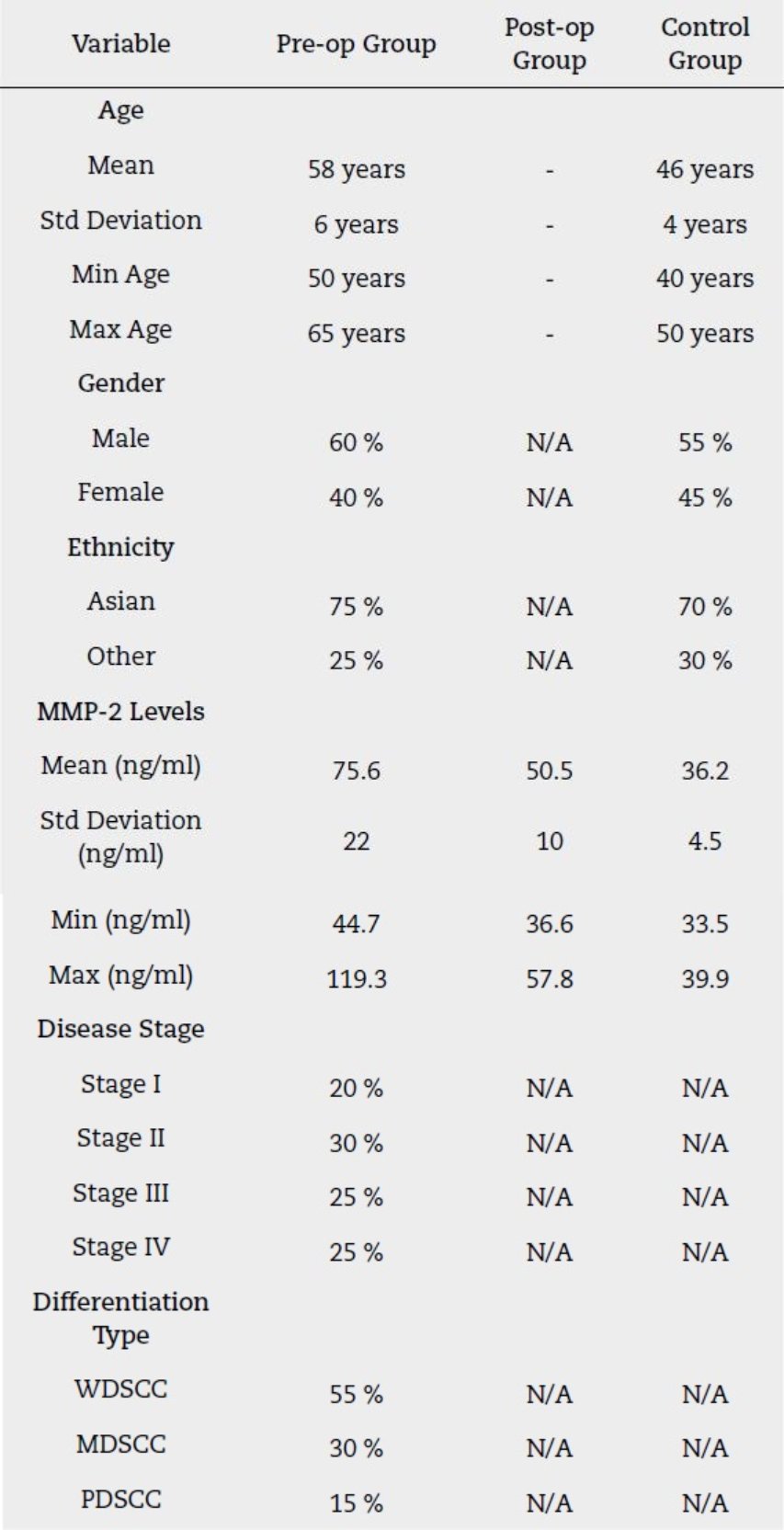
Table 1 descriptive data highlight distinct group characteristic, with elevated MMP-2 levels in the pre-operative group (mean: 75.6 ng/ml) reflecting active disease and tumor aggressiveness. Post-operative levels (mean: 50.5 ng/ml) indicate surgical efficacy but suggest the need for adjunct therapies, while controls showed baseline levels (mean: 36.2 ng/ml). The pre-op group was older (mean age: 58 years) and predominantly male (60 %), with advanced stages (Stage III/IV) and well-differentiated squamous cell carcinoma (55 %). These findings underscore the utility of MMP-2 as a biomarker for OSCC diagnosis, monitoring, and treatment planning.
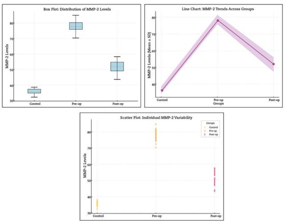
The levels of MMP-2 in saliva were measured using ELISA (Enzyme-Linked Immunosorbent Assay) in three groups: Control, Pre-operative (Pre-op), and Post-operative (Post-op). The Control group, representing healthy individuals, exhibited significantly lower MMP-2 levels (Mean ± SD: 36.20 ± 1.83 ng/ml). These levels serve as a baseline for normal MMP-2 expression in non-cancerous conditions. In contrast, the Pre-op group displayed the highest MMP-2 levels (Mean ± SD: 78.69 ± 2.65 ng/ml), highlighting the biomarker’s association with OSCC. Elevated MMP-2 in this group highlights its role in extracellular matrix degradation and tumor invasion, processes critical for OSCC progression (Table 2, Figure 1).
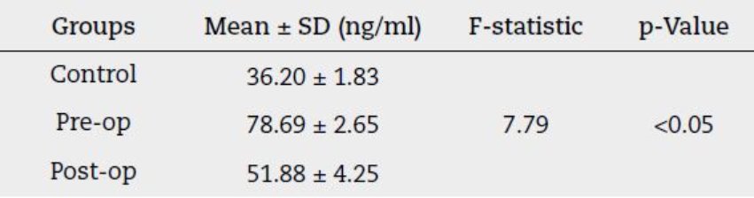
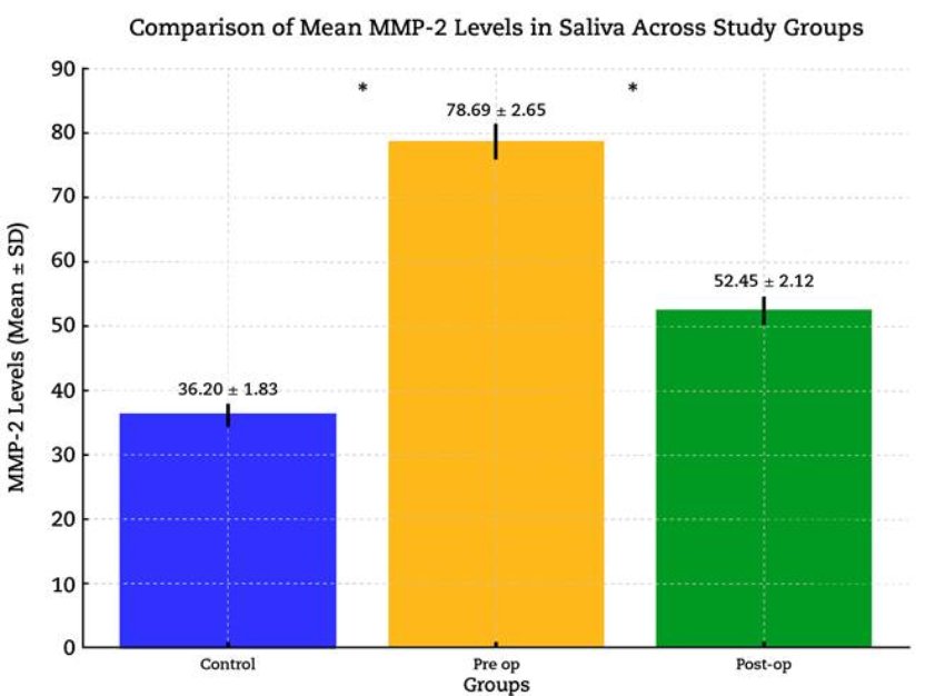
Figure 1. Provides a visual representation of the mean MMP-2 levels, highlighting the stark differences between the control, pre-operative, and post-operative group.
In the Post-op group (Mean ± SD: 51.88 ± 4.25 ng/ml), MMP-2 levels showed a significant decrease compared to the Pre-op group, indicating that the surgery had an effect on tumor-related activity. However, these levels did not fully return to the baseline seen in the Control group, suggesting that MMP-2 expression did not completely resolve. The remaining elevation could be due to ongoing disease activity or inflammation after the surgery.
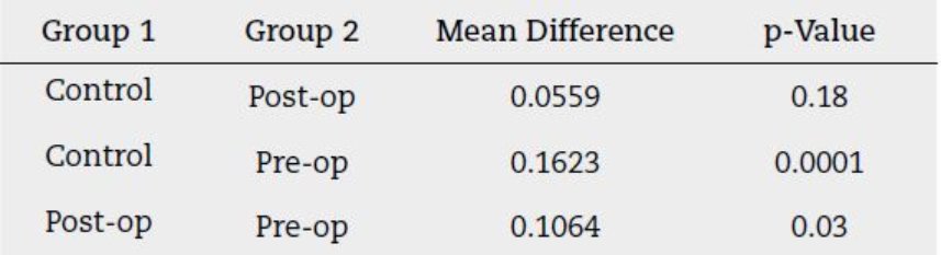
Statistical analysis revealed significant differences in MMP-2 levels across the three groups (F-statistic = 7.79, p < 0.05). Pairwise comparisons confirmed a highly significant difference between the Control and Pre-op groups (p = 0.0001), reinforcing the role of MMP-2 as a reliable marker of OSCC presence. Additionally, the significant difference between the Post-op and Pre-op groups (p = 0.03) demonstrates the reduction in tumor-associated activity following surgery. While the MMP-2 levels in the Post-op group remain higher than in the Control group, the difference is not statistically significant (p = 0.18). This suggests that the levels might trend toward normalization after surgery, but further studies with larger sample sizes are needed to confirm this observation.
The post-hoc Tukey comparison (Table 3) highlights significant differences in MMP-2 levels across the study groups, with the highest levels observed in the Pre-op group. This underscores MMP-2’s role as a biomarker for disease severity, with partial reduction Post-op reflecting surgical efficacy.
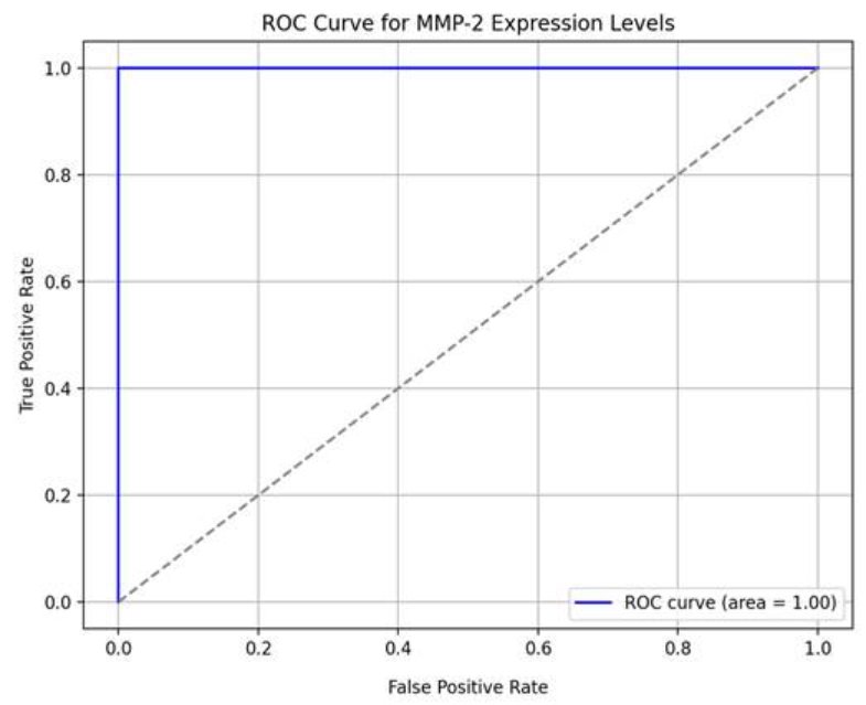
A suggested cutoff value for MMP-2 expression levels, based on the range of 44.68 to 119.31 ng/ml, is 75 ng/ml. This value is slightly above the median of 70.25 ng/ml, which initially stratifies the dataset into low and high expression groups, and offers a practical midpoint within the observed distribution. While the ROC-derived optimal cut-off (0.9174525774375621) appears to be a normalized or transformed value, the proposed cut-off of 75 ng/ml aligns with a straightforward approach that balances simplicity and clinical applicability (Figure 2 and Figure 3). Furthermore, values reported in the literature for MMP-2 expression cut-offs have been observed to fall within a similar range, supporting the biological and clinical relevance of this selection.
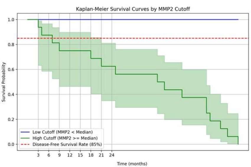
Matrix metalloproteinase-2 (MMP-2) plays a critical role in cancer progression, and its expression is often linked to poor prognosis. The Kaplan-Meier survival analysis compared survival probabilities between patients with low and high MMP-2 levels. For the high MMP-2 group, survival probabilities decreased significantly over time, starting at 1.0 at baseline (0 months), dropping to 0.9375 at 3 months, 0.875 at 4 months, 0.8125 at 7 months, and reaching 0.75 at 9 months (Table 4, Figure 4). These results indicate a steady decline in survival over time for patients with elevated MMP-2 levels. In contrast, the low MMP-2 group showed comparatively better survival probabilities. The steep decline in survival probabilities for the high MMP-2 group suggests that elevated MMP-2 levels are associated with significantly worse outcomes, highlighting its potential as a prognostic marker and a candidate for therapeutic targeting.

DISCUSSION
Matrix Metalloproteinase-2 (MMP-2) has been investigated as a potential biomarker in understanding the pathogenesis, diagnosis, and prognosis of oral squamous cell carcinoma (OSCC)11. This study demonstrated a significant variation in salivary MMP-2 levels across three groups (Control, Pre-operative, and Post-operative), underscoring its diagnostic and prognostic relevance.
The significant elevation of MMP-2 in the Pre-operative group compared to the Control group highlights its robust association with OSCC progression. Elevated MMP-2 is known to degrade extracellular matrix components, facilitating tumor invasion, angiogenesis, and metastasis. This finding is consistent with earlier reports by Monea et al. (2022), who demonstrated that high salivary MMP-2 levels correlate with increased tumor invasiveness and poor clinical outcomes in OSCC patients6,12. Furthermore, the post-operative reduction in MMP-2 levels signifies the effectiveness of surgical intervention in curbing tumor-associated molecular activity. However, the incomplete normalization of MMP-2 levels aligns with studies by Patel BP et al. (2018), suggesting the persistence of residual disease activity or inflammation post-surgery13. This residual elevation underscores the necessity of adjunct therapies such as radiotherapy or targeted molecular treatments to achieve comprehensive tumor eradication14.
Notably, these levels differ from previously reported baseline values for healthy individuals, such as the approximately 2 ng/ml reported by Dalirsani et al. (2019) 15. This discrepancy could arise from differences in study methodologies, population demographics, or assay sensitivity and specificity. Dalirsani et al. observed no significant differences in MMP-2 levels between OSCC patients and healthy controls in head and neck squamous cell carcinoma (HNSCC), suggesting MMP-2 may not always be a definitive biomarker across all populations or settings15. However, our study focuses specifically on OSCC, a distinct subset of HNSCC. It uses a standardized saliva sampling protocol under controlled conditions, which may contribute to higher MMP-2 levels in both patient and control groups.
The elevated MMP-2 levels in the Pre-op group align with its known role in extracellular matrix degradation and tumor invasion. The significant reduction in the Post-op group (p = 0.03) underscores the potential impact of surgical intervention on tumor-associated activity. However, the inability of Post-op levels to return fully to baseline might reflect persistent subclinical disease activity or postsurgical inflammation.
The Kaplan-Meier survival analysis further supports the prognostic value of MMP-2 in OSCC. Patients with high salivary MMP-2 levels exhibited a steady decline in survival probabilities, with significant drops noted at 3 months (93.75 %) and 9 months (75 %). These findings align with data reported by Patel BP et al. (2005), who observed that increased MMP-2 expression was associated with poor survival due to enhanced tumor aggressiveness and metastatic potential16. Conversely, patients with low MMP-2 levels demonstrated comparatively better survival probabilities, underscoring its potential as a prognostic indicator. The survival trends highlight the importance of incorporating MMP-2 level assessments into routine OSCC management to stratify patients into risk categories and tailor treatment approaches accordingly17.
Clinically, the use of ELISA for quantifying salivary MMP-2 offers a non-invasive, sensitive, and specific method for OSCC diagnosis and monitoring. The precise quantification provided by ELISA allows for early detection and tracking of treatment efficacy18. These findings are consistent with recent literature emphasizing the role of salivary biomarkers in enhancing diagnostic accuracy and reducing reliance on invasive tissue biopsies19.
Additionally, the partial reduction of MMP-2 in the post-operative group underscores the need for multi-modal therapeutic strategies. Adjunct therapies targeting MMP-2 activity, such as MMP inhibitors, have shown promise in preclinical studies for reducing tumor progression and improving survival outcomes20. Moreover, long-term follow-up and periodic monitoring of MMP-2 levels could aid in early detection of recurrence, ensuring timely interventions21. Integrating MMP-2 measurements into routine care could enhance OSCC management by aiding early detection and monitoring of treatment outcomes.
Furthermore, the findings of this study emphasize the emerging role of salivary biomarkers like MMP-2 in advancing personalized medicine for OSCC. The non-invasive nature of saliva collection, combined with the high sensitivity of ELISA, makes it an ideal approach for regular patient monitoring22. Recent advancements in salivary diagnostics, such as multiplex assays and point-of-care devices, can further enhance the utility of MMP-2 assessment in clinical settings23,24. Incorporating MMP-2 as part of a salivary biomarker panel, alongside other markers like IL-6 and VEGF, could provide a more comprehensive understanding of tumor biology and improve the predictive accuracy for treatment response and survival outcomes25,26. By leveraging these tools, clinicians can better stratify patients, optimize treatment protocols, and ultimately improve the quality of life and survival rates for OSCC patients. The limitations of this study, including the reduced sample size, limited controls, and lack of detailed information on the evolution of the cases, may affect the generalizability and prognostic inference of the findings. A larger sample size, more comprehensive control group data, and detailed follow-up information would strengthen the conclusions and provide a clearer understanding of the disease progression and treatment outcomes.
Future scope
Salivary MMP-2 has significant potential to enhance the early diagnosis, treatment monitoring, and prognosis of OSCC. Integrating this biomarker into clinical workflows could enable non-invasive detection, guide personalized therapeutic strategies, and improve monitoring of disease progression and recurrence. Advancements in diagnostic technologies and multi-biomarker panels may further optimize its utility, while research into MMP-2-targeted therapies could reduce tumor aggressiveness and recurrence. These efforts collectively aim to improve survival outcomes and quality of life for OSCC patients.
CONCLUSION
This study highlights the potential of salivary MMP-2 as a biomarker for oral squamous cell carcinoma (OSCC). The observed elevation of MMP-2 levels in the pre-operative group highlights its involvement in tumor progression, while the reduction in levels following surgery reflects the effect of the intervention, albeit without a return to baseline. These results indicate a possible role for MMP-2 in the detection and monitoring of OSCC. However, given the study’s limitations, including sample size and scope, further research in larger, more diverse populations is necessary to confirm its diagnostic and prognostic value and establish its clinical utility.

