




 Servicios personalizados
 Inglés (pdf)
 Articulo en XML
 Referencias del artículo
 Traducción automática
 Enviar articulo por email
 Citado por SciELO
  Citado por Google
  Similares en
SciELO
Similares en
SciELO  Similares en Google
 Permalink
INTRODUCTION
The spatial relationship between the mandibular canal and the third molar is a risk indicator for an injury to the inferior alveolar nerve during tooth extraction1. It is defined as the three-dimensional position of the mandibular canal with respect to the third molar, which can be found in buccal, lingual, caudal, intraradicular and interradicular positions and in contact or not with the dental structure2,3,4. Sklavos et al. (5 evaluated in cone beam computed tomography (CBCT) the degree of compression measured at the point of maximum compression between third molar roots and mandibular canal. They found that patients who had a degree of compression where the diameter decreased by a 75 % or greater had increased risk of a postoperative inferior alveolar nerve injury and accounted for 52.17 % of all inferior alveolar nerve injuries. Besides, Klatt et al. (6 found that caudal relationship in contact was associated with the highest risk of a temporal neurological damage. It has been reported that the injury to the inferior alveolar nerve has an incidence of 1.1 % at 7 days after third molar surgery, while the involvement in a time greater than 6 months is 0.5 %7.
Panoramic radiography is the standard imaging test for preoperative evaluation of lower third molar surgery. However, being a two-dimensional radiograph, it may have limitations in assessing the spatial relationship when the mandibular canal is in intimate contact with the third molar8,9. CBCT has greater sensitivity than panoramic radiography for detecting exposure of the inferior alveolar nerve during surgery, with intimate contact with loss of cortical bone of the mandibular canal being the most commonly associated sign. CBCT has a sensitivity of 95.1 % and a specificity of 64.4 %, while panoramic radiography has a sensitivity of 73.9 % and a specificity of 24.8 %10. Although neurosensory deficit does not simply occur after exposure of the neurovascular bundle, it increases the risk of injury to the inferior alveolar nerve11,12. An incidence of sensory disturbance of 20% has been reported after 7 days in cases where there is exposure of the neurovascular bundle12.
Detecting the mandibular canal on panoramic radiographs can be a challenge for the clinician, especially in its anterior and middle section13,14. Even on CBCT, it has been reported that it could not be detected in 18 % of cases at the level of the first molar. The cortex of the mandibular canal may vary according to the degree of bone density within the mandibular body, which can make its evaluation difficult15.
The effective dose represents the radiosensitivity of each organ, that is the risk of tissue damage by ionizing radiation, and allows us to estimate stochastic risks and compare different technologies and procedures16. The effective dose from CBCT is generally higher than intraoral and panoramic radiography but lower than conventional computed tomography (CT) and it is dependent on equipment type and exposure parameters, especially the field of view selected17.
The mandibular canal may present bifid canals along its course. This anatomical variant of the mandibular canal may be formatted by a partial fusion of independent canals present at mandible during latter half of the prenatal period17,18,19. However, it is necessary to clarify the mechanism of forming such anatomical variant18. A meta-analysis has reported the prevalence of bifid canals in 16 % of patients on computed tomography (CT) and CBCT20. In contrast, several studies with CT and CBCT have demonstrated a high frequency of bifid canals per patient with reports of 9.8 % to 66.5 %21. Its clinical relevance lies in the difficulty to detect it with two-dimensional radiographs and has two clinical implications: potential failure to achieve local anesthesia and surgical injuries to the neurovascular bundle20,21,22.
The spatial relationship of the mandibular canal with the third molar is an important factor in a presurgical evaluation, because it can intervene in a potential risk of injury to the inferior alveolar nerve. The present investigation aimed to determine the prevalence of the spatial relationship between the mandibular canal and the third molar in CBCT in the population investigated.
MATERIAL AND METHODS
Study design and population
The present observational, descriptive, cross-sectional study evaluated the cone beam computed tomography (CBCT) scans from the anonymized database of the Department of Imaging of the Faculty of Dentistry of the University of Cuenca (Ecuador) within the period May 2017-January 2023 of patients which were referred to Department of Imaging requiring CBCT imaging as part of their dental treatment. During this period, the universe was composed of 1092 CBCT files.
Ethical approval
The present study was approved by the Research Directorate of the Faculty of Dentistry (DIFO) of the University of Cuenca (Ecuador). This study was conducted in accordance with the ethical foundations of research on human beings based on the Declaration of Helsinki of the WMA (World Medical Association) (21. The database was anonymized to preserve the confidentiality of the data obtained and the patient’s anonymity, to this end, the CBCTs were assigned a code and the information was recorded in digital files that were only accessible to the authors of the study. No data was provided that would allow the patient to be identified. In addition, each patient signed an informed consent prior to being treated at the Faculty of Dentistry.
Eligibility criteria
Inclusion criteria: CBCT corresponds to the Department of Imaging between the period 2017-2023 that shows the mandibular canal and the uni or bilateral lower third molars with complete root formation or in the formation of the apical root third (Nolla stage 9).
Exclusion criteria: Repeated CBCT; pathology in the third molar and the posterior mandibular region; existence of a foreign body artifact such as titanium plates, orthodontic appliances, implants, amalgams, and dental prostheses; motion artifacts; poor quality in CBCT; hypodense trabecular bone that makes it impossible to observe the mandibular canal; a third molar with an abnormal shape and root remains of the third molar which difficult recognize the third molar.
Study method
The CBCT files were evaluated through the i-Dixel Version 6.4.0.5 program. The spatial relationship variable was analyzed in the study population by a single observer after calibration by a specialist in Dental and Maxillofacial Imaging. The Microsoft Excel Version 2102 program was used to record the variables, and the SPSS Statistics Version program was used for the statistical analysis. The CBCT code, right and left side, spatial relationship variable and technical parameters: field of view (FOV) and voxel size were recorded in the Microsoft Excel Version 2102 program. In the registry of variables, the qualitative value of the spatial relationship was noted, therefore, for each position in the buccolingual direction, the distance was described as contact or no contact. In the case of detecting branches of the mandibular canal at the level of the third molar in the transaxial slices, the number and spatial relationship with the third molar were noted. In the statistical analysis, the prevalence (the percentage of each case) and the frequency (the number of each case) were obtained.
Calibration and inter-rater reliability
To avoid inter-observer differences, all CBCTs were evaluated by the same observer. The observer was calibrated by a specialist in Dental and Maxillofacial Imaging to identify the mandibular canal and its relationship to the third molar using the i-Dixel program Version 6.4.0.5, in CBCTs that were not included in the study. The degree of inter-observer agreement was obtained using Cohen’s Kappa index using the SPSS Statistics program Version. A high agreement was found in the buccolingual position of the mandibular canal with the third molar (Kappa = 0.87) and in the distance (Kappa = 1.0) between the two investigators, which suggests that the observer was able to analyze the experimental CBCTs.
Study variables
The radiographic variable analyzed in the CBCT files was the spatial relationship, which is a polychotomous nominal qualitative variable. Spatial relationship is composed by the buccolingual direction and the distance between the mandibular canal and the tooth structure (Figure 1). The operational definitions of the study variables are described below, obtained through a literature review2,3,4,23.
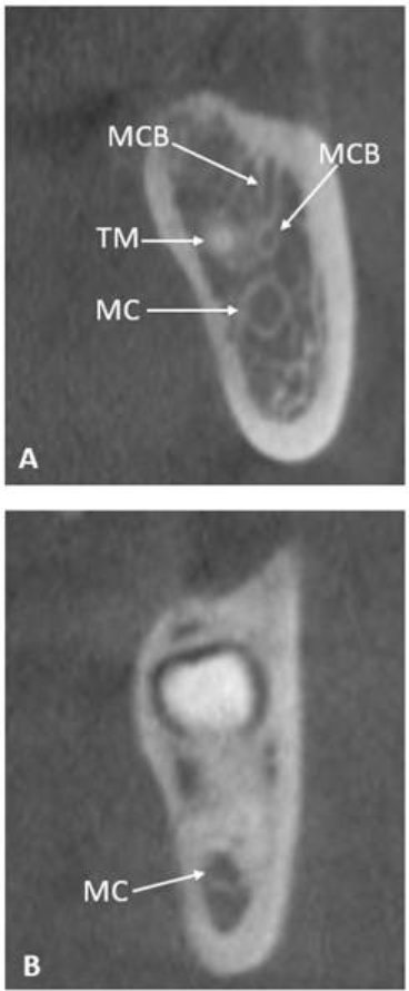
Figure 1. A: mandibular canal (MC) in caudal position without contact with the third molar (TM), mandibular canal branches at the level of the third molar (MCB). B: mandibular canal in interradicular position in contact with the third molar.
Buccal: The mandibular canal is partially or completely in a buccal position to the dental structure during its journey by the third molar.
Lingual: The mandibular canal is partially or completely lingual to the tooth structure during its journey by the third molar.
Caudal: The entire mandibular canal is located below the tooth structure along the entire journey with the third molar.
Interradicular: The mandibular canal is located between the roots of the tooth.
Intraradicular: The mandibular canal passes between converging roots fused at the apex.
Contact: There is no bone between the mandibular canal and the third molar, interruption of the cortex is observed at some point along its journey with the third molar.
No contact: There is bone between the mandibular canal and the third molar, the cortex is preserved without interruptions along the entire journey with the third molar.
Observation method
Images were obtained using a CBCT unit (3D Accuitomo 170, J. Morita, Kyoto, Japan). The i-Dixel software Version 6.4.0.5 was used as the observation tool. Images were evaluated with a slice thickness of 0.1 mm and a slice interval of 0.5 mm. To describe the different spatial relationships of the mandibular canal with the third molar, the direct observation technique was applied to the CBCTs. The observation method was standardized as follows; contrast and brightness were modified to visualize the mandibular canal and the third molar using the BONE 2 option in the Contrast and Color Presets. The position of the anterior nasal spine was evaluated in the axial slices to establish the midsagittal plane, then the third molars were located in the coronal plane and the lower basal edge was aligned with the reference line of the axial section (Figure 2). The cervical region of the mandibular teeth was located in the axial slices.
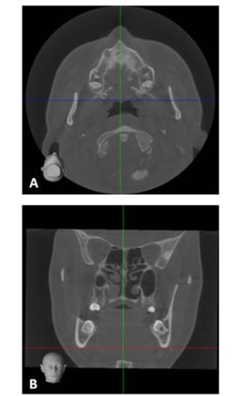
Figure 2. A: evaluation of the anterior nasal spine to establish the midsagittal plane. B: center the third molars and match the lower basal edge with the reference line of the axial section.
The dual-CMPR module was selected and then the option Draw an MPR spline on the axial slice was selected in Tools. This was started on the right side (if applicable). A curve was drawn using points, starting at the posterior and middle of the mandibular ramus, then equidistant points were drawn between the cortical plates at the level of the mandibular molars and continuing to the midline. The process was repeated on the left side by again selecting the option Draw an MPR spline on the axial section. After drawing the curve, the transaxial slices of the bilaterally delimited area were automatically generated (Figure 3).
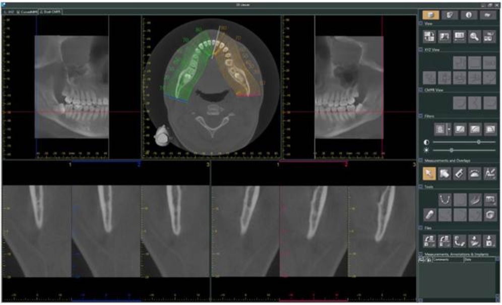
Figure 3. View of the dual-CMPR module showing the transaxial sections of the bilaterally delimited area.
To determine the right or left side was used the reference line on the coronal slices. The transaxial images was evaluated with a slice thickness of 0.1 mm with a slice interval of 0.5 mm. The mandibular canal was recognized as a corticalized hypodense tubular structure in the transaxial slice15. The dental structure is observed as a hyperdense image. Through the transaxial slices were established the relationship of the dental canal with the third molar and whether or not it is in contact.
RESULTS
Prevalence of the spatial relationship of the mandibular canal with the third molar
The universe was composed of 1092 CBCT files. According to the eligibility criteria, the study population comprised 199 CBCT Files. No calculation of the study sample size was performed. In total, 325 lower third molars were evaluated, where 51.4 % were lower right third molars and 48.6 % were lower left third molars. The technical parameters were: FOV of 170 x 120 mm and the voxel size of 0.33 mm in most of the CBCTs analyzed.
The highest prevalence of the spatial relationship between the lower third molar and the mandibular canal was the caudal relationship without contact in 32.0 % (n = 104), followed by the lingual relationship in contact in 22.2 % (n = 72), buccal in contact in 14.8 % (n = 48), caudal in contact in 12.9 % (n = 42), buccal without contact in 8.6 % (n = 28), interradicular in contact in 6.8 % (n = 22), lingual without contact in 1.5 % (n = 5) and intraradicular in contact in 1.2 % (n = 4). Furthermore, mandibular third molars were found in contact with the mandibular canal in 57.8 % and without contact in 42.4 % (Table 1, Figure 4). In the interradicular and intraradicular spatial relationship, the canal was always found in contact with the third molar.
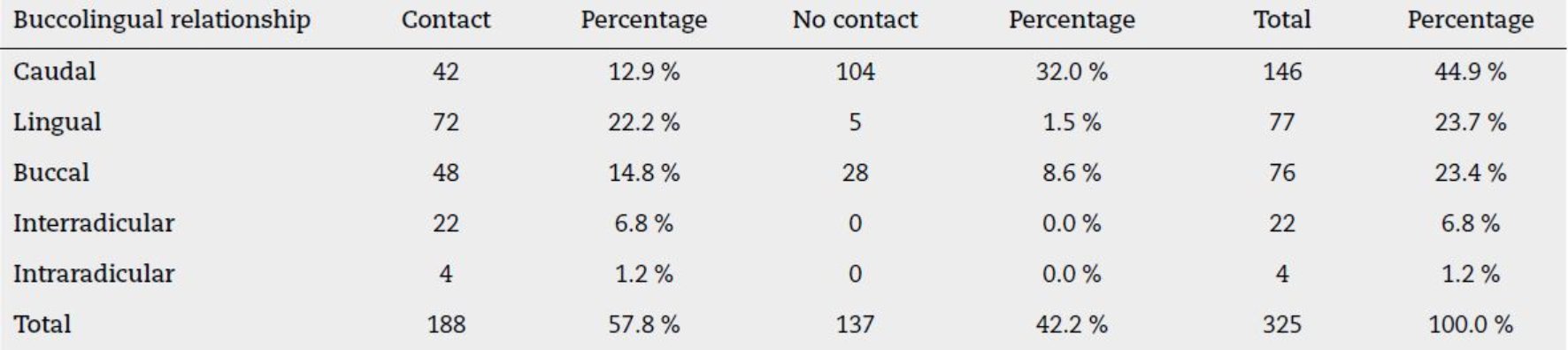
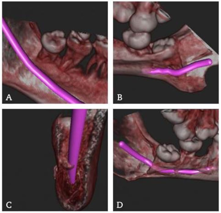
Figure 4. Three-dimensional reconstruction of the mandibular canal path with the third molar. A: spatial relationship in a caudal direction without contact. B: spatial relationship in a lingual direction in contact. C: spatial relationship in a buccal direction in contact. D: spatial relationship in an intraradicular direction.
Another imaging finding was the presence of mandibular canal branches in 28.9 % at the level of third molars, where 27.7 % of the cases presented bifid mandibular canal, while 1.2 % presented trifid mandibular canal. The highest prevalence of the spatial relationship of the mandibular canal branche with the third molar was the buccal relationship without contact in 22.5 % (n = 22), followed by the cranial relationship without contact in 20.4 % (n = 20), caudal in contact in 13.3 % (n = 13), buccal in contact in 12.2 % (n = 12), lingual in contact in 7.1 % (n = 7), cranial in contact in 5.1 % (n = 5), caudal without contact in 5.1 % (n = 5) and lingual without contact in 2.0 % (n = 2) (Table 2).
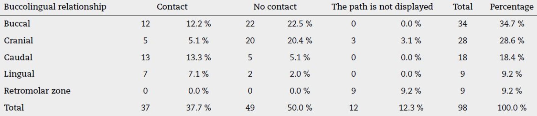
The lower third molars were found without contact with the mandibular canal branches in 50 % (n = 49), while they were found in contact in 37.7 % (n = 37) and observation of the path was impossible in 12.3 % (n = 12) of the cases due to loss of cortex of the canal, therefore it could not be determined whether or not it was in contact with the dental structure. Some branches of the mandibular canal were observed in the retromolar area towards the third molar; however, the route was not visualized due to the loss of the cortex of the canal (Table 2).
DISCUSSION
The spatial relationship of the third molar with the mandibular canal has been shown to have high variability, which may be due to the ethnicity of the patients and the size of the study population 24,25.
Yamada et al. (3 in their CBCT investigation, evaluated the spatial relationship of 112 impacted third molars at the closest point to the mandibular canal from three examined areas and found that in 36.6 % of the cases, the mandibular canal was caudal in contact to the third molar. Lübbers et al. (26 evaluated 707 impacted third molars by CBCT and reported that in 52.8 % of the cases, the mandibular canal was buccal to the third molar. In both studies, to be part of the study population, the mandibular canal was overlapping on the third molar in panoramic radiographs and the preoperative CBCT was evaluated.
In the study by Wang et al. (24 137 impacted third molars were evaluated in CBCT and found that in 60.6 % of the cases the mandibular canal was caudal without contact with the third molar. Quirino de Almeida Barros et al. (27 evaluated 173 thirds molars in CBCT, and found that in 28.3 % of the cases, the mandibular canal was mainly located caudal in contact with the third molar. In these studies, the study population were patients with impacted third molars.
Previous studies present limitations in the study methodology, such as non-specific operational definitions of study variables, lack of calibration and inter-observer reliability, and non-specific eligibility criteria. In addition, imaging studies of anatomical structures must have an observation method to align the CBCT in the same axial, sagittal and coronal plane. Further studies with homogeneity in methodology should be carried out.
In the present study, 325 third molars were evaluated in CBCT and the most prevalent spatial relationship of the mandibular canal with the third molar was the caudal relationship without contact in 32.0 % of the cases. Study population were patients in whom the reason for the radiological examination was unknown, which allows for internal validity at a local level. In the results of the present study most of the third molars were found in contact with the third molar, which is similar with the study by Yamada et al. (3, Lübbers et al. (26 and Quirino de Almeida Barros et al. (27.
Interruption of the mandibular canal cortex on CBCT, the interradicular and lingual position of the mandibular canal has been associated with intraoperative exposure of the inferior alveolar nerve during third molar surgery 11,28. In the present study, it was demonstrated that in the intra and interradicular position, there was always direct contact between the third molar and the mandibular canal, while in the lingual position the majority of the cases were in contact.
Yamada et al. (3 found mandibular canal branches in 94.6 % of the cases at the level of third molars. In contrast, the present study found mandibular canal branches in 28.9 % of the cases at the level of third molars. This difference may be due to the ethnicity of the patients, operational definition of the study variables, size of the study population, selection criteria and voxel. Voxel size is an important factor for the recognition of mandibular canal branches. The smaller the voxel size, the better the resolution of CBCT, and the better the recognition of branches of the mandibular canal, thus there is a positive association between the prevalence of branches of the mandibular canal and the voxel size of a CBCT18. In the present study, the majority of CBCTs examined had a voxel size of 0.33 mm.
Preoperative assessment by computerized tomography or cone beam computed tomography is based on the presence of mandibular canal wall for its localization, which may be further affected by decreased bone trabeculation15. In the present study, it demonstrated the difficulty of evaluating branches of the mandibular canal due to the loss of the cortex, making it impossible to observe their course in 12.3 % of cases. Magnetic resonance imaging (MRI) is a valid alternative for preoperative assessment radiation-free of the inferior alveolar nerve and accesory inferior alveolar nerves29,30. Special “black bone” MRI sequences such as 3D Double Echo Steady State and 3D Short Tau Inversion Recovery sequences providing high-resolution and high-contrast images that allow simultaneous visualization of the inferior alveolar nerve tissue within osseous limits of the mandibular canal29,31,32. Besides, Al-Haj Husain et al.33 demonstrated that in 3D Double Echo Steady State MRI, the observation of the entire course of nerve lingual from the foramen ovale to the mandibular third molar region was possible in 35 % of the cases. The decision to choose any imaging modality in mandibular third molar surgery has to be based on his indications, contraindications, limitations and their respective advantages and disadvantages29. In recent years, deep learning-based evaluation had demonstrated high sensitivity and specificity for detection and evaluation of the spatial relationship between mandibular canal and third molar on CBCT images which could be utilized for preoperative planning34,35.
This study presents some limitations. First, the study universe was obtained from single database therefore further studies with homogeneity in methodology should be carried out. This study presents an observation method and operational definitions of the study variables that can be replicated for the investigation of the spatial relationship of the mandibular canal with the lower third molar. Second, gender and age were not investigated due to the database anonymization protocol. Third, It is not possible to conclude that the tubular bone structures arising from the mandibular canal contain branches of the inferior alveolar nerve. Finally, the present study evaluated mandibular canal branches that were found at the level of the third molar excluding those branches that were found only in the retromolar region and in front of the third molar, which could increase their prevalence.
CONCLUSIONS
The most prevalent spatial relationship of the mandibular canal with the third molar was the caudal relationship without contact, in 32.0 % of the cases. However, most of the third molars were found in contact with the third molar, in 57.8 % of the cases. Anatomical variations of mandibular canal were found in 28.9 % of third molars only at the level of the third molars, presenting in most cases a bifid mandibular canal. Its most prevalent spatial relationship was the buccal relationship without contact. Most branches of the mandibular canal were found without contact with the third molar, and its complete visualization was impossible in 12.3 % of cases due to the loss of the cortex of the canal.

