




 Servicios personalizados

 Inglés (pdf)
 Articulo en XML
 Referencias del artículo

 Traducción automática
 Enviar articulo por email
 Citado por SciELO
 Citado por Google
 Similares en
SciELO
 Similares en Google

 Permalink

INTRODUCTION
Salivary glands are exocrine glands responsible for saliva secretion. Humans have three major salivary glands (parotid, submandibular, and sublingual). In addition, more than 800 minor salivary glands are distributed everywhere, including the sinonasal cavity and oral cavity, and might involve the pharynx, larynx, trachea, lungs, and middle ear cavity1,2,3. However, it is mostly presented along the submucosa of the lip, cheek, soft or hard palate, and floor of the mouth. Tumors, benign or malignant, affect the major and minor salivary glands at different rates. Minor salivary gland tumors represent < 25 % of intraoral salivary neoplasms with the majority of tumors being malignant. These tumors have explicit characteristics regarding their frequency, distribution, and clinical behavior4,5. Despite the relatively low frequency of occurrence of these lesions, they represent a diverse group of neoplasms with a vast range of histological types and growth modes. They may appear at any age, mostly occurring for benign lesions in the fourth decade of life6,7. Pleomorphic adenoma of the minor salivary gland is mostly present in the hard palate. With a wide variety of histological characteristics and development patterns, minor salivary gland tumors are a diverse category of neoplasms. Incomplete surgical excision of these tumors has a significant recurrence rate, and malignant transformation must be taken into account8. According to reported literatures; the majority of women are affected by minor salivary gland tumors3,7, which are primarily found on the palate, lips, cheek mucosa, tongue, and floor of the mouth. They are glandular in origin in the head and neck and typically manifest as a mobile, slowly growing, firm swelling that is painless and does not cause ulceration of the mucosa on top9. Since lasers are extremely effective scalpels for the excision of tumors in oral and maxillofacial surgery with the greatest amount of tissue preservation, they are increasingly the treatment of choice for both patients and clinicians. However, surgical management of orofacial tumors presents significant challenges due to their proximity to sensitive areas of the mouth and face. The maxillofacial surgeon selected the surgical manipulation modalities based on the clinical examination and compiled diagnostic data, taking into account the least amount of deformity, minimal scarring, and quick recovery10,11. The objective was to document the clinicopathological features of pleomorphic adenoma of the palatal minor salivary gland excised utilizing diode lasers at 980 nm and 940 nm.
PATIENTS AND METHODS
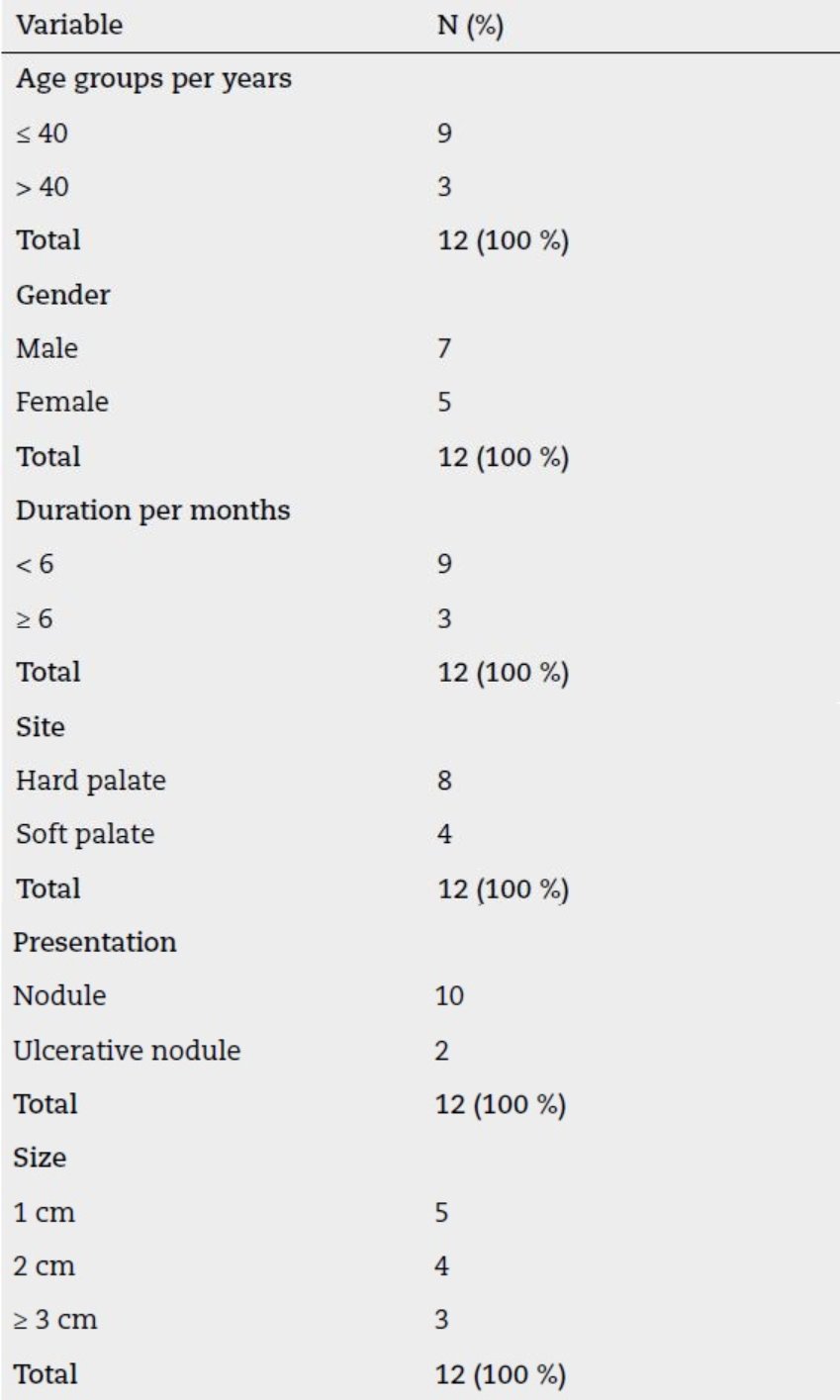
This prospective clinicopathological study included patients who attended the department of Maxillofacial Surgery, Ramadi Teaching Hospital and Private Clinic at Ramadi, Anbar Health Directorate with soft tissue swelling that involved the soft and/or hard palate. The patients’ names, age, gender, tumor location, and symptoms were among the demographic information. In order to assess the lesions in terms of location, shape, color of the overlying tissue, size, surface texture, consistency, fluctuation, and examinations of locoregional lymph nodes, both intraoral and extraoral clinical examinations were carried out by careful inspection and gentle digital palpation of the soft and bony tissues (Table 1). The medical laboratory investigations were requested as needed depending on the medical history. The radiographical examination was conducted using conventional radiography, computed tomography (CT), ultrasonic examination with fine-needle aspiration cytology (FNAC) for initial diagnosis of the referred case; It is a straightforward and acceptable procedure for examining patients with pathological disorders, such as inflammations and masses, after anesthesia. It involves using an 18-gauge needle attached to a 5-milliliter syringe in accordance with specified cytological protocols.
All cases were prepared for laser surgical procedures including safety goggles should be used to protect the skin and eyes during laser surgery that uses a diode laser with a wavelength of 940 nm or 980 nm and a power of 1.4-1.8 W in CP1 mode with a fiber optic delivery system. The surgical procedures were carried out under local anesthesia (2 % lidocaine injection) for complete ablation of the mass with a safe margin of 1 mm and deep to the underlying bone. Each patient received a prescription for medication, including metronidazole, non-steroidal anti-inflammatory analgesics, and appropriate antibiotics, along with postoperative instructions and intraoral antisepsis using Listerine mouthwash for 30 seconds throughout postoperative period (Figure 1).
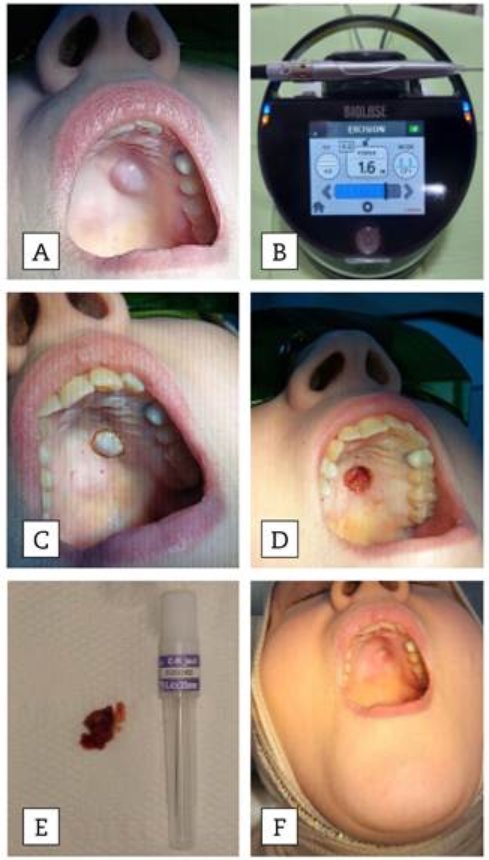
Figure 1. A: preoperative view. B: laser parameter installation. C: laser application and demarcation. D: laser ablation of the tumor. E: Gross specimen of the tumor. F: postoperative view showing complete healing.
In order to confirm the diagnosis, two independent oral pathologists and a general pathologist examined the entire set of samples at the Histopathology department using paraffin embedding blocks and stained them with hematoxylin and eosin (H & E) for histopathological and immunohistochemical (IHC) analysis. Serial sections were produced for each formalin-fixed paraffin-embedded specimen in the manner described below: For routine Haematoxylin and Eosin staining (H&E), sections of each block of the investigated sample with a thickness of 5 µm were placed on glass slides for histopathological assessment.
For each specific antibody (P63, S100), additional 5μm sections were obtained and mounted on positively charged microscope slides using the suggested dilution. According to the manufacturer’s data sheets and earlier research, tumor cells with clear brown cytoplasmic staining were regarded as positive in actin, and cytoplasmic or cytoplasmic/nuclear cells were regarded as positive in S100 and P63 immunostaining patterns. Myoepithelial cells, ductal cells, and chondromyxoid regions were assessed by immunohistochemistry in pleomorphic adenoma. If the analyzed slides show any positive expression of tumor cells, the case is deemed positive; if not, it is deemed negative (Figure 2).
Every clinical and histological technique was carried out in compliance with the Helsinki Declaration of 1975, as amended in 2008, and the ethical guidelines of the relevant institutional or regional council on human experimentation.
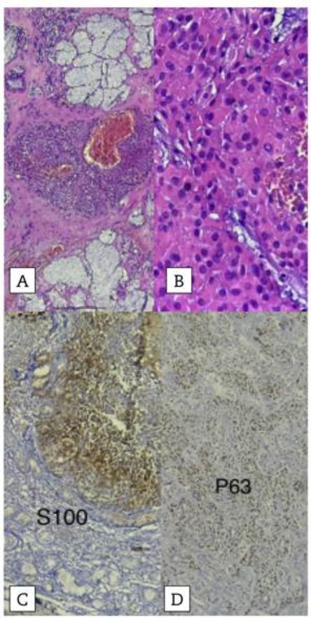
Figure 2. A and B: histopathological confirmation H&E (10X, 40X) reveals a classic pleomorphic adenoma consisting of nests of squamous epithelia with prominent intercellular bridges, a sheet of characteristic plasmacytoid myoepithelial cells, a thin layer of dense fibrous capsule, a number of keratin pearls, and nests of myoepithelial cells in a dense hyalinized stroma. C and D: immunhistochemical expression of S100 and P63 showing posaitive expression in myoepithelial cells’ nuclei of the normal glandular parenchyma.
Patients were closely monitored throughout their follow-up visits for two years after the end of treatment. Their total happiness with the treatment was documented during this time, as were any issues that arose. Following the last treatment, patients were closely observed at 6-month intervals during follow-up appointments. Throughout the follow-up time, their general level of happiness was noted, along with any issues that came up. Ethical approvals for the study were obtained from the University of Anbar / Ethical Approval Committee (181 on August 20, 2024). All data and materials used and analysed during the current study are available from the corresponding author. All the data were statistically analyzed using SPSS software version 26 as non-parametric data and tested by descriptive frequencies for each studied case and variable alone, followed by a ratio-statistics test for each separately.
RESULTS
Twelve patient data sheets that involved pleomorphic adenoma of the minor salivary gland were treated by laser ablation using diode laser 940 nm or 980nm. Regarding age and gender, with age range from 34 years old to 72 years old, mean age 47 years old, SD ± 0.5. This study showed that pleomorphic adenoma is most prevalent was 9 (75 %) in age above 40 years old with a male predilection including 7 (58 %) were males and 5 (42 %) were females with male to female ratio of 1:1.1. About 8 (67 %) of the cases were located in the hard palate and 4 (33 %) were located in soft palate. 5 cases size 1 cm, 4 cases size 2 cm, and 3 cases size ≥ 3 cm), about 10 (83 %) presented as mass while 2 (17 %) presented as ulcerative nodule, no patient reported recurrence of pleomorphic adenomas during the follow up period of two years post laser surgery and there was complete healing with good function and aesthetic outcome.
DISCUSSION
The field of craniofacial surgery is seeing an increase in the use of laser technology due to its rapid development. Therefore, it becomes crucial for the surgeon to learn about it in order to carry out surgical treatments more successfully and efficiently. Finding the circumstances in which a laser would be more effective than traditional methods and applying them is crucial. Additionally, the surgeon needs to understand safety concerns and adhere to safety protocols. The laser has significant antibacterial efficacy, diminishing the necessity for antibiotics and the incidence of infection complications, even in instances of secondary intention wound healing. In this clinicopathological investigation, the use of laser allowed for the surgical operation to be performed in an outpatient environment, economic resources as well as saving time10,11.
The WHO’s pathological classification of salivary gland tumors has been adopted globally and has proven to be the most practical in day-to-day practice. Few papers concentrate solely on minor salivary gland tumors, while the majority of studies on salivary neoplasms cover both the major and minor salivary glands12,13.
Rare neoplasms of the upper aerodigestive tract are tumors that start in the minor salivary glands. Although pleomorphic adenomas are typically well-defined or encapsulated, tumor extension into the capsule is a frequent occurrence, and occasionally tumor lobules may seem entirely distinct from the main tumor mass. The most recent benign salivary gland tumor is called pleomorphic adenoma. Differentiated morphologically by a two-phase paradigm that incorporates the components of mesenchymal and epithelial14,15. The modulated myoepithelial cells might play an essential role in its histologic distinction by forming an extracellular matrix.
Pleomorphic adenoma usually presents clinically as a sluggish-growing, painless lump that might present for several years, according to physical examination, cytology, and histopathology and CT scans are helpful techniques to determine the site and size of the neoplasm and its extension to nearby areas, especially bone 16 . Most of the affected patients in this study were in fourth decay of life which showed that the pleomorphic adenoma of minor salivary glands occurred in the third and fourth decades. The surgical approach by laser ablation for treated cases with a 1mm safe margin, and since these tumors do not invade the periosteum, there was no request to shave the bone. In this study, the recurrence rate after excision of the lesion was negative 17,18.
Pleomorphic adenoma’s histopathological characteristics include a varied epithelial pattern inside a loosely fibrous stroma that might be mucoid, chondroid, or myxoid. Ductal structures, which are frequently bilayered, are prevalent, and the epithelium is typically organized in sheets or strands. The diagnosis was supported by tubular and ductular patterns lined by single to double layered cuboidal cells with a large darkly stained nucleus in the center and eosinophilic cytoplasm interspersed between large myxoid areas. Lesions in the minor glands are frequently more solid or cellular than those seen in the major glands, and the myoepithelial cells are frequently polygonal with a pale eosinophilic cytoplasm giving an epithelioid or plasmacytoid phenotype. There is no connective tissue capsule16,19,20.
By distinguishing benign from malignant disease, fine-needle aspiration cytology helps in surgical planning and provides more precise patient counseling. The preoperative diagnosis of pleomorphic adenoma is more accurately made by FNAC and an correct histological diagnosis requires excisional biopsy, and meticulous preoperative planning is required to determine the optimal surgical strategy for achieving total excision. The standard of therapy for suspected pleomorphic adenomas of the palate is to completely remove the lesion, just like with any other pleomorphic adenoma. When the lesion is enucleated or the capsule is dissected and the tumor spills intraoperatively, a high recurrence rate is noted. If the bone is invaded, it is highly advised that it be removed7,21.
The most frequent tumor in the salivary glands is pleomorphic adenoma, which most often develops in the parotid gland and, among smaller salivary glands, most typically in the palatine gland. The parotid gland is said to be the most prevalent site of Pleomorphic adenoma occurrence, while the palate is the most common intraoral location. It can be difficult to get the correct diagnosis. S100 and P63 proteins were first thought to be the most widely used markers for myopeithelial cells since they were found in healthy salivary glands using fine needle aspiration cytology and immunohistochemistry. Both S100 and P63 protein expression in the current investigation was primarily luminal and negatively expressed by myopeithelial cells 13,22,23 .
CONCLUSION
During the quick follow-up visits, the use of diode laser ablation at wavelengths of 980 and 940 nm, along with a safety margin, yielded satisfactory results in terms of both operative and aesthetic impacts. The preferred course of treatment is complete wide local surgical excision. Longer patient follow-up is necessary to identify late recurrences. Because they are in charge of critically analyzing the patient’s comprehensive clinical, radiographic, and histological examination, oral pathologists significantly aid in the diagnosis of oral lesions.

