




 Servicios personalizados
 Inglés (pdf)
 Articulo en XML
 Referencias del artículo
 Traducción automática
 Enviar articulo por email
 Citado por SciELO
  Citado por Google
  Similares en
SciELO
Similares en
SciELO  Similares en Google
 Permalink
INTRODUCTION
Arthroplasty has improved the quality of life of millions of patients and is highly effective, although implant-related complications can arise (1). Prosthetic joint infection (PJI) is one of the most important complications with a high morbidity and mortality rates, and associated costs. The incidence of PJI varies across countries from 0.5 % up to 2 % (2). Staphylococci are the most common etiological agents associated with PJI, with Staphylococcus aureus (30-40 %) and S. epidermidis being 2 of the main microorganisms involved (3). PJI results from the ability of microorganisms to develop a biofilm, a conglomerate of bacterial cells of, at least, 1 species that adheres to a surface or interface with complex socio-microbiological interactions among them (4).
Immediately after implantation with and without a perioperative PJI, the implantation surface is coated with plasma proteins that direct cellular adhesion and activation. Last step in the prosthesis implantation process is a foreign body reaction, that occurs when the inflammatory and wound healing responses have occurred including macrophages and foreign body giant cells (FBGCs) (9). These FBGCs are present on the surface of the implants throughout the life of the implant and produce stress cracks and oxidative damage (7). These macrophages and FBGCs secrete cytokines that in the early stages will modulate neutrophils and lymphocytes recruitment and activation (8). IL-4 and IL-13, both secreted by CD4+ T cells, are key cytokines for the formation of FGBCs involved in material degradation and implant rejection (9). In each one of the 4 phases for cell fusion, similar and different mechanisms for the formation of FBGCs or osteoclasts (multinucleated bone cells with the function of resorbing bone) have been described (10). At the end of this process, adherent macrophages and FBGCs induce biomaterial degradation with ensuing clinical device failure (5). Therefore, it is still being studied whether different materials respond differently to FBGCs adherence and how this biomaterial surface can be modulated to alter the presence and activity of adherent macrophages and FBGCs (11).
Purinergic signalling is also activated in cell fusion, especially in inflammation. Pharmacologically, 2 families of purinergic receptors are known: P1 receptors selective for adenosine, and P2 receptors selective for purine and pyrimidine nucleotides and dinucleotides (11,12). Adenosine is generated extra- and intra-cellularly by the hydrolysis of adenine nucleotides and performs its physiological functions through the activation of G-protein coupled transmembrane receptors (A1, A2A, A2B and A3), whose tissue distribution, pharmacological profile and effects are different (12). Purine and pyrimidine nucleotides activate 2 types of P2 receptors: P2X receptors—ionotropic, ion-linked channels—and P2Y receptors—metabotropic, G protein-coupled (11). The role of several P1 and P2 receptors in bone cells is well-known. The adenosine A1 receptor is critical and essential for differentiation and function of osteoclasts (13), while the adenosine A2A receptor induces an inhibition of osteoclast formation and function both in vitro (18) and in the osteolysis model (16). The adenosine A2B receptor is involved in osteoblast differentiation and function, with a more ambiguous role for the A2A receptor (16,17). Activation of adenosine A2A receptor both directly (by specific agonist) or indirectly (by drugs that increase extracellular adenosine levels by blocking its transport) promotes bone formation at levels similar to BMP-2 (bone morphogenic protein 2) (17). Among P2 receptors, P2X7 is especially relevant as its expression is positively regulated by pro-inflammatory cytokines (18). The P2X7 receptor is activated and is involved in cell fusion, where it intervenes in the late phases of membrane fusion and establishes union bridges between the cytoplasm of adjacent cells (19).
In orthopaedic surgery and traumatology, the most widely used medical alloys are Ti-6Al-4V and Co-Cr-Mo. Ti-6Al-4V alloy has favorable biocompatibility, mechanical properties and corrosion resistance (20). Furthermore, Co-Cr-Mo alloy exhibits high biocompatibility levels, with exceptional properties including abrasion, cracking-corrosion, pitting and wear resistance, malleability, and a high fatigue resistance and ductility (21).
Surprisingly, we know nothing of the effect that the presence of bacteria, such as S. aureus may or may not have during the formation process of these FBGCs. Therefore, we aim to investigate if variations in the metal alloys of the implants would change the recruitment of macrophages to the implantation area and, therefore, the formation of FBGCs since they would modulate the cell fusion process. More specifically, we believe that a differential activation of the purinergic system may be involved, and especially the P2X7 receptor and adenosine A1 and A2A receptors, as they are involved in the fusion of macrophages to osteoclasts.
METHODS
MATERIALS
An 18-mm diameter bar of ELI grade Ti-6Al-4V manufactured to ASTM F136-02, was supplied by Surgical Co., SAU (Valencia, Spain). The rod was cut into 2 mm thick disc specimens and ground through successive grades of SiC paper up to 2000 grade. To achieve a smooth surface with a final controlled roughness, chemical-mechanical polishing was then carried out in a commercial colloidal silica suspension (OP-S Suspension. 0.25 μm from Struers, Copenhagen, Denmark) with hydrogen peroxide with a volume ratio of 9:1.
A rod of Co-Cr-Mo alloy with a diameter of 19 mm was supplied by Carpenter Technology Corp. (Philadelphia, PA, USA), which was melted and tested in accordance with ASTM F1537-11. The rod was cut into 2 mm thick discs. One side of each disc was ground on silicon carbide paper and polished to a 3 μm diamond finish. The chemical composition of both alloys has been described elsewhere (22).
CELL LINES
Immortal murine macrophage line RAW264.7 (ATCC, LGC Standards S.L.U., Barcelona, Spain) were used. Cell culture was maintained in alpha MEM with 10 % FBS and 1 % penicillin/streptomycin and maintained in a humidified chamber at 37 °C, in 95 % air and 5 % CO2.
The S. aureus strain chosen was a small colony variant strain (Sa35) isolated from a total hip PJI of a 79 years-old woman identified in the Clinical Microbiology Department at the Hospital Universitario Fundación Jiménez Díaz (Madrid, Spain). Several colonies of Sa35 grown on chocolate-blood agar were inoculated in an 8-mL tube of brain-heart infusion (BHI, BD, New Jersey, United States) at 37 °C for 24 h, then centrifuged at 3500 rpm at 22 °C for 10 min. The supernatant was removed, and the pellet was washed 3 times with sterile 0.9 % NaCl saline solution (SS) (B. Braun, Melsungen, Germany) and then resuspended and diluted in SS to get a bacterial solution (approximately, 1.61 ± 0.22·109 CFU/mL). The bacterial solution was autoclaved at 121 °C for 20 min. Both types of alloy coupons were incubated in 5 mL of this 1:100-bacterial solution in a sterile nontreated six-well plate (Thermo Fisher Scientific, Waltham, MA, USA) at 37 °C for 24 h to allow bacterial adhesion. Afterwards, the supernatant was discarded, and the metal samples were used in the next described experiment.
FOREIGN BODY GIANT CELLS (FBGCS) FORMATION
To determine the optimal conditions for infection by S. aureus in the RAW264.7 cells, proliferation (alamar blue, Bio-Rad, Madrid, Spain) was performed on the cells at 72 hours, in 3 different culture medias: a) alpha MEM with 10 % FBS without penicillin/streptomicin; b) RPMI with 5 % FBS without penicillin/streptomycin; and c) RPMI with 50 % bovine serum albumin (BSA) (n = 5).
A total of 10,000 RAW264.7 cells were differentiated with 20 ng/mL of IL-4 in the presence of adhered unviableS. aureus (1.61 ± 0.22·107, 106 and 105, 106 and 105 CFU/mL) for 5 days for giant cell formation. In some of the wells the co-culture will be performed in the presence of the material of the 2 above-mentioned alloys: Ti-6Al-4V and Co-Cr-Mo, and other well will be incubated with selective adenosine A2A agonist (CGS21680) and antagonist (ZM241385) 1 μM each. Cells were stained using the quick panoptic stain (PanReac Applichem, ITW Reagents, Barcelona, Spain) a non-vital, differential stain based on the May Grünwald-Giemsa staining. To check FBGCs formation in the metal alloys, phalloidin staining was performed when RAW264.7 cells were differentiated with 20 ng/mL of IL-4 in the presence of adhered unviable S. aureus (1.61 ± 0.22·107 CFU/mL) for 5 days. Briefly, cells were fixed with 4 % PFA, blocked in PBS BSA 1 % and 0.1 % Triton X-100 for 30 minutes and stained Alexa Fluor 555-phalloidin (Invitrogen, Fisher Scientific, Madrid, Spain) for 30 minutes.
NUCLEOTIDE CONCENTRATION DETERMINATION VIA HIGH-PERFORMANCE LIQUID CHROMATOGRAPHY (HPLC)
A total of 10,000 RAW264.7 cells were differentiated with 20 ng/mL of IL-4 in the presence of adhered unviable S. aureus (1.61 ± 0.22·107 CFU/mL) alone or in the presence of Ti-6Al-4V and Co-Cr-Mo for 3 or 5 days and supernatant were collected (n = 8 each). Samples were treated with EHNA and dipyridamole 1 μM each to avoid adenosine degradation/cellular uptaken. Protein denaturation and HPLC analysis were performed as described previously by Vivero-Lopez et al. (23). Briefly, a heat shock step was performed for 2 min at 98 °C, and samples were centrifuged at 13 000 g for 10 min at 4 °C. Supernatants were collected and stored at -80 °C until use. Inosine, adenosine, AMP, ADP and ATP concentrations were determined via high-performance liquid chromatography (HPLC) using a liquid chromatography with a reversed phase column (Agilent 1100 Series Liquid Chromatography) and a UV detector set at 254 nm. The buffer (0.1 mol/l KH2PO4 pH 7.5, 18 % acetonitrile) was run at 1.5 mL/min for 20 min. Compounds were identified and quantified by their retention times and peak areas of known standards, calibrated via spectrophotometry. The results are expressed as the mean ± standard error of the mean (SEM). All results were corrected according to heat shock lost and calculated as a percentage of basal cells.
CYTOKINE AND CHEMOKINE ARRAY
A total of 10,000 RAW264.7 cells were differentiated with 20 ng/mL of IL-4 in the presence of adhered unviable S. aureus (1.61 ± 0.22·107 CFU/mL) alone or in the presence of Ti-6Al-4V and Co-Cr-Mo alloys for 5 days and supernatant were collected (n = 5-6 each). Cytokines were measured using the Mouse Cytokine Array Q1 (QAM-CYT-1-4, RayBiotech, CliniSciences, Madrid, Spain) which, simultaneously, detects 20 cytokines. The cytokines concentration was measured by fluorescence using GenePix 4000B following the manufacturer´s recommendations.
RNA ISOLATION AND RT-PCR
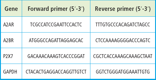
Total RNA was isolated from cultures using TRIzol reagent based on the manufacturer's protocol. RNA (1 μg) was reverse transcribed with a High Capacity cDNA Reverse Transcription Kit (2.5 U/μL), with RNase Inhibitor 1 U/μL, Random Hexamers 2.5 U/μL, MgCl2 5 mM, PCR buffer II 1× and dNTPs 1 mM (Applied Biosystems, Foster City, CA, USA). Relative analysis of gene expression was performed via real-time RT-PCR on a Step One Plus with Power UP SYBR Green MasterMix (Applied Biosystems). The primers used are listed in table I. Gene expression levels were calculated with the ΔΔCt method.
RESULTS
THE PRESENCE OF ADHERED UNVIABLE S. AUREUS INDUCES AN INCREASE IN THE FORMATION OF FBGCS THAT IS ABROGATED IN THE PRESENCE OF METAL ALLOYS
To understand the role of S. aureus in the formation of FGBCs, we first tested the most suitable media for the growth of RAW264.7 cells and S. aureus. Figure 1A illustrates how RAW264.7 cells proliferated adequately when they were seeded in αMEM medium with 10 % FBS and without antibiotics, while RPMI media with 5 % FBS but without antibiotics and RPMI with 50 % BSA, were not adequate for RAW264.7 cells proliferation. Afterwards, we tested the number of RAW264.7 cells and the concentration and IL-4 necessary for the correct differentiation of FBGCs. Figure 1B illustrates that the best formation of FBGCs was achieved with 10,000 RAW264.7 cells and 20 ng/mL of IL-4. Finally, we tested whether the presence of S. aureus modified FBGCs formation. We observed that the presence of S. aureus induced an increase in the formation of FBGCs in a concentration-dependent manner (54 ± 6 % increase for 1.61 ± 0.22·107 CFU/mL, p < 0.0001).
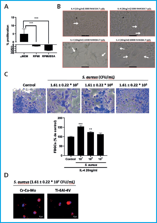
Figura 1. Adherent unviable S. aureus induces formation of FBGCs and presence of metal alloys inhibiting it. A. Optimal culture media condition determination. B. Determination of Raw264.7 cell number and IL-4 concentration optimal for culture. White arrows indicate the presence of giant cells. C. Panoptic staining representative images and quantification for FBGCs formation with adherent unviable S. aureus. D. Representative images for FBGCs formation with adherent unviable S. aureus in the presence of IL-4 and Ti-6Al-4V and Co-Cr-Mo alloys. All images were taken at 40x magnification. Data are presented as the mean ± SEM. ***p < 0.0001 and **p < 0.001 vs control. One-way ANOVA and Bonferroni post-hoc test.
Therefore, we decided to conduct all experiments in αMEM with 10 % FBS and without antibiotics, 10,000 RAW264.7 cells, 20 ng/mL of IL-4 and 1.61 ± 0.22·107 CFU/mL.
When RAW264.7 cells and S. aureus where co-cultured in the presence of IL-4 and the metal alloys, immunofluorescence staining with phalloidin demonstrated that both Ti-6Al-4V and Co-Cr-Mo alloys reduced the formation of FBGCs in the presence of 1.61 ± 0.22·107 CFU/mL S. aureus (Fig. 1D).
S. AUREUS INDUCES THE SECRETION OF ADENOSINE INTO THE EXTRACELLULAR MEDIUM WHILE METAL ALLOYS TI-6AL-4V AND CO-CR-MO INCREASE SUCH CONCENTRATION
The HPLC study of extracellular nucleotide concentrations reflected a significant increase in inosine in the presence of S. aureus on days 3 and 5 of differentiation with IL-4 20 ng/mL (p < 0.0001, n = 8), which were reduced significantly when co-culture was performed in the presence of Ti-6Al-4V alloy on days 3 and 5 of differentiation and in the presence of Co-Cr-Mo alloy at 5 days (p < 0.0001, n = 8) (Fig. 2A). Regarding extracellular concentrations of adenosine, we observed that no significant changes occurred in the presence of IL-4 20 ng/mL alone, or S. aureus on days 3 or 5 of differentiation. However, both Ti-6Al-4V and Co-Cr-Mo alloys induced a significant increase in extracellular adenosine levels vs both the basal and S. aureus at both time points (p < 0.0001 vs S. aureus, n = 8) (Fig. 2B). Extracellular levels of AMP increased significantly on day 5 of differentiation in all cases (p < 0.001, p < 0.0001, n = 8) without significant differences between groups (Fig. 2C). No significant changes in the extracellular concentrations of ADP or ATP were found (p = ns, n = 8) (Fig. 2 D-E).
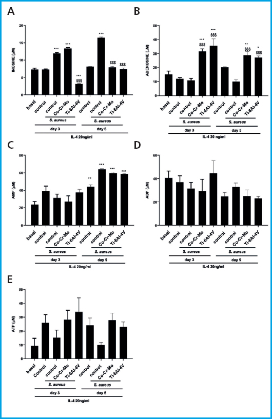
Figura 2. Nucleotide analysis via HPLC on days 3 and 5 of differentiation in the presence of adherent unviable S. aureus and metal alloys. A. Extracellular concentrations of inosine. B. Extracellular concentrations of adenosine. C. Extracellular concentrations of AMP. D. Extracellular concentrations of ADP. E. Extracellular concentrations of ATP. Data are presented as the mean ± SEM. ***p < 0.0001 vs basal; **p < 0.001 vs -basal; $$$p < 0.0001 vs S. aureus. One-way ANOVA and Bonferroni post-hoc test.
ADENOSINE A1 AND A2A RECEPTORS AND P2X7 RECEPTORS EXPRESSION IS MODULATED BY S. AUREUS AND THE PRESENCE OF METAL ALLOYS
Following the increase in extracellular adenosine levels, we then tried to understand whether purinergic receptors involved in fusogenic functions (adenosine A1 and A2A receptors and P2X7 receptors) are also involved in the formation of FBGCs in the presence of S. aureus and metal alloys. The expression of mRNA for adenosine A1 receptor in RAW264.7 cells increased at 3 and 5 days in the presence of 20 ng/mL IL-4 and was enhanced on day 5 of differentiation in the presence of S. aureus although this increase was not significant (p = ns, n = 3-4) (Fig. 3A). Although the Ti-6Al-4V alloy increased A1 receptor mRNA expression, this increase was significant on day 5 of differentiation (11120.6 ± 6630 fold change S. aureus + Ti-6Al-4V vs 11.7 ± 8.073 fold change S. aureus, p < 0.05, n = 3-4) (Fig. 3A). A similar trend was seen for the Cr-Co-Mo alloy, although in this case, the increased expression was not significant (p = ns, n = 3-4) (Fig. 3A).
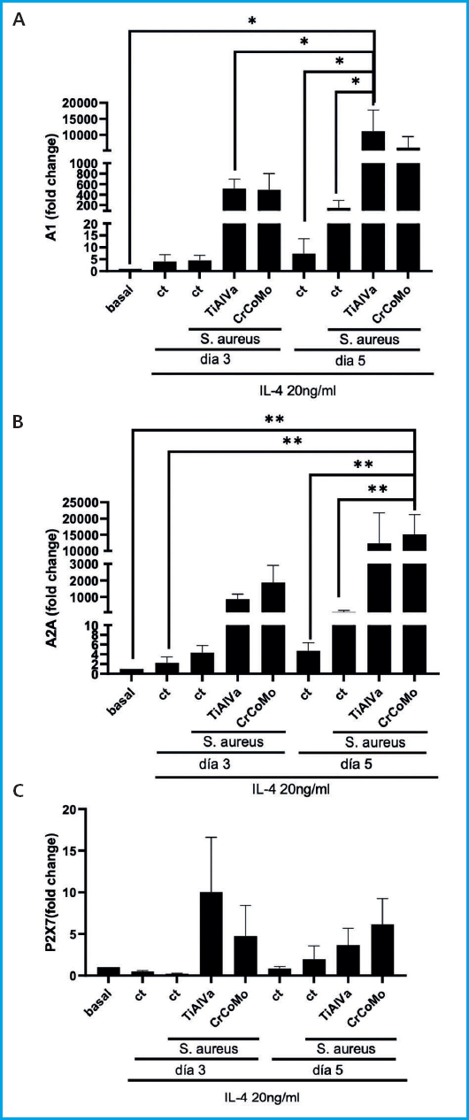
Figura 3. Purinergic receptor mRNA expression on days 3 and 5 of differentiation in the presence ofadherent unviable S. aureus and metal alloys. A. mRNA expression for adenosine A1 receptor in the presence of adherent unviable S. aureus and Ti-6Al-4V and Co-Cr-Mo alloys. B. mRNA expression for adenosine A2A receptor in the presence of adherent unviable S. aureus and Ti-6Al-4V and Co-Cr-Mo alloys. C. mRNA expression for P2X7 receptor in the presence of adherent unviable S. aureus and Ti-6Al-4V and Co-Cr-Mo alloys. Data are presented as the mean ± SEM. *p < 0.05. One-way ANOVA and Bonferroni post-hoc test.
A similar tendency was observed for adenosine A2A receptor in RAW264.7 cells. A2A receptor mRNA expression increased at 3 and 5 days in the presence of 20 ng/mL IL-4 and was enhanced on day 5 of differentiation in the presence of S. aureus although this increase was not significant (p = ns, n = 3-4) (Fig. 3B). Although the Ti-6Al-4V alloy increased A2A receptor mRNA expression, this increase was not significant (p = ns, n = 3-4). Furthermore, the Co-Cr-Mo alloy significantly increased A2A receptor mRNA expression on day 5 of differentiation (15114 ± 6082-fold change S. aureus + Co-Cr-Mo vs 8.17 ± 2.95-fold change S. aureus, p < 0.05, n = 3-4) (Fig. 3B).
The expression of the P2X7 ATP receptor does not increase significantly in the presence of IL-4 20 ng/mL, both on days 3 and 5 of differentiation. However, it does increase in the presence of S. aureus, with this increase being further enhanced by both metal alloys. Nevertheless, this increase was not found to be statically significant (p = ns, n = 3-4) (Fig. 3C).
ADENOSINE A2A RECEPTOR ACTIVATION INHIBITS S. AUREUS-INDUCED FBGC FORMATION
Incubation of RAW264.7 cells with the adenosine A2A receptor agonist CGS21680 1 μM, inhibited FBGCs formation induced by IL-4 20 ng/mL (reduction of 37.89 ± 1.97 % vs 100 % FBGCs in IL-4, p < 0.0001, n = 6), being reversed in the presence of the A2A receptor antagonist ZM241385 1 μM (Fig. 4). In the presence of S. aureus, there was an increased in FBGCs formation (increase of 70.67 ± 2.33 % vs 100 % FBGCs in IL-4, p < 0.0001, n = 6) as described above that was significantly reduced in the presence of CGS21680 1 μM (decrease of 43.17 ± 3.7 % vs IL-4 and S. aureus, p < 0.0001, n = 6) which is also reversed in the presence of ZM241385 1 μM (Fig. 4).
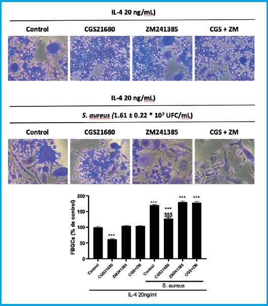
Figura 4. The formation of FBGCs in the presence of S. aureus is decreased by the adenosine A2A receptor agonist CGS21680. Panoptic staining representative images and quantification for FBGCs formation. RAW264.7 cells treated with IL-4 20 ng/mL and adenosine A2A receptor agonist CGS21680 1 μM and antagonist ZM241385 1 μM alone or in the presence of adherent unviable S. aureus. All images were taken at 40x magnification. Data are presented as the mean ± SEM. ***p < 0.0001 vs control; $$$p < 0.0001 vs S. aureus. One-way ANOVA and Bonferroni post-hoc test.
S. AUREUS AND METAL ALLOYS SLIGHTLY CHANGE THE CYTOKINES PROLIFE SECRETION OF RAW264.7 CELLS
Afterwards, we tried to identify changes in cytokine secretion due to presence of S. aureus and metal alloys. For this purpose, we performed a cytokine array that measures 20 cytokines and chemokines (Table II). When we analyzed the expression of pro-inflammatory cytokines, we found that the presence of S. aureus only increased the expression of IL-6 and TNFα vs both with basal conditions and control conditions in the presence of IL-4 20 ng/mL (137.2 ± 23.12 pg/mL S. aureus vs 2.28 ± 1.23 pg/mL IL-4 for IL-6; and 781.1 ± 289.9 pg/mL S. aureus vs 7.051 ± 3.287 pg/mL IL-4 for TNFα, p < 0.0001 and p < 0.05 respectively, n = 5-6) (Table II). When RAW264.7 cells were differentiated in the presence of IL-4 20 ng/mL and the Co-Cr-Mo alloy, no differences in pro-inflammatory cytokines were observed vs control conditions in the presence of IL-4 20 ng/mL (Table II). However, when RAW264.7 cells were differentiated in the presence of S. aureus and the Co-Cr-Mo alloy, we observed a significant increase in IL-1α, IL-1β and IL-6 expression vs all conditions (336.6 ± 139.5 pg/mL S. aureus + Co-Cr-Mo vs 22.98 ± 10.5 pg/mL S. aureus for IL-1α; 114.5 ± 23.21 pg/mL S. aureus + Co-Cr-Mo vs 2.636 ± 1.895 pg/mL S. aureus for IL-1β; and 925.9 ± 283.621 pg/mL S. aureus + Co-Cr-Mo vs 137.2 ± 23.12 pg/mL S. aureus for IL-6, p < 0.05, p < 0.0001 and p < 0.0001 respectively, n = 5-6) (Table II). In a similar way, when RAW264.7 cells were differentiated in the presence of IL-4 20 ng/mL and the Ti-6Al-4V alloy, no differences in pro-inflammatory cytokines were observed vs control conditions in the presence of IL-4 20 ng/mL (Table II). Moreover, when RAW264.7 cells were differentiated in the presence of S. aureus and the Ti-6Al-4V alloy, we observed a significant increase in IL-1β and IL-6 expression when vs all conditions (209.5 ± 52,88 pg/mL S. aureus + Ti-6Al-4V vs 2.636 ± 1.895 pg/mL S. aureus for IL-1β; and 3366 ± 818.3 pg/mL S. aureus + Ti-6Al-4V vs 137.2 ± 23.12 pg/mL S. aureus for IL-6, p < 0.0001 and p < 0.0001 respectively, n = 5-6) (Table II).
Table II. Inflammatory cytokines level measured using the mouse cytokine array Q1.
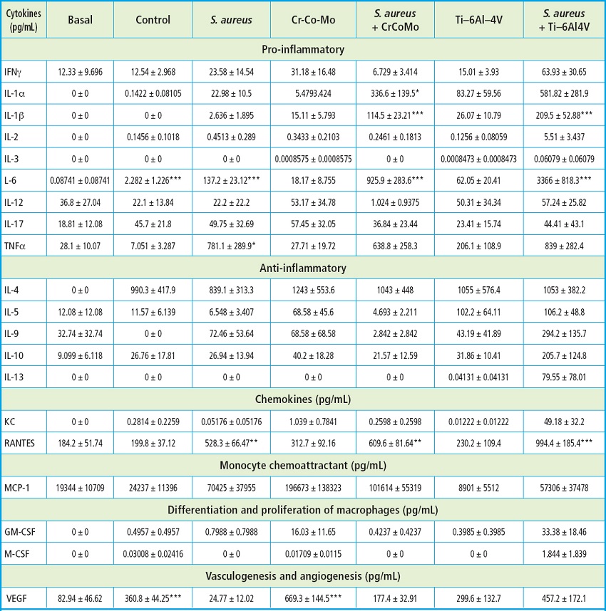
Cytokines and chemokines in pg/mL. Data are express as mean ± SEM. *p < 0.05, **p < 0.001, and ***p < 0.0001. One-way ANOVA and Bonferroni post-hoc test.
No changes were observed among conditions in any anti-inflammatory cytokine (Table II).
RANTES, a classical chemotactic cytokine also known as CCL5, was increased in the presence of S. aureus (528.3 ± 66.47 pg/mL S. aureus vs 199.8 ± 37.12 pg/mL IL-4, p < 0.001, n = 5-6) and enhanced when co-culture was performed in the presence of metal alloys (609.6 ± 81.64 pg/mL S. aureus + Co-Cr-Mo vs 528.3 ± 66.47 pg/mL S. aureus and 994.4 ± 185,4 pg/mL S. aureus + Ti-6Al-4V vs 528,3 ± 66,47 pg/mL S. aureus, p < 0.001 and p < 0.0001 respectively, n = 5-6) (Table II).
Finally, we found that the presence of IL-4 20 ng/mL induced an increased expression of VEGF (360.8 ± 44.25 pg/mL IL4 vs 82.94 ± 46.62 pg/mL basal, p < 0.0001, n = 5-6) that was not significantly reduced in the presence of S. aureus but was significantly increased in the presence of both IL-4 and the Co-Cr-Mo alloy (669.3 ± 144.5 pg/mL Co-Cr-Mo vs 82.94 ± 46,62 pg/mL basal, p < 0.0001, n = 5-6) (Table II).
DISCUSSION
In this manuscript we have demonstrated that the presence of Ti-6Al-4V and Co-Cr-Mo alloys required purinergic system activation in macrophages to inhibit FBGCs formation induced by adhered unviable S. aureus.
As we can conclude from our data, adhered unviable S. aureus can form FBGCs although non-infectious activity. Although the use of non-viable S. aureus represents a limitation of our study, it is worth noting that during S. aureus biofilm formation bacterial lysis plays a key role (24), so this study may reveal what role these non-viable cells play in the development of FBGCs. S. aureus can attach to biotic surfaces through electrostatic and hydrophobic interactions under static conditions, even if it is not viable (25). These adhered unviable staphylococci which are not forming a biofilm will be detected by the Toll-like receptors and induce the phagocytic cell activation (26). Macrophage fusion to form a FBGC occurs in response to tissue injury and the presence of any biomaterial (7), whose physical features (ie, substrate stiffness, topography, and surface chemistry) will determine such response. Interestingly, this foreign body response is also regulated by Toll-like receptors (27). Our results illustrate that unviable S. aureus can induce the formation of FBGCs. Furthermore, it is plausible that the presence of metal alloys Ti-6Al-4V and Co-Cr-Mo would be necessary for the inhibition of that formation of FBGCs even in the presence of unviable S. aureus.
Fibrotic encapsulation emerged as a crucial tissue response to foreign objects inadvertently becoming lodged in the body. The immune capacity to successfully block access to the rest of the body by fibrotic encapsulation is pivotal since these foreign items pose numerous hazards, including the potential to spread infection (28). If fibrotic encapsulation were to take place on the S. aureus-infected implant, the infection foci would have access to fewer nutrients from the periprosthetic tissues. Hence, the inhibition of the development of FBGCs by those metal alloys may inhibit the fibrous encapsulation of these staphylococci-covered alloys (29), thus favoring the maintenance of the staphylococcal infection which would not have any fibrotic capsule impairing the arrival of nutrients into their immediate environment.
HPLC analysis showed increased adenosine extracellular levels induced by metal alloys that were not exert neither by IL-4 alone nor in combination with adhered unviable S. aureus. This adenosine increased correlates with the expression of adenosine receptors showed. Although both metal alloys increased in a similar manner adenosine levels, and both have the same trend in adenosine A1 and A2A receptors mRNA increased, Ti-6Al-4V only significantly increased mRNA levels for adenosine A1 receptor meanwhile Co-Cr-Mo only significantly induced adenosine A2A receptor. None of the conditions induce P2X7 changes, in correlation with no changes of ATP levels. Considering that both FBGCs and osteoclasts share some mechanism during their formation process (10), it is plausible that adenosine receptors have a similar role in both FBGCS and osteoclast formation. It has been extensively demonstrated that adenosine A2A receptor inhibits osteoclast formation (14,15). The increase in this receptor together with the rise in adenosine levels induced by metal alloys indicate that similar role might be award to this receptor in FGBCs formation, primary in the presence of Co-Cr-Mo alloys.
On the other hand, it has been demonstrated that adenosine A1 receptors are involved in fusion of human peripheral blood monocytes into giant cells although ith as not been fully established (30). It is known that adenosine A1 receptor induces osteoclast differentiation (13), and blockade or deletion of the receptor suppresses osteoclast differentiation in vitro and in vivo (31). Furthermore, it has been demonstrated that adenosine A1 receptor on both primary murine and human osteoclast precursors is constitutively active, with selective antagonists working as an inverse agonists at A1 receptor (32), which might be the reason why changes in adenosine A1 receptor mRNA expression in IL-4 and S. aureus alone groups vs basal conditions are not significant.
Although the increase in A1 receptor in the presence of metal alloys needs to be address better, such an increase might be a compensatory mechanism induced by S. aureus as it has been observed that A1 receptor stimulation is essential for the formation of giant cells in vitro in the syncytia in giant cell arteritis, a characteristic where inflammation is crucial (33). Our data indicate that the presence of metal alloys induces a significant increase in IL-1β and IL-6, 2 key pro-inflammatory cytokines. This increase may be involved in the rise of the adenosine A1 receptor. However, the inflammatory role of adenosine A1 receptor is controversial. As it has been observed in some scenarios, A1 receptor can induce anti-inflammatory effects, including renal ischemia reperfusion, lungs (where the absence of adenosine A1 receptor induce leukocyte migration and increased cytokines), and sepsis where A1KO mice exhibit a higher degree of renal dysfunction and higher release of pro-inflammatory cytokines (34). These data indicate that the presence of endogenous A1 receptor is necessary to protect vs exacerbation of the disease and organ dysfunction. It is plausible that same as it occurs in sepsis, the imbalance in inflammation induced by S. aureus promotes the activation of A1 receptor by metal alloys in our system to counteract the hyper-inflammatory scenario, being in this situation not only the adenosine A2A receptor—the only anti-inflammatory receptor—but also the A1 receptor. Therefore, more data are needed to try to understand metal alloys induced A1 receptor expression in this system.
Finally, one of the limitations of this study was the impossibility of using live S. aureus bacteria in our assays. Although different bacterial infection assays were performed (data not shown), the results obtained were erratic and the amount of FBGCs remaining adhered after infection was too small to perform the assays conducted in this study. It would be convenient to conduct experiments with viable bacteria, with validation of the appropriate moments of infection, co-culture methods (adherence or non-adherence), etc.
In conclusion, the increased levels of adenosine would be responsible for the inhibition of cell fusion and the following reduction of S. aureus-induced FBGCs. Also relevant is the activation of the adenosine A2A receptor induced by the presence of Co-Cr-Mo alloys.

